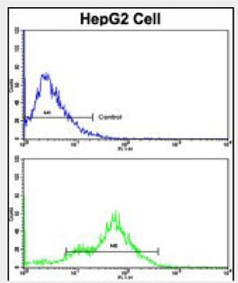
 FCM/FACS (Flow Cytometry)
(Flow cytometric analysis of HepG2 cells using CEACAM Antibody (N-term)(bottom histogram) compared to a negative control cell (top histogram). FITC-conjugated goat-anti-rabbit secondary antibodies were used for the analysis.)
FCM/FACS (Flow Cytometry)
(Flow cytometric analysis of HepG2 cells using CEACAM Antibody (N-term)(bottom histogram) compared to a negative control cell (top histogram). FITC-conjugated goat-anti-rabbit secondary antibodies were used for the analysis.)
Rabbit anti-Human CEACAM Polyclonal Antibody | anti-CEACAM1 antibody
CEACAM Antibody (N-term)
FCM/FACS (Flow Cytometry)
(Flow cytometric analysis of HepG2 cells using CEACAM Antibody (N-term)(bottom histogram) compared to a negative control cell (top histogram). FITC-conjugated goat-anti-rabbit secondary antibodies were used for the analysis.)
FCM/FACS (Flow Cytometry)
(Flow cytometric analysis of HepG2 cells using CEACAM Antibody (N-term)(bottom histogram) compared to a negative control cell (top histogram). FITC-conjugated goat-anti-rabbit secondary antibodies were used for the analysis.)
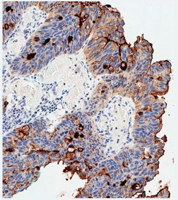
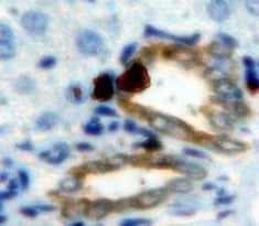
IHC (Immunohistochemisry)
(Immunohistochemical analysis of paraffin-embedded Human colon carcinoma tissue using AP7364a performed on the Leica® BOND RXm. Tissue was fixed with formaldehyde at room temperature, antigen retrieval was by heat mediation with a EDTA buffer (pH9. 0). Samples were incubated with primary antibody(1:500) for 1 hours at room temperature. A undiluted biotinylated CRF Anti-Polyvalent HRP Polymer antibody was used as the secondary antibody.)
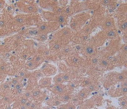
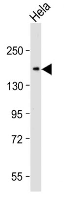
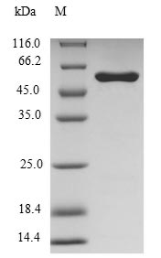
WB (Western Blot)
(Anti-CEACAM Antibody (N-term) at 1:2000 dilution + Hela whole cell lysate Lysates/proteins at 20 ug per lane.Secondary Goat Anti-Rabbit IgG, (H+L), Peroxidase conjugated at 1/10000 dilution.Predicted band size : 58 kDaBlocking/Dilution buffer: 5% NFDM/TBST.)
WB (Western Blot)
(Anti-CEACAM Antibody (N-term) at 1:2000 dilution + Hela whole cell lysate Lysates/proteins at 20 ug per lane.Secondary Goat Anti-Rabbit IgG, (H+L), Peroxidase conjugated at 1/10000 dilution.Predicted band size : 58 kDaBlocking/Dilution buffer: 5% NFDM/TBST.)
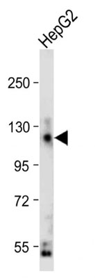
WB (Western Blot)
(Anti-CEACAM Antibody (N-term) at 1:2000 dilution + HepG2 whole cell lysate Lysates/proteins at 20 ug per lane.Secondary Goat Anti-Rabbit IgG, (H+L), Peroxidase conjugated at 1/10000 dilution.Predicted band size : 58 kDaBlocking/Dilution buffer: 5% NFDM/TBST.)
WB (Western Blot)
(Anti-CEACAM Antibody (N-term) at 1:2000 dilution + HepG2 whole cell lysate Lysates/proteins at 20 ug per lane.Secondary Goat Anti-Rabbit IgG, (H+L), Peroxidase conjugated at 1/10000 dilution.Predicted band size : 58 kDaBlocking/Dilution buffer: 5% NFDM/TBST.)
Li,C., Exp. Cell Res. 315 (7), 1225-1233 (2009)
Nouvion,A.L., Med Sci (Paris) 25 (3), 247-252 (2009)
NCBI and Uniprot Product Information
Customer Reviews
Loading reviews...
Share Your Experience
Similar Products


Product Notes
The CEACAM1 ceacam1 (Catalog #AAA285413) is an Antibody produced from Rabbit and is intended for research purposes only. The product is available for immediate purchase. The immunogen sequence is 50-77. The CEACAM Antibody (N-term) reacts with Human and may cross-react with other species as described in the data sheet. AAA Biotech's CEACAM can be used in a range of immunoassay formats including, but not limited to, FCM/FACS (Flow Cytometry), IHC (Immunohistochemistry), ELISA, WB (Western Blot). Researchers should empirically determine the suitability of the CEACAM1 ceacam1 for an application not listed in the data sheet. Researchers commonly develop new applications and it is an integral, important part of the investigative research process. It is sometimes possible for the material contained within the vial of "CEACAM, Polyclonal Antibody" to become dispersed throughout the inside of the vial, particularly around the seal of said vial, during shipment and storage. We always suggest centrifuging these vials to consolidate all of the liquid away from the lid and to the bottom of the vial prior to opening. Please be advised that certain products may require dry ice for shipping and that, if this is the case, an additional dry ice fee may also be required.Precautions
All products in the AAA Biotech catalog are strictly for research-use only, and are absolutely not suitable for use in any sort of medical, therapeutic, prophylactic, in-vivo, or diagnostic capacity. By purchasing a product from AAA Biotech, you are explicitly certifying that said products will be properly tested and used in line with industry standard. AAA Biotech and its authorized distribution partners reserve the right to refuse to fulfill any order if we have any indication that a purchaser may be intending to use a product outside of our accepted criteria.Disclaimer
Though we do strive to guarantee the information represented in this datasheet, AAA Biotech cannot be held responsible for any oversights or imprecisions. AAA Biotech reserves the right to adjust any aspect of this datasheet at any time and without notice. It is the responsibility of the customer to inform AAA Biotech of any product performance issues observed or experienced within 30 days of receipt of said product. To see additional details on this or any of our other policies, please see our Terms & Conditions page.Item has been added to Shopping Cart
If you are ready to order, navigate to Shopping Cart and get ready to checkout.