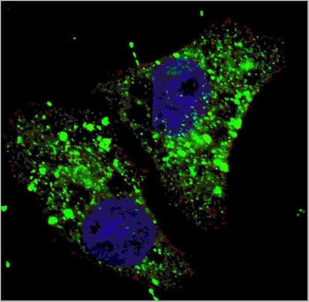
 IF (Immunofluorescence)
(Fluorescent image of U251 cells stained with cleaved LC3A antibody. U251 cells were treated with Chloroquine (50 uM,16h), then fixed with 4% PFA (20 min), permeabilized with Triton X-100 (0.2%, 30 min). Cells were then incubated with AAA28658 cleaved LC3A primary antibody (1:100, 2 h at room temperature). For secondary antibody, Alexa Fluor 488 conjugated donkey anti-rabbit antibody (green) was used (1:1000, 1h). Nuclei were counterstained with Hoechst 33342 (blue) (10 ug/ml, 5 min). LC3 immunoreactivity is localized to autophagic vacuoles in the cytoplasm of U251 cells.)
IF (Immunofluorescence)
(Fluorescent image of U251 cells stained with cleaved LC3A antibody. U251 cells were treated with Chloroquine (50 uM,16h), then fixed with 4% PFA (20 min), permeabilized with Triton X-100 (0.2%, 30 min). Cells were then incubated with AAA28658 cleaved LC3A primary antibody (1:100, 2 h at room temperature). For secondary antibody, Alexa Fluor 488 conjugated donkey anti-rabbit antibody (green) was used (1:1000, 1h). Nuclei were counterstained with Hoechst 33342 (blue) (10 ug/ml, 5 min). LC3 immunoreactivity is localized to autophagic vacuoles in the cytoplasm of U251 cells.)
Rabbit Cleaved LC3A Polyclonal Antibody | anti-MAP1LC3A antibody
Cleaved LC3A Antibody
IF (Immunofluorescence)
(Fluorescent image of U251 cells stained with cleaved LC3A antibody. U251 cells were treated with Chloroquine (50 uM,16h), then fixed with 4% PFA (20 min), permeabilized with Triton X-100 (0.2%, 30 min). Cells were then incubated with AAA28658 cleaved LC3A primary antibody (1:100, 2 h at room temperature). For secondary antibody, Alexa Fluor 488 conjugated donkey anti-rabbit antibody (green) was used (1:1000, 1h). Nuclei were counterstained with Hoechst 33342 (blue) (10 ug/ml, 5 min). LC3 immunoreactivity is localized to autophagic vacuoles in the cytoplasm of U251 cells.)
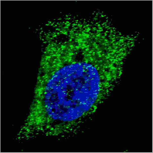
IF (Immunofluorescence)
(Fluorescent image of U251 cells stained with cleaved LC3A antibody. U251 cells were fixed with 4% PFA (20 min), permeabilized with Triton X-100 (0.2%, 30 min). Cells were then incubated with AAA28658 cleaved LC3A primary antibody (1:100, 2 h at room temperature). For secondary antibody, Alexa Fluor 488 conjugated donkey anti-rabbit antibody (green) was used (1:1000, 1h). Nuclei were counterstained with Hoechst 33342 (blue) (10 ug/ml, 5 min). LC3 immunoreactivity is localized to autophagic vacuoles in the cytoplasm of U251 cells.)
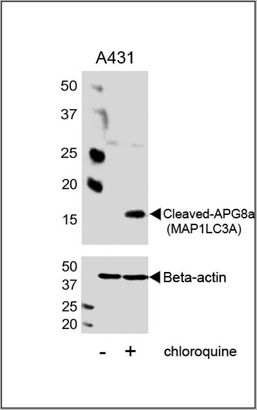
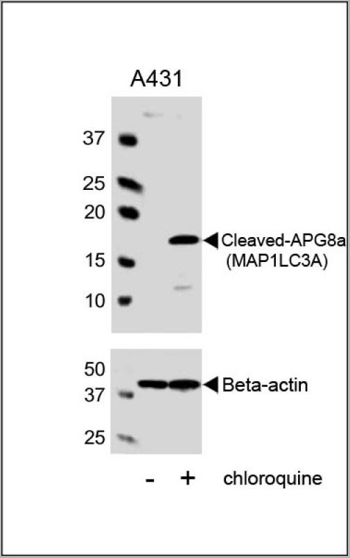
WB (Western Blot)
(Western blot analysis of lysates from A431 cell line, untreated or treated with chloroquine, 100ng/ml, using Cleaved-APG8a (MAP1LC3A) (upper) or Beta-actin (lower).)
WB (Western Blot)
(Western blot analysis of lysates from A431 cell line, untreated or treated with chloroquine, 100ng/ml, using Cleaved-APG8a (MAP1LC3A) (upper) or Beta-actin (lower).)
WB (Western Blot)
(Western blot analysis of lysates from A431 cell line, untreated or treated with chloroquine, 100ng/ml, using Cleaved-APG8a (MAP1LC3A) (upper) or Beta-actin (lower).)
WB (Western Blot)
(Western blot analysis of lysates from A431 cell line, untreated or treated with chloroquine, 100ng/ml, using Cleaved-APG8a (MAP1LC3A) (upper) or Beta-actin (lower).)
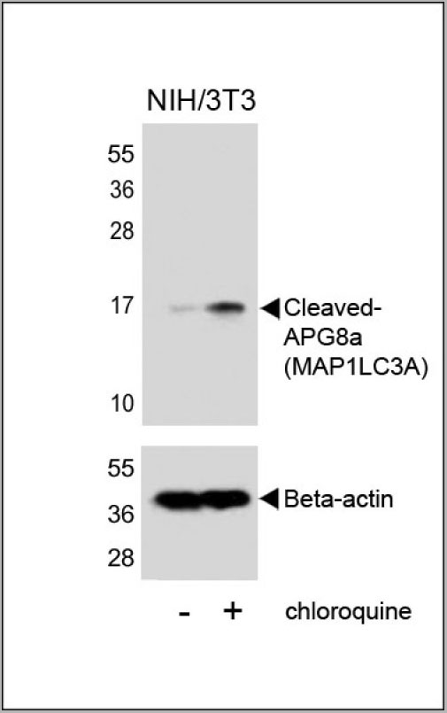
WB (Western Blot)
(Western blot analysis of lysates from NIH/3T3 cells, untreated or treated with chloroquine, using Cleaved-APG8a (MAP1LC3A) (upper) or Beta-actin (lower).)
WB (Western Blot)
(Western blot analysis of lysates from NIH/3T3 cells, untreated or treated with chloroquine, using Cleaved-APG8a (MAP1LC3A) (upper) or Beta-actin (lower).)
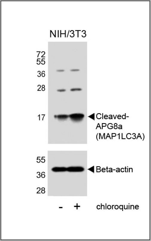
WB (Western Blot)
(Western blot analysis of lysates from NIH/3T3 cells, untreated or treated with chloroquine, using Cleaved-APG8a (MAP1LC3A)(RB52604)(upper) or Beta-actin (lower).)
WB (Western Blot)
(Western blot analysis of lysates from NIH/3T3 cells, untreated or treated with chloroquine, using Cleaved-APG8a (MAP1LC3A)(RB52604)(upper) or Beta-actin (lower).)
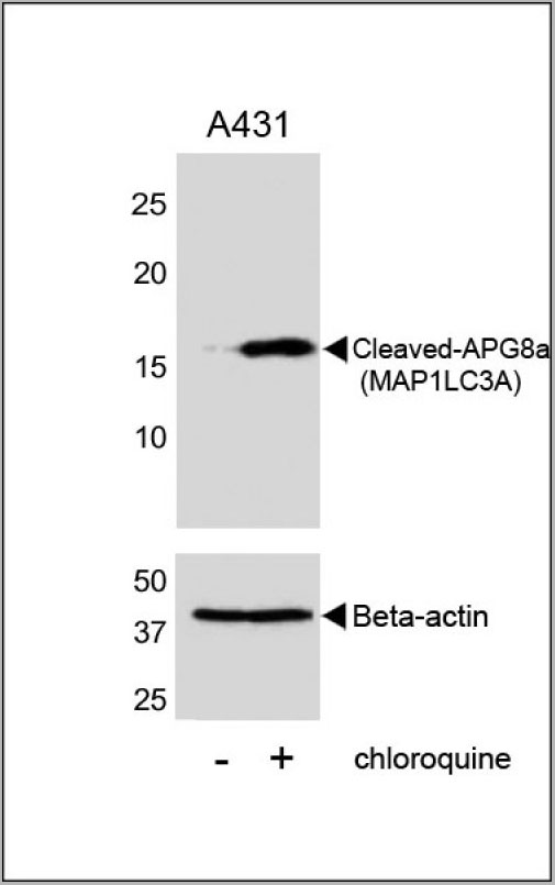
WB (Western Blot)
(Western blot analysis of lysates from A431 cell line, untreated or treated with chloroquine, 100ng/ml, using Cleaved-APG8a (MAP1LC3A) Antibody (upper) or Beta-actin (lower).)
WB (Western Blot)
(Western blot analysis of lysates from A431 cell line, untreated or treated with chloroquine, 100ng/ml, using Cleaved-APG8a (MAP1LC3A) Antibody (upper) or Beta-actin (lower).)
1.Baehrecke EH. Nat Rev Mol Cell Biol. 6(6):505-10. (2005)
2.Lum JJ, et al. Nat Rev Mol Cell Biol. 6(6):439-48. (2005)
3.Greenberg JT. Dev Cell. 8(6):799-801. (2005)
4. Levine B. Cell. 120(2):159-62. (2005)
5.Shintani T and Klionsky DJ. Science. 306(5698):990-5. (2004)
6.Tanida I., et al. Int. J. Biochem. Cell Biol. 36:2503-2518(2004)
7.He H., et al. J. Biol. Chem. 278:29278-29287(2003)
8.Tanida I., et al. J. Biol. Chem. 279:36268-36276(2004)
References for U251 cell line:
1. Westermark B.; Pontén J.; Hugosson R. (1973)." Determinants for the establishment of permanent tissue culture lines from human gliomas". Acta Pathol Microbiol Scand A. 81:791-805. [PMID: 4359449].
2. Pontén, J.,Westermark B. (1978)." Properties of Human Malignant Glioma Cells in Vitro". Medical Biology 56: 184-193.[PMID: 359950].
3. Geng Y.;Kohli L.; Klocke B.J.; Roth K.A.(2010). "Chloroquine-induced autophagic vacuole accumulation and cell death in glioma cells is p53 independent". Neuro Oncol. 12(5): 473-481.[ PMID: 20406898].
NCBI and Uniprot Product Information
Similar Products


Product Notes
The MAP1LC3A map1lc3a (Catalog #AAA28658) is an Antibody produced from Rabbit and is intended for research purposes only. The product is available for immediate purchase. The immunogen sequence is 89-120. The Cleaved LC3A Antibody reacts with Human, mouse (Predicted Reactivity: Rat, Bovine) and may cross-react with other species as described in the data sheet. AAA Biotech's Cleaved LC3A can be used in a range of immunoassay formats including, but not limited to, Western Blot (WB), ELISA (EIA), Immunohistochemistry (IHC), Immunofluorescence (IF), Immunocytochemistry (ICC). WB~~1:1000. Researchers should empirically determine the suitability of the MAP1LC3A map1lc3a for an application not listed in the data sheet. Researchers commonly develop new applications and it is an integral, important part of the investigative research process. It is sometimes possible for the material contained within the vial of "Cleaved LC3A, Polyclonal Antibody" to become dispersed throughout the inside of the vial, particularly around the seal of said vial, during shipment and storage. We always suggest centrifuging these vials to consolidate all of the liquid away from the lid and to the bottom of the vial prior to opening. Please be advised that certain products may require dry ice for shipping and that, if this is the case, an additional dry ice fee may also be required.Precautions
All products in the AAA Biotech catalog are strictly for research-use only, and are absolutely not suitable for use in any sort of medical, therapeutic, prophylactic, in-vivo, or diagnostic capacity. By purchasing a product from AAA Biotech, you are explicitly certifying that said products will be properly tested and used in line with industry standard. AAA Biotech and its authorized distribution partners reserve the right to refuse to fulfill any order if we have any indication that a purchaser may be intending to use a product outside of our accepted criteria.Disclaimer
Though we do strive to guarantee the information represented in this datasheet, AAA Biotech cannot be held responsible for any oversights or imprecisions. AAA Biotech reserves the right to adjust any aspect of this datasheet at any time and without notice. It is the responsibility of the customer to inform AAA Biotech of any product performance issues observed or experienced within 30 days of receipt of said product. To see additional details on this or any of our other policies, please see our Terms & Conditions page.Item has been added to Shopping Cart
If you are ready to order, navigate to Shopping Cart and get ready to checkout.