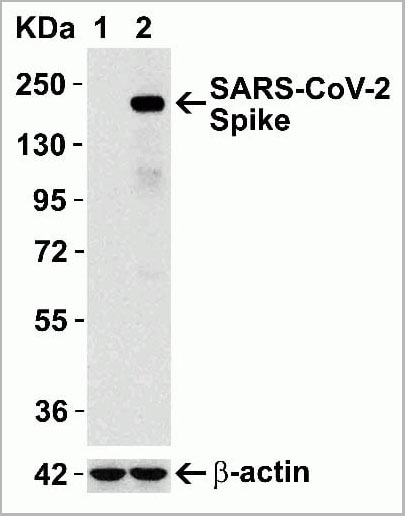


 WB (Western Blot)
(Figure 12 Overexpression Validation in Spike Transfected 293 Cells Loading: 15 ug per lane of 293 cell lysate. Antibodies: SARS-CoV-2 (COVID-19) Spike, AAA10931 (1 ug/mL), 1h incubation at RT in 5% NFDM/TBST. Secondary: Goat anti rabbit IgG HRP conjugate at 1:10000 dilution. Lane 1: WT293 cells and Lane 2: SARS-CoV-2 Spike overexpressed 293 cells.)
WB (Western Blot)
(Figure 12 Overexpression Validation in Spike Transfected 293 Cells Loading: 15 ug per lane of 293 cell lysate. Antibodies: SARS-CoV-2 (COVID-19) Spike, AAA10931 (1 ug/mL), 1h incubation at RT in 5% NFDM/TBST. Secondary: Goat anti rabbit IgG HRP conjugate at 1:10000 dilution. Lane 1: WT293 cells and Lane 2: SARS-CoV-2 Spike overexpressed 293 cells.)
Rabbit COVID 19 Spike Protein Coronavirus Polyclonal Antibody | anti-COVID-19 antibody
SARS-CoV-2 (COVID-19, 2019-nCoV) Spike Antibody
The immunogen is located within the last 50 amino acids of SARS-CoV-2 (COVID-19, 2019-nCoV) Spike protein.
WB (Western Blot)
(Figure 12 Overexpression Validation in Spike Transfected 293 Cells Loading: 15 ug per lane of 293 cell lysate. Antibodies: SARS-CoV-2 (COVID-19) Spike, AAA10931 (1 ug/mL), 1h incubation at RT in 5% NFDM/TBST. Secondary: Goat anti rabbit IgG HRP conjugate at 1:10000 dilution. Lane 1: WT293 cells and Lane 2: SARS-CoV-2 Spike overexpressed 293 cells.)
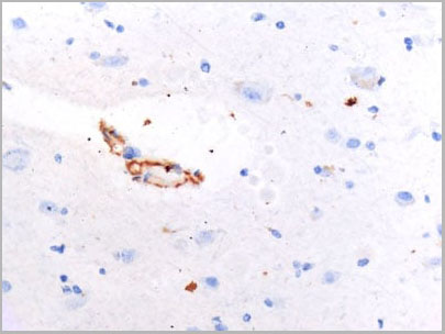
IHC (Immunohistochemistry)
(Figure 11 IHC Validation of SARS-CoV2 Spike in COVID-19 Patient Brain Spike protein was detected by anti-spike antibodies (AAA10931, 0.2 ug/mL) in the brain of the COVID-19 patient confirmed by PCR. (Courtesy of Dr. Nuovo Gerard J., OSU))
IHC (Immunohistochemistry)
(Figure 11 IHC Validation of SARS-CoV2 Spike in COVID-19 Patient Brain Spike protein was detected by anti-spike antibodies (AAA10931, 0.2 ug/mL) in the brain of the COVID-19 patient confirmed by PCR. (Courtesy of Dr. Nuovo Gerard J., OSU))
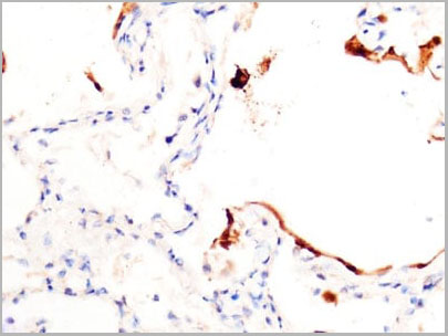
IHC (Immunohistochemistry)
(Figure 10 IHC Validation of SARS-CoV2 Spike in COVID-19 Patient Lung Strong spike signal was detected by anti-spike antibodies (AAA10931, 0.2 ug/mL) in the lung of the COVID-19 patient confirmed by PCR. (Courtesy of Dr. Nuovo Gerard J., OSU))
IHC (Immunohistochemistry)
(Figure 10 IHC Validation of SARS-CoV2 Spike in COVID-19 Patient Lung Strong spike signal was detected by anti-spike antibodies (AAA10931, 0.2 ug/mL) in the lung of the COVID-19 patient confirmed by PCR. (Courtesy of Dr. Nuovo Gerard J., OSU))
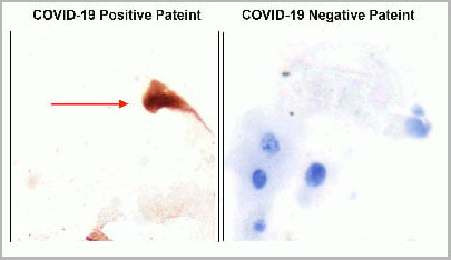
IHC (Immunohistchemistry)
(Figure 9 IHC Validation of SARS-CoV2 Spike in the Nasopharyngeal Swab Sample of the COVID-19 Patient Strong spike signal was detected by anti-spike antibodies (AAA10931, 0.2 ug/mL) in the nasopharyngeal swab sample of the COVID-19 patient and no spike signal was observed in the sample of the COVID-19 negative patient. COVID-19 cases were confirmed by PCR. (Courtesy of Dr. Nuovo Gerard J., OSU))
IF (Immunofluorescence)
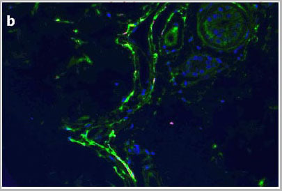
(Figure 8 IF Validation of SARS-CoV2 Spike in COVID-19 Patient Skin (Magro et al., 2020) C4d is highlighted green while COVID-19 spike protein detected by anti-spike antibodies (AAA10931, 0.2 ug/mL) shows a red staining pattern; a yellow signal is discernible indicative of co-localization of C4d and viral protein within the microvasculature.)
IF (Immunofluorescence)
(Figure 7 IF Validation of SARS-CoV2 Spike in COVID-19 Patient Lung (Magro et al., 2020) SARS-CoV2 spike protein (red, panel C) detected by anti-spike antibodies (AAA10931, 0.2 ug/mL) colocalized with C4d (green in panel d, merged in yellow). Spike protein (red, panel g) was also colocalized with C5b-9 (green in panel f&h, merged in yellow))
IHC (Immunohistchemistry)
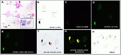
(Figure 6 IHC/IF Validation in COVID-19 Patient Sample (Nuovo et al., 2020) Detection of SARS-CoV-2 proteins in nasopharyngeal swab cell preparations. B. An intense signal for covid-19 spike protein tested by SARS-CoV-2 spike antibodies (AAA10931) was observed in the glandular cells. F-H. Co-expression of spike detected by spike antibodies (AAA10931, 0.2 ug/mL) and envelope proteins detected by envelope antibodies (, 2 ug/mL) of SARSCoV- 2 (F) documented localization of each protein to glandular cells (G, yellow). No signal was seen in oral swabs of positive cases (H). Both the spike and envelope protein detected by anti-spike antibodies (AAA10931) and anti-envelope antibodies (,) produced a signal in the nasopharyngeal swabs of the three cases and no signal was evident in the nasopharyngeal swabs of the seven controls.)
IHC (Immunohistchemistry)
(Figure 6 IHC/IF Validation in COVID-19 Patient Sample (Nuovo et al., 2020) Detection of SARS-CoV-2 proteins in nasopharyngeal swab cell preparations. B. An intense signal for covid-19 spike protein tested by SARS-CoV-2 spike antibodies (AAA10931) was observed in the glandular cells. F-H. Co-expression of spike detected by spike antibodies (AAA10931, 0.2 ug/mL) and envelope proteins detected by envelope antibodies (, 2 ug/mL) of SARSCoV- 2 (F) documented localization of each protein to glandular cells (G, yellow). No signal was seen in oral swabs of positive cases (H). Both the spike and envelope protein detected by anti-spike antibodies (AAA10931) and anti-envelope antibodies (,) produced a signal in the nasopharyngeal swabs of the three cases and no signal was evident in the nasopharyngeal swabs of the seven controls.)
IHC (Immunohistochemistry)
(Figure 5 Immunohistochemistry Validation of SARSCoV-2 (COVID-19) Spike in COVID-19 Patient Lung Immunohistochemical analysis of paraffin-embedded COVID-19 patient lung tissue using anti-SARS-CoV-2 (COVID-19) Spike S2 antibody (AAA10931, 0.5 ug/mL). Tissue was fixed with formaldehyde and blocked with 10% serum for 1 h at RT; antigen retrieval was by heat mediation with a citrate buffer (pH6). Samples were incubated with primary antibody overnight at 4°C. A goat anti-rabbit IgG H&L (HRP) at 1/250 was used as secondary. Counter stained with Hematoxylin. Strong spike protein signal wasobserved in macrophages of COVID-19 patient lung.)
IHC (Immunohistochemistry)
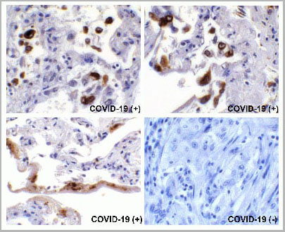
(Figure 4 Immunohistochemistry Validation of SARSCoV-2 (COVID-19) Spike in COVID-19 Patient Lung Immunohistochemical analysis of paraffin-embedded COVID-19 patient lung tissue using anti-SARS-CoV-2 (COVID-19) Spike S2 antibody (AAA10931, 0.5 ug/mL). Tissue was fixed with formaldehyde and blocked with 10% serum for 1 h at RT; antigen retrieval was by heat mediation with a citrate buffer (pH6). Samples were incubated with primary antibody overnight at 4°C. A goat anti-rabbit IgG H&L (HRP) at 1/250 was used as secondary. Counter stained with Hematoxylin. Strong spike protein signal was observed in macrophages and airway epithelium of COVID-19 patient lung, but not in non-COVID-19 patient lung.)
IHC (Immunohistochemistry)
(Figure 4 Immunohistochemistry Validation of SARSCoV-2 (COVID-19) Spike in COVID-19 Patient Lung Immunohistochemical analysis of paraffin-embedded COVID-19 patient lung tissue using anti-SARS-CoV-2 (COVID-19) Spike S2 antibody (AAA10931, 0.5 ug/mL). Tissue was fixed with formaldehyde and blocked with 10% serum for 1 h at RT; antigen retrieval was by heat mediation with a citrate buffer (pH6). Samples were incubated with primary antibody overnight at 4°C. A goat anti-rabbit IgG H&L (HRP) at 1/250 was used as secondary. Counter stained with Hematoxylin. Strong spike protein signal was observed in macrophages and airway epithelium of COVID-19 patient lung, but not in non-COVID-19 patient lung.)
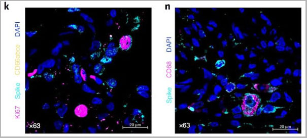
IF (Immunofluorescence)
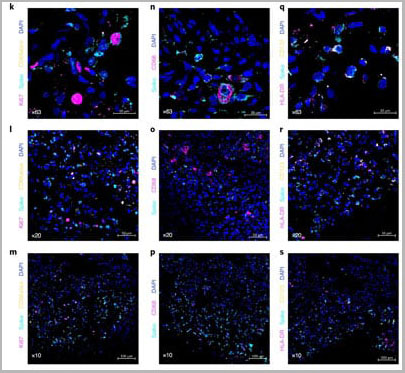
(Figure 3 Immunofluorescent Validation of AAA10931 in SARS-CoV-2 Infected Lung Tissue (Singh et al., Nature Microbiology, 2021) Multilabel confocal immunofluorescence microscopy of formalin-fixed paraffin-embedded lung sections from rhesus macaques infected with SARS-CoV-2. SARS-CoV-2 spike-specific antibodies, AAA10931. (k-s) (turquoise); Ki67 (magenta) and neutrophil marker CD66abce (yellow) (k-m); pan-macrophage marker CD68 (magenta) (n-p); HLA-DR (magenta) and pDC marker CD123 (yellow) (q–s) and DAPI (blue).)
IF (Immunofluorescence)
(Figure 3 Immunofluorescent Validation of AAA10931 in SARS-CoV-2 Infected Lung Tissue (Singh et al., Nature Microbiology, 2021) Multilabel confocal immunofluorescence microscopy of formalin-fixed paraffin-embedded lung sections from rhesus macaques infected with SARS-CoV-2. SARS-CoV-2 spike-specific antibodies, AAA10931. (k-s) (turquoise); Ki67 (magenta) and neutrophil marker CD66abce (yellow) (k-m); pan-macrophage marker CD68 (magenta) (n-p); HLA-DR (magenta) and pDC marker CD123 (yellow) (q–s) and DAPI (blue).)
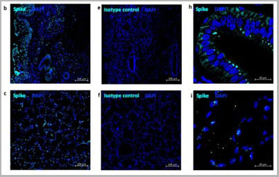
IF (Immunofluorescence)
(Figure 2 Immunofluorescent Validation of AAA10931 in SARS-CoV-2 Infected Nose and Tonsil (Singh et al., Nature Microbiology, 2021) Multi-label confocal immunofluorescence microscopy of nasal epithelium (20X-b, 63xh) and tonsil (20X-c,63X-i) from rhesus macaques infected with SARS-CoV-2 with SARS-CoV-2 spike-specific antibodies, AAA10931. (turquoise), DAPI (blue). Rabbit IgG isotype control antibody was used to stain the tissues to rule out any nonspecific staining (e, f).)
IF (Immunofluorescence)
(Firure 1. Immunofluorescent Validation of AAA10931 in SARS-CoV-2 Infected Lung Tissue (Singh et al., Nature Microbiology, 2021)Multilabel confocal immunofluorescence microscopy of formalin-fixed paraffin-embedded lung sections from rhesus macaques infected with SARS-CoV-2. SARS-CoV-2 spike-specific antibodies, AAA10931. (k, n) (turquoise); Ki67 (magenta) and neutrophil marker CD66abce (yellow) (k); pan-macrophage marker CD68 (magenta) (n) and DAPI (blue).)
2) Hui et al. Int J Infect Dis. 2020;91:264-266.
3) Belouzard et al. Viruses. 2012;4(6):1011-33.
4) Lee et al. J Virol. 2006;80(8):4079-87.
5) Wan et al. J Virol. 2020.
6) Wrapp et al. Science. 2020.
NCBI and Uniprot Product Information
Similar Products


Product Notes
The COVID-19 (Catalog #AAA10931) is an Antibody produced from Rabbit and is intended for research purposes only. The product is available for immediate purchase. The SARS-CoV-2 (COVID-19, 2019-nCoV) Spike Antibody reacts with Virus and may cross-react with other species as described in the data sheet. AAA Biotech's COVID 19 Spike Protein Coronavirus can be used in a range of immunoassay formats including, but not limited to, ELISA, Immunofluorescence (IF), Immunohistochemistry (IHC), Western Blot (WB). Optimal dilutions for each application to be determined by the researcher. Researchers should empirically determine the suitability of the COVID-19 for an application not listed in the data sheet. Researchers commonly develop new applications and it is an integral, important part of the investigative research process. It is sometimes possible for the material contained within the vial of "COVID 19 Spike Protein Coronavirus, Polyclonal Antibody" to become dispersed throughout the inside of the vial, particularly around the seal of said vial, during shipment and storage. We always suggest centrifuging these vials to consolidate all of the liquid away from the lid and to the bottom of the vial prior to opening. Please be advised that certain products may require dry ice for shipping and that, if this is the case, an additional dry ice fee may also be required.Precautions
All products in the AAA Biotech catalog are strictly for research-use only, and are absolutely not suitable for use in any sort of medical, therapeutic, prophylactic, in-vivo, or diagnostic capacity. By purchasing a product from AAA Biotech, you are explicitly certifying that said products will be properly tested and used in line with industry standard. AAA Biotech and its authorized distribution partners reserve the right to refuse to fulfill any order if we have any indication that a purchaser may be intending to use a product outside of our accepted criteria.Disclaimer
Though we do strive to guarantee the information represented in this datasheet, AAA Biotech cannot be held responsible for any oversights or imprecisions. AAA Biotech reserves the right to adjust any aspect of this datasheet at any time and without notice. It is the responsibility of the customer to inform AAA Biotech of any product performance issues observed or experienced within 30 days of receipt of said product. To see additional details on this or any of our other policies, please see our Terms & Conditions page.Item has been added to Shopping Cart
If you are ready to order, navigate to Shopping Cart and get ready to checkout.