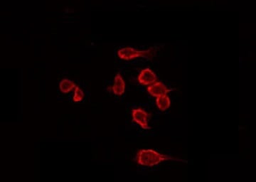
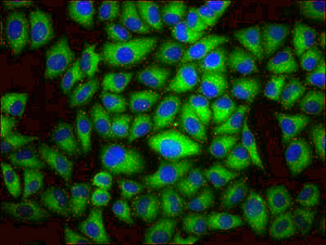
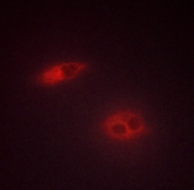
 IF (Immunofluorescence)
(AAA320797 staining Hela by IF/ICC. The sample were fixed with PFA and permeabilized in 0.1% Triton X-100, then blocked in 10% serum for 45 minutes at 25 degree C. The primary antibody was diluted at 1/200 and incubated with the sample for 1 hour at 37 degree C. An Alexa Fluor 594 conjugated goat anti-rabbit IgG (H+L) Ab, diluted at 1/600, was used as the secondary antibody.)
IF (Immunofluorescence)
(AAA320797 staining Hela by IF/ICC. The sample were fixed with PFA and permeabilized in 0.1% Triton X-100, then blocked in 10% serum for 45 minutes at 25 degree C. The primary antibody was diluted at 1/200 and incubated with the sample for 1 hour at 37 degree C. An Alexa Fluor 594 conjugated goat anti-rabbit IgG (H+L) Ab, diluted at 1/600, was used as the secondary antibody.)
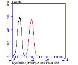
Rabbit anti-Human, Mouse Dysferlin Polyclonal Antibody | anti-DYSF antibody
Dysferlin Antibody
Gene Names
DYSF; MMD1; FER1L1; LGMD2B; LGMDR2
Reactivity
Human, Mouse
Applications
ELISA, Immunocytochemistry, Immunofluorescence, Western Blot
Purity
The antiserum was purified by peptide affinity chromatography using SulfoLink Coupling Resin.
Synonyms
Dysferlin, Antibody; Dysferlin Antibody; DMAT; DYSF; DYSF_HUMAN; Dysferlin; Dysferlin limb girdle muscular dystrophy 2B (autosomal recessive); Dysferlin limb girdle muscular dystrophy 2B; Dystrophy associated fer 1 like 1; Dystrophy associated fer 1 like protein; Dystrophy associated fer1 like 1; Dystrophy associated fer1 like protein; Dystrophy-associated fer-1-like protein; Fer 1 like protein 1; Fer-1-like protein 1; Fer1 like protein 1; FER1L1; FLJ00175; FLJ90168; LGMD 2B; LGMD2B; Limb girdle muscular dystrophy 2B (autosomal recessive); Limb girdle muscular dystrophy 2B; Miyoshi myopathy; MM; MMD1; anti-DYSF antibody
Host
Rabbit
Reactivity
Human, Mouse
Clonality
Polyclonal
Isotype
IgG
Specificity
Dysferlin antibody detects endogenous levels of total Dysferlin
Purity/Purification
The antiserum was purified by peptide affinity chromatography using SulfoLink Coupling Resin.
Form/Format
Liquid
Phosphate buffered saline, pH 7.4, 150mM NaCl, 0.02% sodium azide and 50% glycerol.
Phosphate buffered saline, pH 7.4, 150mM NaCl, 0.02% sodium azide and 50% glycerol.
Concentration
1mg/ml (varies by lot)
Sequence Length
2081
Applicable Applications for anti-DYSF antibody
ELISA, ICC (Immunocytochemistry), IF (Immunofluorescence), WB (Western Blot)
Immunogen
A synthesized peptide derived from human Dysferlin
Subcellular Location
Cell Membrane > Sarcolemma. Cytoplasmic Vesicle Membrane. Colocalizes, during muscle differentiation, with BIN1 in the T-tubule system of myotubules and at the site of contact between two myotubes or a myoblast and a myotube. Wounding of myotubes led to its focal enrichment to the site of injury and to its relocalization in a Ca(2+)-dependent manner toward the plasma membrane. Colocalizes with AHNAK, AHNAK2 and PARVB at the sarcolemma of skeletal muscle. Detected on the apical plasma membrane of the syncytiotrophoblast. Reaches the plasmma membrane through a caveolin-independent mechanism. Retained by caveolin at the plasmma membrane (By similarity). Colocalizes, during muscle differentiation, with CACNA1S in the T-tubule system of myotubules (By similarity). Accumulates and colocalizes with fusion vesicles at the sarcolemma disruption sites.
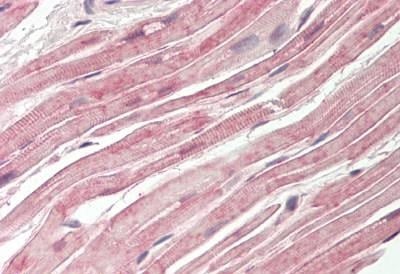
Tissue Specificity
Expressed in skeletal muscle, myoblast, myotube and in the syncytiotrophoblast (STB) of the placenta (at protein level). Ubiquitous. Highly expressed in skeletal muscle. Also found in heart, brain, spleen, intestine, placenta and at lower levels in liver, lung, kidney and pancreas.
Predicted Cross Reactivity
Pig, Bovine, Horse, Sheep, Rabbit, Dog
Similarity
Pig (100%), Bovine (100%), Horse (100%), Sheep (100%), Rabbit (100%), Dog (100%)
Conjugation
Unconjugated
Preparation and Storage
Store at -20 degree C. Stable for 12 months from date of receipt.
IF (Immunofluorescence)
(AAA320797 staining Hela by IF/ICC. The sample were fixed with PFA and permeabilized in 0.1% Triton X-100, then blocked in 10% serum for 45 minutes at 25 degree C. The primary antibody was diluted at 1/200 and incubated with the sample for 1 hour at 37 degree C. An Alexa Fluor 594 conjugated goat anti-rabbit IgG (H+L) Ab, diluted at 1/600, was used as the secondary antibody.)
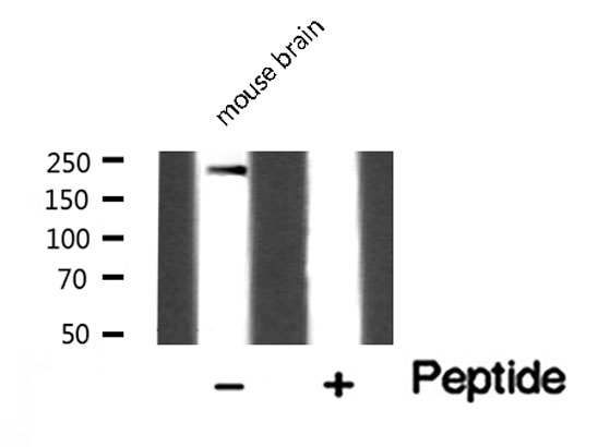
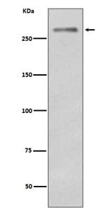
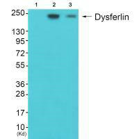
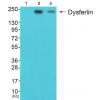
WB (Western Blot)
(Western blot analysis of Dysferlin expression in Mouse brain lysate)
WB (Western Blot)
(Western blot analysis of Dysferlin expression in Mouse brain lysate)
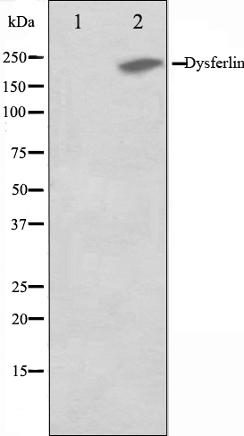
WB (Western Blot)
(Western blot analysis on K562 cell lysate using Dysferlin Antibody, The lane on the left is treated with the antigen-specific peptide.)
WB (Western Blot)
(Western blot analysis on K562 cell lysate using Dysferlin Antibody, The lane on the left is treated with the antigen-specific peptide.)
Related Product Information for anti-DYSF antibody
Description: DYSF Key calcium ion sensor involved in the Ca(2+)-triggered synaptic vesicle-plasma membrane fusion. Plays a role in the sarcolemma repair mechanism of both skeletal muscle and cardiomyocytes that permits rapid resealing of membranes disrupted by mechanical stress. Belongs to the ferlin family. Interacts with CACNA1S. Interacts with ANXA1; the interaction is Ca(2+)-and injury state-dependent.
Function: Key calcium ion sensor involved in the Ca2+-triggered synaptic vesicle-plasma membrane fusion. Plays a role in the sarcolemma repair mechanism of both skeletal muscle and cardiomyocytes that permits rapid resealing of membranes disrupted by mechanical stress (By similarity).
Subunit Structure: Interacts with CACNA1S. Interacts with ANXA1; the interaction is Ca2+-and injury state-dependent. Interacts with ANXA2; the interaction is Ca2+-and injury state-dependent. Interacts with CACNA1S and PARVB. Interacts with TRIM72/MG53; interaction is required for transport to sites of cell injury during repair patch formation (By similarity). Interacts with RIPOR2; this interaction occurs during early myogenic differentiation (PubMed:24687993). Interacts with CAV3 and PARVB. Interacts with AHNAK; the interaction is direct and Ca2+-independent. Interacts with AHNAK2; the interaction is direct and Ca2+-independent.
Similarity: All seven C2 domains associate with lipid membranes in a calcium-dependent manner. Domains C2 1 and 3 have the highest affinity for calcium, the C2 domain 1 seems to be largely unstructured in the absence of bound ligands. The C2 domain 1 from isoform 14 does not bind calcium in the absence of bound phospholipid (PubMed:24239457, PubMed:24461013). Belongs to the ferlin family.
NCBI and Uniprot Product Information
NCBI GI #
NCBI GeneID
NCBI Accession #
NCBI GenBank Nucleotide #
UniProt Accession #
Molecular Weight
Observed: 240 kDa
Predicted: 238 kDa
Predicted: 238 kDa
NCBI Official Full Name
dysferlin isoform 2
NCBI Official Synonym Full Names
dysferlin
NCBI Official Symbol
DYSF
NCBI Official Synonym Symbols
MMD1; FER1L1; LGMD2B; LGMDR2
NCBI Protein Information
dysferlin
UniProt Protein Name
Dysferlin
UniProt Gene Name
DYSF
UniProt Synonym Gene Names
FER1L1
Customer Reviews
Loading reviews...
Share Your Experience
Similar Products


Product Notes
The DYSF dysf (Catalog #AAA320797) is an Antibody produced from Rabbit and is intended for research purposes only. The product is available for immediate purchase. The Dysferlin Antibody reacts with Human, Mouse and may cross-react with other species as described in the data sheet. AAA Biotech's Dysferlin can be used in a range of immunoassay formats including, but not limited to, ELISA, ICC (Immunocytochemistry), IF (Immunofluorescence), WB (Western Blot). Researchers should empirically determine the suitability of the DYSF dysf for an application not listed in the data sheet. Researchers commonly develop new applications and it is an integral, important part of the investigative research process. It is sometimes possible for the material contained within the vial of "Dysferlin, Polyclonal Antibody" to become dispersed throughout the inside of the vial, particularly around the seal of said vial, during shipment and storage. We always suggest centrifuging these vials to consolidate all of the liquid away from the lid and to the bottom of the vial prior to opening. Please be advised that certain products may require dry ice for shipping and that, if this is the case, an additional dry ice fee may also be required.Precautions
All products in the AAA Biotech catalog are strictly for research-use only, and are absolutely not suitable for use in any sort of medical, therapeutic, prophylactic, in-vivo, or diagnostic capacity. By purchasing a product from AAA Biotech, you are explicitly certifying that said products will be properly tested and used in line with industry standard. AAA Biotech and its authorized distribution partners reserve the right to refuse to fulfill any order if we have any indication that a purchaser may be intending to use a product outside of our accepted criteria.Disclaimer
Though we do strive to guarantee the information represented in this datasheet, AAA Biotech cannot be held responsible for any oversights or imprecisions. AAA Biotech reserves the right to adjust any aspect of this datasheet at any time and without notice. It is the responsibility of the customer to inform AAA Biotech of any product performance issues observed or experienced within 30 days of receipt of said product. To see additional details on this or any of our other policies, please see our Terms & Conditions page.Item has been added to Shopping Cart
If you are ready to order, navigate to Shopping Cart and get ready to checkout.