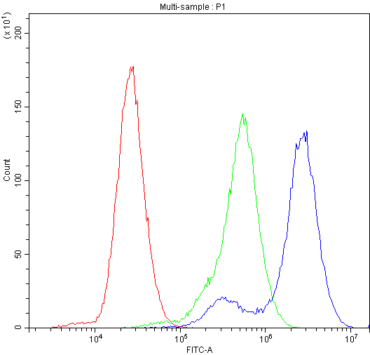
 FCM/FACS (Flow Cytometry)
(Figure 3. Flow Cytometry analysis of HEPG2 cells using anti-Factor I antibody (AAA46611).Overlay histogram showing HEPG2 cells stained with AAA46611 (Blue line).The cells were blocked with 10% normal goat serum. And then incubated with rabbit anti-Factor I Antibody (AAA46611,1ug/1x10^6 cells) for 30 min at 20 degree C. DyLight®488 conjugated goat anti-rabbit IgG (5-10ug/1x10^6 cells) was used as secondary antibody for 30 minutes at 20 degree C. Isotype control antibody (Green line) was rabbit IgG (1ug/1x106) used under the same conditions. Unlabelled sample (Red line) was also used as a control.)
FCM/FACS (Flow Cytometry)
(Figure 3. Flow Cytometry analysis of HEPG2 cells using anti-Factor I antibody (AAA46611).Overlay histogram showing HEPG2 cells stained with AAA46611 (Blue line).The cells were blocked with 10% normal goat serum. And then incubated with rabbit anti-Factor I Antibody (AAA46611,1ug/1x10^6 cells) for 30 min at 20 degree C. DyLight®488 conjugated goat anti-rabbit IgG (5-10ug/1x10^6 cells) was used as secondary antibody for 30 minutes at 20 degree C. Isotype control antibody (Green line) was rabbit IgG (1ug/1x106) used under the same conditions. Unlabelled sample (Red line) was also used as a control.)
Rabbit Factor I Polyclonal Antibody | anti-CFI antibody
Anti-Factor I Antibody
No cross reactivity with other proteins.
No cross reactivity with other proteins.
FCM/FACS (Flow Cytometry)
(Figure 3. Flow Cytometry analysis of HEPG2 cells using anti-Factor I antibody (AAA46611).Overlay histogram showing HEPG2 cells stained with AAA46611 (Blue line).The cells were blocked with 10% normal goat serum. And then incubated with rabbit anti-Factor I Antibody (AAA46611,1ug/1x10^6 cells) for 30 min at 20 degree C. DyLight®488 conjugated goat anti-rabbit IgG (5-10ug/1x10^6 cells) was used as secondary antibody for 30 minutes at 20 degree C. Isotype control antibody (Green line) was rabbit IgG (1ug/1x106) used under the same conditions. Unlabelled sample (Red line) was also used as a control.)
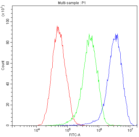
FCM/FACS (Flow Cytometry)
(Figure 2. Flow Cytometry analysis of U-87 cells using anti-Factor I antibody (AAA46611).Overlay histogram showing U-87 cells stained with AAA46611 (Blue line).The cells were blocked with 10% normal goat serum. And then incubated with rabbit anti-Factor I Antibody (AAA46611,1ug/1x10^6 cells) for 30 min at 20 degree C. DyLight®488 conjugated goat anti-rabbit IgG (5-10ug/1x10^6 cells) was used as secondary antibody for 30 minutes at 20 degree C. Isotype control antibody (Green line) was rabbit IgG (1ug/1x106) used under the same conditions. Unlabelled sample (Red line) was also used as a control.)
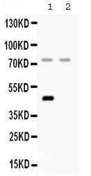
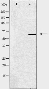
WB (Western Blot)
(Figure 1. Western blot analysis of Factor I using anti- Factor I antibody (AAA46611).Electrophoresis was performed on a 5-20% SDS-PAGE gel at 70V (Stacking gel) / 90V (Resolving gel) for 2-3 hours. The sample well of each lane was loaded with 50ug of sample under reducing conditions.Lane 1: rat liver tissue lysates,Lane 2: HELA whole cell lysates.After Electrophoresis, proteins were transferred to a Nitrocellulose membrane at 150mA for 50-90 minutes. Blocked the membrane with 5% Non-fat Milk/ TBS for 1.5 hour at RT. The membrane was incubated with rabbit anti- Factor I antigen affinity purified polyclonal antibody at 0.5ug/mL overnight at 4 degree C, then washed with TBS-0.1%Tween 3 times with 5 minutes each and probed with a goat anti-rabbit IgG-HRP secondary antibody at a dilution of 1:10000 for 1.5 hour at RT. The signal is developed using an Enhanced Chemiluminescent detection (ECL) kit with Tanon 5200 system. A specific band was detected for Factor I at approximately 75KD; 45KD. The expected band size for Factor I is at 66KD.)
Background: Complement factor I, also known as C3b/C4b inactivator, is a protein that in humans is encoded by the CFI gene. This gene encodes a serine proteinase that is essential for regulating the complement cascade. The encoded preproprotein is cleaved to produce both heavy and light chains, which are linked by disulfide bonds to form a heterodimeric glycoprotein. This heterodimer can cleave and inactivate the complement components C4b and C3b, and it prevents the assembly of the C3 and C5 convertase enzymes. Defects in this gene cause complement factor I deficiency, an autosomal recessive disease associated with a susceptibility to pyogenic infections. Mutations in this gene have been associated with a predisposition to atypical hemolytic uremic syndrome, a disease characterized by acute renal failure, microangiopathic hemolytic anemia and thrombocytopenia. Primary glomerulonephritis with immune deposits and age-related macular degeneration are other conditions associated with mutations of this gene.
2. Goldberger G, Bruns GA, Rits M, Edge MD, Kwiatkowski DJ (Jul 1987). "Human complement factor I: analysis of cDNA-derived primary structure and assignment of its gene to chromosome 4". The Journal of Biological Chemistry 262 (21): 10065-71.
NCBI and Uniprot Product Information
Customer Reviews
Loading reviews...
Share Your Experience
Similar Products

Product Notes
The CFI cfi (Catalog #AAA46611) is an Antibody produced from Rabbit and is intended for research purposes only. The product is available for immediate purchase. The Anti-Factor I Antibody reacts with Human, Rat No cross reactivity with other proteins. and may cross-react with other species as described in the data sheet. AAA Biotech's Factor I can be used in a range of immunoassay formats including, but not limited to, WB (Western Blot). Researchers should empirically determine the suitability of the CFI cfi for an application not listed in the data sheet. Researchers commonly develop new applications and it is an integral, important part of the investigative research process. It is sometimes possible for the material contained within the vial of "Factor I, Polyclonal Antibody" to become dispersed throughout the inside of the vial, particularly around the seal of said vial, during shipment and storage. We always suggest centrifuging these vials to consolidate all of the liquid away from the lid and to the bottom of the vial prior to opening. Please be advised that certain products may require dry ice for shipping and that, if this is the case, an additional dry ice fee may also be required.Precautions
All products in the AAA Biotech catalog are strictly for research-use only, and are absolutely not suitable for use in any sort of medical, therapeutic, prophylactic, in-vivo, or diagnostic capacity. By purchasing a product from AAA Biotech, you are explicitly certifying that said products will be properly tested and used in line with industry standard. AAA Biotech and its authorized distribution partners reserve the right to refuse to fulfill any order if we have any indication that a purchaser may be intending to use a product outside of our accepted criteria.Disclaimer
Though we do strive to guarantee the information represented in this datasheet, AAA Biotech cannot be held responsible for any oversights or imprecisions. AAA Biotech reserves the right to adjust any aspect of this datasheet at any time and without notice. It is the responsibility of the customer to inform AAA Biotech of any product performance issues observed or experienced within 30 days of receipt of said product. To see additional details on this or any of our other policies, please see our Terms & Conditions page.Item has been added to Shopping Cart
If you are ready to order, navigate to Shopping Cart and get ready to checkout.