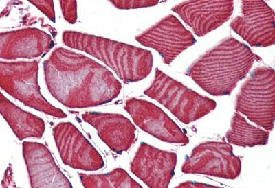
 IHC (Immunohistochemistry)
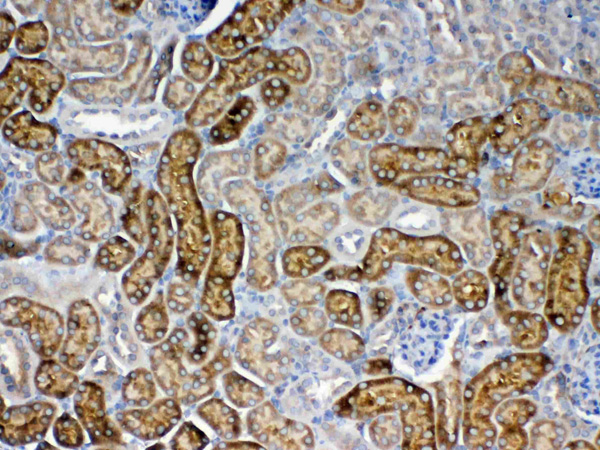
(Figure 5. IHC analysis of FZD3 using anti-FZD3 antibody (AAA124724).FZD3 was detected in paraffin-embedded section of rat kidney tissue. Heat mediated antigen retrieval was performed in citrate buffer (pH6, epitope retrieval solution) for 20 mins. The tissue section was blocked with 10% goat serum. The tissue section was then incubated with 1ug/ml rabbit anti-FZD3 Antibody (AAA124724) overnight at 4 degree C. Biotinylated goat anti-rabbit IgG was used as secondary antibody and incubated for 30 minutes at 37 degree C. The tissue section was developed using Strepavidin-Biotin-Complex (SABC) with DAB as the chromogen.)
IHC (Immunohistochemistry)
(Figure 5. IHC analysis of FZD3 using anti-FZD3 antibody (AAA124724).FZD3 was detected in paraffin-embedded section of rat kidney tissue. Heat mediated antigen retrieval was performed in citrate buffer (pH6, epitope retrieval solution) for 20 mins. The tissue section was blocked with 10% goat serum. The tissue section was then incubated with 1ug/ml rabbit anti-FZD3 Antibody (AAA124724) overnight at 4 degree C. Biotinylated goat anti-rabbit IgG was used as secondary antibody and incubated for 30 minutes at 37 degree C. The tissue section was developed using Strepavidin-Biotin-Complex (SABC) with DAB as the chromogen.)
Rabbit FZD3/Frizzled 3 Polyclonal Antibody | anti-FZD3 antibody
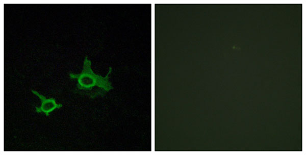
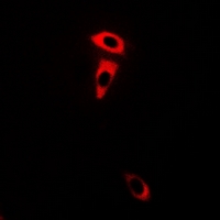
Anti-FZD3/Frizzled 3 antibody
No cross reactivity with other proteins.
No cross reactivity with other proteins.
IHC (Immunohistochemistry)
(Figure 5. IHC analysis of FZD3 using anti-FZD3 antibody (AAA124724).FZD3 was detected in paraffin-embedded section of rat kidney tissue. Heat mediated antigen retrieval was performed in citrate buffer (pH6, epitope retrieval solution) for 20 mins. The tissue section was blocked with 10% goat serum. The tissue section was then incubated with 1ug/ml rabbit anti-FZD3 Antibody (AAA124724) overnight at 4 degree C. Biotinylated goat anti-rabbit IgG was used as secondary antibody and incubated for 30 minutes at 37 degree C. The tissue section was developed using Strepavidin-Biotin-Complex (SABC) with DAB as the chromogen.)
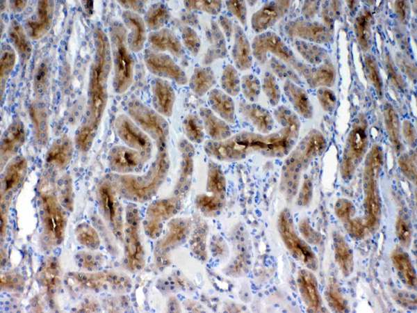
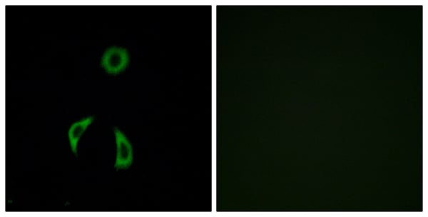
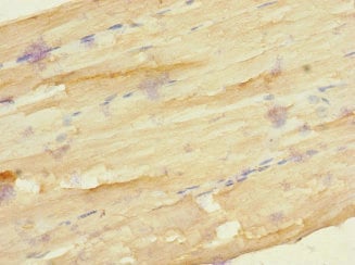
IHC (Immunohistochemistry)
(Figure 4. IHC analysis of FZD3 using anti-FZD3 antibody (AAA124724).FZD3 was detected in paraffin-embedded section of mouse kidney tissue. Heat mediated antigen retrieval was performed in citrate buffer (pH6, epitope retrieval solution) for 20 mins. The tissue section was blocked with 10% goat serum. The tissue section was then incubated with 1ug/ml rabbit anti-FZD3 Antibody (AAA124724) overnight at 4 degree C. Biotinylated goat anti-rabbit IgG was used as secondary antibody and incubated for 30 minutes at 37 degree C. The tissue section was developed using Strepavidin-Biotin-Complex (SABC) with DAB as the chromogen.)
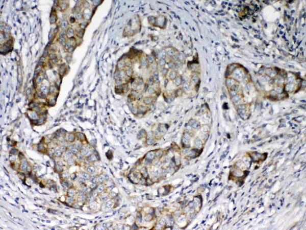
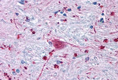
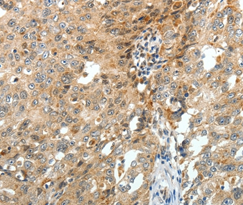
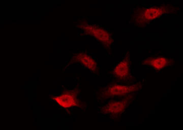
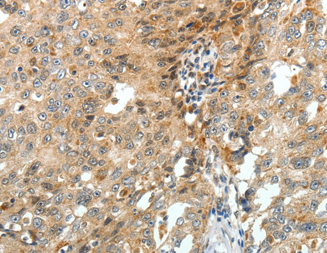
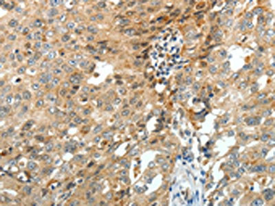
IHC (Immunohistochemisry)
(Figure 3. IHC analysis of FZD3 using anti-FZD3 antibody (AAA124724).FZD3 was detected in paraffin-embedded section of human mammary cancer tissue. Heat mediated antigen retrieval was performed in citrate buffer (pH6, epitope retrieval solution) for 20 mins. The tissue section was blocked with 10% goat serum. The tissue section was then incubated with 1ug/ml rabbit anti-FZD3 Antibody (AAA124724) overnight at 4 degree C. Biotinylated goat anti-rabbit IgG was used as secondary antibody and incubated for 30 minutes at 37 degree C. The tissue section was developed using Strepavidin-Biotin-Complex (SABC) with DAB as the chromogen.)
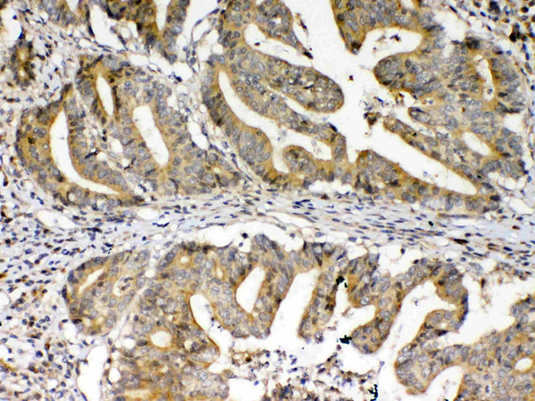
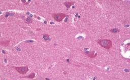
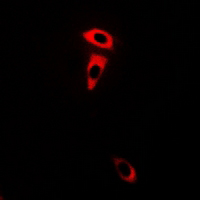
IHC (Immunohiostchemistry)
(Figure 2. IHC analysis of FZD3 using anti-FZD3 antibody (AAA124724).FZD3 was detected in paraffin-embedded section of human colon cancer tissue. Heat mediated antigen retrieval was performed in citrate buffer (pH6, epitope retrieval solution) for 20 mins. The tissue section was blocked with 10% goat serum. The tissue section was then incubated with 1ug/ml rabbit anti-FZD3 Antibody (AAA124724) overnight at 4 degree C. Biotinylated goat anti-rabbit IgG was used as secondary antibody and incubated for 30 minutes at 37 degree C. The tissue section was developed using Strepavidin-Biotin-Complex (SABC) with DAB as the chromogen.)
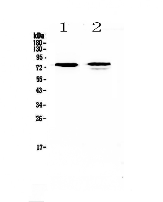
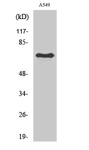
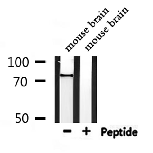
WB (Western Blot)
(Figure 1. Western blot analysis of FZD3 using anti-FZD3 antibody (AAA124724).Electrophoresis was performed on a 5-20% SDS-PAGE gel at 70V (Stacking gel) / 90V (Resolving gel) for 2-3 hours. The sample well of each lane was loaded with 50ug of sample under reducing conditions.Lane 1: human SK-OV-3 cell lysates,Lane 2: human Jurkat cell lysates.After Electrophoresis, proteins were transferred to a Nitrocellulose membrane at 150mA for 50-90 minutes. Blocked the membrane with 5% Non-fat Milk/ TBS for 1.5 hour at RT. The membrane was incubated with rabbit anti-FZD3 antigen affinity purified polyclonal antibody at 0.5ug/mL overnight at 4 degree C, then washed with TBS-0.1%Tween 3 times with 5 minutes each and probed with a goat anti-rabbit IgG-HRP secondary antibody at a dilution of 1:10000 for 1.5 hour at RT. The signal is developed using an Enhanced Chemiluminescent detection (ECL) kit with Tanon 5200 system. A specific band was detected for FZD3 at approximately 76KD. The expected band size for FZD3 is at 76KD.)
Protein Function: Receptor for Wnt proteins. Most of frizzled receptors are coupled to the beta-catenin canonical signaling pathway, which leads to the activation of disheveled proteins, inhibition of GSK- 3 kinase, nuclear accumulation of beta-catenin and activation of Wnt target genes. A second signaling pathway involving PKC and calcium fluxes has been seen for some family members, but it is not yet clear if it represents a distinct pathway or if it can be integrated in the canonical pathway, as PKC seems to be required for Wnt-mediated inactivation of GSK-3 kinase. Both pathways seem to involve interactions with G-proteins. Activation by Wnt5A stimulates PKC activity via a G-protein-dependent mechanism. Involved in transduction and intercellular transmission of polarity information during tissue morphogenesis and/or in differentiated tissues. Plays a role in controlling early axon growth and guidance processes necessary for the formation of a subset of central and peripheral major fiber tracts. Required for the development of major fiber tracts in the central nervous system, including: the anterior commissure, the corpus callosum, the thalamocortical, corticothalamic and nigrostriatal tracts, the corticospinal tract, the fasciculus retroflexus, the mammillothalamic tract, the medial lemniscus, and ascending fiber tracts from the spinal cord to the brain. In the peripheral nervous system, controls axon growth in distinct populations of cranial and spinal motor neurons, including the facial branchimotor nerve, the hypoglossal nerve, the phrenic nerve, and motor nerves innervating dorsal limbs. Involved in the migration of cranial neural crest cells. May also be implicated in the transmission of sensory information from the trunk and limbs to the brain. Controls commissural sensory axons guidance after midline crossing along the anterior-posterior axis in the developing spinal cord in a Wnt-dependent signaling pathway. Together with FZD6, is involved in the neural tube closure and plays a role in the regulation of the establishment of planar cell polarity (PCP), particularly in the orientation of asymmetric bundles of stereocilia on the apical faces of a subset of auditory and vestibular sensory cells located in the inner ear. Promotes neurogenesis by maintaining sympathetic neuroblasts within the cell cycle in a beta-catenin-dependent manner (By similarity).
NCBI and Uniprot Product Information
Customer Reviews
Loading reviews...
Share Your Experience
Similar Products

Product Notes
The FZD3 fzd3 (Catalog #AAA124724) is an Antibody produced from Rabbit and is intended for research purposes only. The product is available for immediate purchase. The Anti-FZD3/Frizzled 3 antibody reacts with Human, Mouse, Rat No cross reactivity with other proteins. and may cross-react with other species as described in the data sheet. AAA Biotech's FZD3/Frizzled 3 can be used in a range of immunoassay formats including, but not limited to, WB (Western Blot), IHC (Immunohistochemistry). Researchers should empirically determine the suitability of the FZD3 fzd3 for an application not listed in the data sheet. Researchers commonly develop new applications and it is an integral, important part of the investigative research process. It is sometimes possible for the material contained within the vial of "FZD3/Frizzled 3, Polyclonal Antibody" to become dispersed throughout the inside of the vial, particularly around the seal of said vial, during shipment and storage. We always suggest centrifuging these vials to consolidate all of the liquid away from the lid and to the bottom of the vial prior to opening. Please be advised that certain products may require dry ice for shipping and that, if this is the case, an additional dry ice fee may also be required.Precautions
All products in the AAA Biotech catalog are strictly for research-use only, and are absolutely not suitable for use in any sort of medical, therapeutic, prophylactic, in-vivo, or diagnostic capacity. By purchasing a product from AAA Biotech, you are explicitly certifying that said products will be properly tested and used in line with industry standard. AAA Biotech and its authorized distribution partners reserve the right to refuse to fulfill any order if we have any indication that a purchaser may be intending to use a product outside of our accepted criteria.Disclaimer
Though we do strive to guarantee the information represented in this datasheet, AAA Biotech cannot be held responsible for any oversights or imprecisions. AAA Biotech reserves the right to adjust any aspect of this datasheet at any time and without notice. It is the responsibility of the customer to inform AAA Biotech of any product performance issues observed or experienced within 30 days of receipt of said product. To see additional details on this or any of our other policies, please see our Terms & Conditions page.Item has been added to Shopping Cart
If you are ready to order, navigate to Shopping Cart and get ready to checkout.