Rabbit anti-Human, Mouse HDAC5 Polyclonal Antibody | anti-HDAC5 antibody
HDAC5 Antibody
Phosphate buffered saline, pH 7.4, 150mM NaCl, 0.02% sodium azide and 50% glycerol.
IHC: 1:50-1:200
IF/ICC: 1:100-1:500
WB (Western Blot)
(Western blot analysis of HDAC5 expression in mouse brain tissue lysates, The lane on the right is treated with the antigen-specific peptide.)
IHC (Immunohistchemistry)
(AAA31097 at 1/50 staining human colon cancer tissue sections by IHC-P. The tissue was formaldehyde fixed and a heat mediated antigen retrieval step in citrate buffer was performed. The tissue was then blocked and incubated with the antibody for 1.5 hours at 22 degree C. An HRP conjugated goat anti-rabbit antibody was used as the secondary.)
IHC (Immunohistochemistry)
(AAA31097 at 1/200 staining human colon cancer tissue sections by IHC-P. The tissue was formaldehyde fixed and a heat mediated antigen retrieval step in citrate buffer was performed. The tissue was then blocked and incubated with the antibody for 1.5 hours at 22 degree C. An HRP conjugated goat anti-rabbit antibody was used as the secondary.)
IHC (Immunohistochemistry)
(AAA31097 at 1/50 staining human colon cancer tissue sections by IHC-P. The tissue was formaldehyde fixed and a heat mediated antigen retrieval step in citrate buffer was performed. The tissue was then blocked and incubated with the antibody for 1.5 hours at 22 degree C. An HRP conjugated goat anti-rabbit antibody was used as the secondary.)
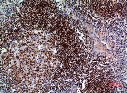
IHC (Immunohistochemistry)
(AAA31097 at 1/100 staining human breast carcinoma tissues sections by IHC-P. The tissue was formaldehyde fixed and a heat mediated antigen retrieval step in citrate buffer was performed. The tissue was then blocked and incubated with the antibody for 1.5 ho)
IF (Immunofluorescence)
(AAA31097 staining HepG2 by IF/ICC. The sample were fixed with PFA and permeabilized in 0.1% Triton X-100, then blocked in 10% serum for 45 minutes at 25 degree C. The primary antibody was diluted at 1/200 and incubated with the sample for 1 hour at 37 degree C. An Alexa Fluor 594 conjugated goat anti-rabbit IgG (H+L) Ab, diluted at 1/600, was used as the secondary antibody.)
WB (Western Blot)
(Western blot analysis of HDAC5 expression in HepG2 whole cell lysates, The lane on the left is treated with the antigen-specific peptide.)
Function: Responsible for the deacetylation of lysine residues on the N-terminal part of the core histones (H2A, H2B, H3 and H4). Histone deacetylation gives a tag for epigenetic repression and plays an important role in transcriptional regulation, cell cycle progression and developmental events. Histone deacetylases act via the formation of large multiprotein complexes. Involved in muscle maturation by repressing transcription of myocyte enhancer MEF2C. During muscle differentiation, it shuttles into the cytoplasm, allowing the expression of myocyte enhancer factors. Involved in the MTA1-mediated epigenetic regulation of ESR1 expression in breast cancer.
Subunit Structure: Interacts with AHRR, BAHD1, BCOR, HDAC7, HDAC9, CTBP1, MEF2C, NCOR2, NRIP1, PHB2 and a 14-3-3 chaperone protein. Interacts with BCL6, DDIT3/CHOP, GRK5, KDM5B and MYOCD. Interacts with EP300 in the presence of TFAP2C. Interacts with ANKRA2. Interacts with CUL7 (as part of the 3M complex); negatively regulated by ANKRA2. Interacts with ZBTB7B; the interaction allows the recruitment of HDAC4 on CD8 loci for deacetylation and possible inhibition of CD8 genes expression (By similarity).
Post-translational Modifications: Phosphorylated by AMPK, CaMK1, SIK1 and PRKD1 at Ser-259 and Ser-498. The phosphorylation is required for the export to the cytoplasm and inhibition. Phosphorylated by the PKC kinases PKN1 and PKN2, impairing nuclear import. Phosphorylated by GRK5, leading to nuclear export of HDAC5 and allowing MEF2-mediated transcription (By similarity). Ubiquitinated. Polyubiquitination however does not lead to its degradation.
Similarity: The nuclear export sequence mediates the shuttling between the nucleus and the cytoplasm. Belongs to the histone deacetylase family. HD type 2 subfamily.
NCBI and Uniprot Product Information
Predicted: 122 kDa