


 WB (Western Blot)
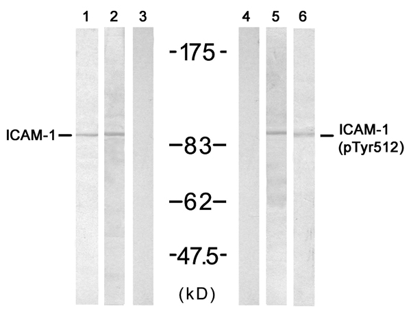
(Western blot analysis of extracts from various samples, using Phospho-ICAM-1 (Tyr512) Antibody. Lane 1: VERO cells, treated with blocking peptideLane 2: VERO cellsLane 3: A2780 cells.)
WB (Western Blot)
(Western blot analysis of extracts from various samples, using Phospho-ICAM-1 (Tyr512) Antibody. Lane 1: VERO cells, treated with blocking peptideLane 2: VERO cellsLane 3: A2780 cells.)
Rabbit ICAM-1 Polyclonal Antibody | anti-ICAM-1 antibody
Phospho-ICAM-1 (Tyr512) Antibody
WB (Western Blot)
(Western blot analysis of extracts from various samples, using Phospho-ICAM-1 (Tyr512) Antibody. Lane 1: VERO cells, treated with blocking peptideLane 2: VERO cellsLane 3: A2780 cells.)
WB (Western Blot)
(Western blot analysis of extracts from various samples, using Phospho-ICAM-1 (Tyr512) Antibody. Lane 1: VERO cells, treated with blocking peptideLane 2: VERO cellsLane 3: A2780 cells.)
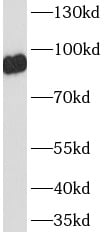
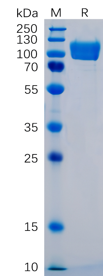
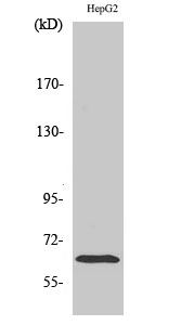
WB (Western Blot)
(Western blot analysis of ICAM-1 phosphorylation expression in TNF-alpha treated HeLa whole cell lysates, The lane on the left is treated with the antigen-specific peptide.)
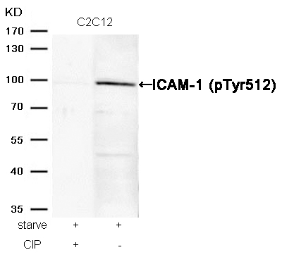
WB (Western Blot)
(Western blot analysis of ICAM-1 phosphorylation expression in TNF-alpha treated HeLa whole cell lysates, The lane on the left is treated with the antigen-specific peptide.)
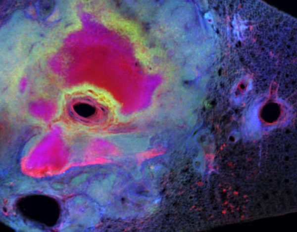
IHC (Immunohistchemistry)
(AAA30979 at 1/100 staining human liver tissue sections by IHC-P. The tissue was formaldehyde fixed and a heat mediated antigen retrieval step in citrate buffer was performed. The tissue was then blocked and incubated with the antibody for 1.5 hours at 22 degree C. An HRP conjugated goat anti-rabbit antibody was used as the secondary.)
IHC (Immunohistochemistry)
(AAA30979 at 1/100 staining human appendiceal tissue sections by IHC-P. The tissue was formaldehyde fixed and a heat mediated antigen retrieval step in citrate buffer was performed. The tissue was then blocked and incubated with the antibody for 1.5 hours at 22 degree C. An HRP conjugated goat anti-rabbit antibody was used as the secondary.)
IHC (Immunohistochemistry)
(AAA30979 at 1/100 staining human skin tissue sections by IHC-P. The tissue was formaldehyde fixed and a heat mediated antigen retrieval step in citrate buffer was performed. The tissue was then blocked and incubated with the antibody for 1.5 hours at 22 degree C. An HRP conjugated goat anti-rabbit antibody was used as the secondary.)
IHC (Immunohistochemistry)
(AAA30979 at 1/100 staining mouse brain tissue sections by IHC-P. The tissue was formaldehyde fixed and a heat mediated antigen retrieval step in citrate buffer was performed. The tissue was then blocked and incubated with the antibody for 1.5 hours at 22 degree C. An HRP conjugated goat anti-rabbit antibody was used as the secondary.)
IHC (Immunohistochemistry)
(AAA30979 at 1/100 staining mouse kidney tissue sections by IHC-P. The tissue was formaldehyde fixed and a heat mediated antigen retrieval step in citrate buffer was performed. The tissue was then blocked and incubated with the antibody for 1.5 hours at 22 degree C. An HRP conjugated goat anti-rabbit antibody was used as the secondary.)
NCBI and Uniprot Product Information
Customer Reviews
Loading reviews...
Share Your Experience
Similar Products


Product Notes
The ICAM-1 icam1 (Catalog #AAA30979) is an Antibody produced from Rabbit and is intended for research purposes only. The product is available for immediate purchase. The Phospho-ICAM-1 (Tyr512) Antibody reacts with Human, Mouse, Monkey and may cross-react with other species as described in the data sheet. AAA Biotech's ICAM-1 can be used in a range of immunoassay formats including, but not limited to, WB (Western Blot), IHC (Immunohistochemistry), IF (Immunofluorescence), ICC (Immunocytochemistry), ELISA. WB 1:500-1:2000, IHC 1:50-1:200, IF/ICC 1:100-1:500, ELISA(peptide) 1:20000-1:40000. Researchers should empirically determine the suitability of the ICAM-1 icam1 for an application not listed in the data sheet. Researchers commonly develop new applications and it is an integral, important part of the investigative research process. It is sometimes possible for the material contained within the vial of "ICAM-1, Polyclonal Antibody" to become dispersed throughout the inside of the vial, particularly around the seal of said vial, during shipment and storage. We always suggest centrifuging these vials to consolidate all of the liquid away from the lid and to the bottom of the vial prior to opening. Please be advised that certain products may require dry ice for shipping and that, if this is the case, an additional dry ice fee may also be required.Precautions
All products in the AAA Biotech catalog are strictly for research-use only, and are absolutely not suitable for use in any sort of medical, therapeutic, prophylactic, in-vivo, or diagnostic capacity. By purchasing a product from AAA Biotech, you are explicitly certifying that said products will be properly tested and used in line with industry standard. AAA Biotech and its authorized distribution partners reserve the right to refuse to fulfill any order if we have any indication that a purchaser may be intending to use a product outside of our accepted criteria.Disclaimer
Though we do strive to guarantee the information represented in this datasheet, AAA Biotech cannot be held responsible for any oversights or imprecisions. AAA Biotech reserves the right to adjust any aspect of this datasheet at any time and without notice. It is the responsibility of the customer to inform AAA Biotech of any product performance issues observed or experienced within 30 days of receipt of said product. To see additional details on this or any of our other policies, please see our Terms & Conditions page.Frequently Asked Questions
What is ICAM-1 used as a marker for in inflammation or immune studies?
ICAM-1 is used as a marker to show inflammation or immune system activation. When cells are stressed, infected, or inflamed, they make more ICAM-1. Scientists measure it to understand how strong the immune response is in tissues or cells.
Can an ICAM-1 antibody be used to detect endothelial cell activation?
Yes. ICAM-1 antibodies are often used to detect activated endothelial cells. During inflammation, these cells ramp up ICAM-1 production. This helps immune cells stick to tissues and migrate in.
Is ICAM-1 expression upregulated during infection or inflammatory conditions?
Yes, during infection or inflammation, ICAM-1 levels increase significantly. This happens because the body wants immune cells to move quickly to the affected area. Higher ICAM-1 helps white blood cells attach to blood vessels and enter inflamed tissues.
Can this ICAM-1 antibody be used for flow cytometry or immunofluorescence?
Yes, many ICAM-1 antibodies work for immunofluorescence and flow cytometry. These techniques let scientists visualize or measure ICAM-1 on cell surfaces, aiding studies of inflammation, immune activation, and cell interactions.
how does ICAM-1 expression change after cytokine stimulation (e.g., TNF-α, IL-1β)?
Cytokines like TNF-α or IL-1β quickly boost ICAM-1 expression after stimulation. These danger signals prompt cells to upregulate ICAM-1, helping immune cells attach and reach inflamed or damaged areas.
Is ICAM-1 a reliable marker for studying leukocyte adhesion and migration?
Yes, ICAM-1 is a reliable and widely used marker for studying how white blood cells stick to blood vessels and move into tissues. Because it directly helps immune cells attach and migrate, it is very useful in inflammation research.
Item has been added to Shopping Cart
If you are ready to order, navigate to Shopping Cart and get ready to checkout.