


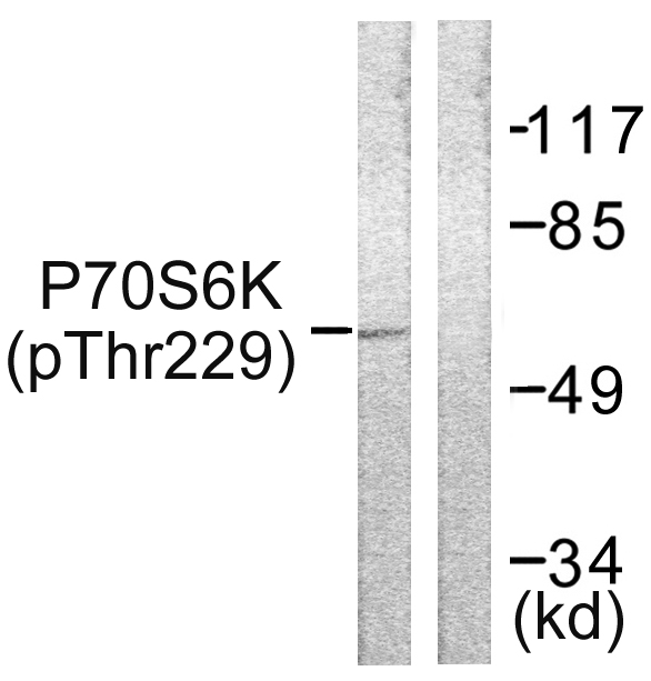
 WB (Western Blot)
(Western blot analysis of p70 S6 Kinase expression in mouse muscle tissue lysates, The lane on the right is treated with the antigen-specific peptide.)
WB (Western Blot)
(Western blot analysis of p70 S6 Kinase expression in mouse muscle tissue lysates, The lane on the right is treated with the antigen-specific peptide.)
Rabbit p70 S6 Kinase Polyclonal Antibody | anti-RPS6KB1 antibody
p70 S6 Kinase Antibody
Phosphate buffered saline, pH 7.4, 150mM NaCl, 0.02% sodium azide and 50% glycerol.
IHC: 1:50-1:200
IF/ICC: 1:100-1:500
WB (Western Blot)
(Western blot analysis of p70 S6 Kinase expression in mouse muscle tissue lysates, The lane on the right is treated with the antigen-specific peptide.)
WB (Western Blot)
(Western blot analysis of p70 S6 Kinase expression in mouse muscle tissue lysates, The lane on the right is treated with the antigen-specific peptide.)
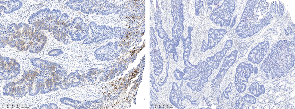
IHC (Immunohistochemistry)
(AAA31088 at 1/100 staining Human gastric tissue by IHC-P. The sample was formaldehyde fixed and a heat mediated antigen retrieval step in citrate buffer was performed. The sample was then blocked and incubated with the antibody for 1.5 hours at 22 degree C. An HRP conjugated goat anti-rabbit antibody was used as the secondary.)
IF (Immunofluorescence)
(AAA31088 staining MCF-7 cells by IF/ICC. The sample were fixed with PFA and permeabilized in 0.1% Triton X-100, then blocked in 10% serum for 45 minutes at 25 degree C. The primary antibody was diluted at 1/200 and incubated with the sample for 1 hour at 37 degree C. An Alexa Fluor 594 conjugated goat anti-rabbit IgG (H+L) antibody, diluted at 1/600, was used as secondary antibody.)
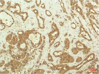
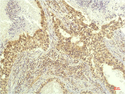
IHC (Immunohistochemistry)
(AAA31088 at 1/100 staining human brain tissues sections by IHC-P. The tissue was formaldehyde fixed and a heat mediated antigen retrieval step in citrate buffer was performed. The tissue was then blocked and incubated with the antibody for 1.5 hours at 55 degree C)
IHC (Immunohistochemistry)
(AAA31088 at 1/100 staining human brain tissues sections by IHC-P. The tissue was formaldehyde fixed and a heat mediated antigen retrieval step in citrate buffer was performed. The tissue was then blocked and incubated with the antibody for 1.5 hours at 55 degree C)
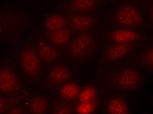
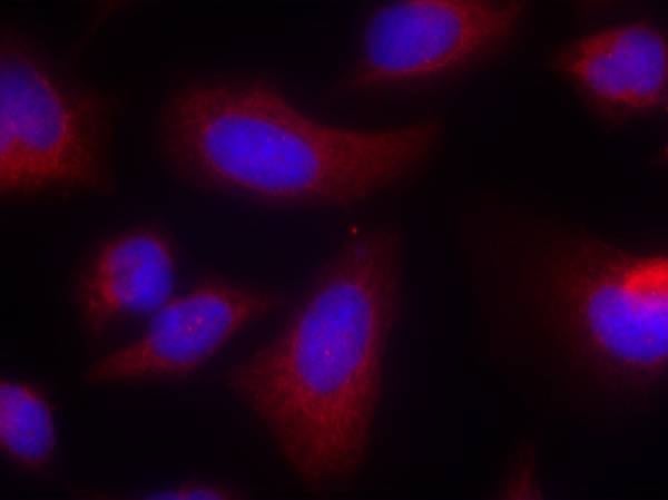
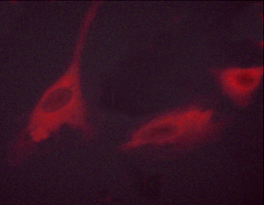
IF (Immunofluorescence)
(AAA31088 staining NIH-3T3 by IF/ICC. The sample were fixed with PFA and permeabilized in 0.1% Triton X-100, then blocked in 10% serum for 45 minutes at 25 degree C. The primary antibody was diluted at 1/200 and incubated with the sample for 1 hour at 37 degree C. An Alexa Fluor 594 conjugated goat anti-rabbit IgG (H+L) Ab, diluted at 1/600, was used as the secondary antibody.)
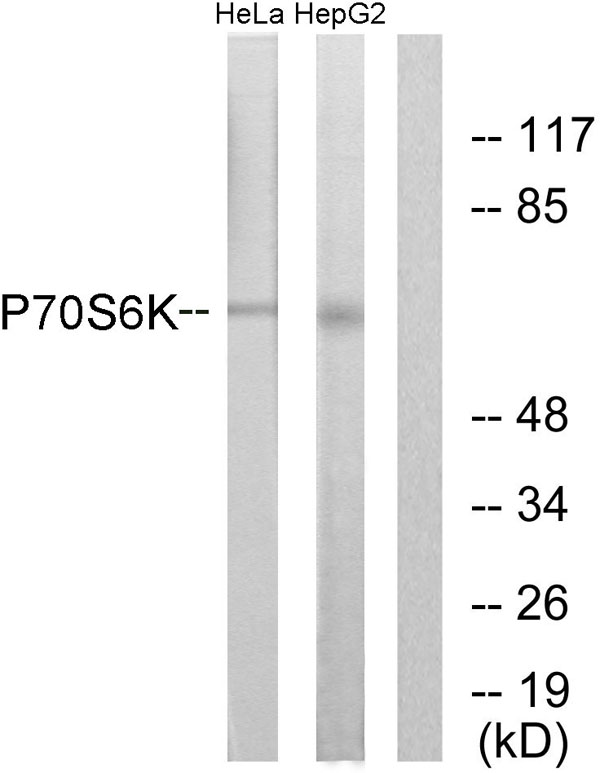
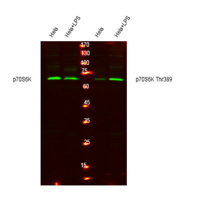
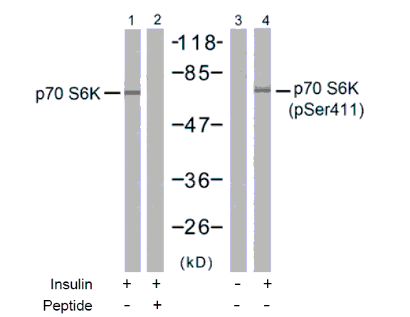
WB (Western Blot)
(Western blot analysis of p70 S6 Kinase expression in Jurkat whole cell lysates, The lane on the left is treated with the antigen-specific peptide.)
WB (Western Blot)
(Western blot analysis of p70 S6 Kinase expression in Jurkat whole cell lysates, The lane on the left is treated with the antigen-specific peptide.)
Function: Serine/threonine-protein kinase that acts downstream of mTOR signaling in response to growth factors and nutrients to promote cell proliferation, cell growth and cell cycle progression. Regulates protein synthesis through phosphorylation of EIF4B, RPS6 and EEF2K, and contributes to cell survival by repressing the pro-apoptotic function of BAD. Under conditions of nutrient depletion, the inactive form associates with the EIF3 translation initiation complex. Upon mitogenic stimulation, phosphorylation by the mammalian target of rapamycin complex 1 (mTORC1) leads to dissociation from the EIF3 complex and activation. The active form then phosphorylates and activates several substrates in the pre-initiation complex, including the EIF2B complex and the cap-binding complex component EIF4B. Also controls translation initiation by phosphorylating a negative regulator of EIF4A, PDCD4, targeting it for ubiquitination and subsequent proteolysis. Promotes initiation of the pioneer round of protein synthesis by phosphorylating POLDIP3/SKAR. In response to IGF1, activates translation elongation by phosphorylating EEF2 kinase (EEF2K), which leads to its inhibition and thus activation of EEF2. Also plays a role in feedback regulation of mTORC2 by mTORC1 by phosphorylating RICTOR, resulting in the inhibition of mTORC2 and AKT1 signaling. Mediates cell survival by phosphorylating the pro-apoptotic protein BAD and suppressing its pro-apoptotic function. Phosphorylates mitochondrial URI1 leading to dissociation of a URI1-PPP1CC complex. The free mitochondrial PPP1CC can then dephosphorylate RPS6KB1 at Thr-412, which is proposed to be a negative feedback mechanism for the RPS6KB1 anti-apoptotic function. Mediates TNF-alpha-induced insulin resistance by phosphorylating IRS1 at multiple serine residues, resulting in accelerated degradation of IRS1. In cells lacking functional TSC1-2 complex, constitutively phosphorylates and inhibits GSK3B. May be involved in cytoskeletal rearrangement through binding to neurabin. Phosphorylates and activates the pyrimidine biosynthesis enzyme CAD, downstream of MTOR (PubMed:11500364, PubMed:12801526, PubMed:14673156, PubMed:15071500, PubMed:15341740, PubMed:16286006, PubMed:17052453, PubMed:17053147, PubMed:17936702, PubMed:18952604, PubMed:19085255, PubMed:19720745, PubMed:19935711, PubMed:19995915, PubMed:23429703). Following activation by mTORC1, phosphorylates EPRS and thereby plays a key role in fatty acid uptake by adipocytes and also most probably in interferon-gamma-induced translation inhibition (PubMed:28178239).
Subunit Structure: Interacts with PPP1R9A/neurabin-1 (By similarity). Interacts with RPTOR (PubMed:12150926). Interacts with IRS1 (PubMed:18952604). Interacts with EIF3B and EIF3C (PubMed:16286006). Interacts with TRAF4 (PubMed:12801526). Interacts with POLDIP3 (PubMed:15341740). Interacts (via N-terminus) with IER5 (PubMed:26496226).
Post-translational Modifications: Phosphorylation at Thr-412 is regulated by mTORC1. The phosphorylation at this site is maintained by an agonist-dependent autophosphorylation mechanism (By similarity). Activated by phosphorylation at Thr-252 by PDPK1. Dephosphorylation by PPP1CC at Thr-412 in mitochondrion.
Similarity: The autoinhibitory domain is believed to block phosphorylation within the AGC-kinase C-terminal domain and the activation loop.The TOS (TOR signaling) motif is essential for activation by mTORC1. Belongs to the protein kinase superfamily. AGC Ser/Thr protein kinase family. S6 kinase subfamily.
NCBI and Uniprot Product Information
Predicted: 60 kDa
Customer Reviews
Loading reviews...
Share Your Experience
Similar Products


Product Notes
The RPS6KB1 rps6kb1 (Catalog #AAA31088) is an Antibody produced from Rabbit and is intended for research purposes only. The product is available for immediate purchase. The p70 S6 Kinase Antibody reacts with Human, Mouse, Rat and may cross-react with other species as described in the data sheet. AAA Biotech's p70 S6 Kinase can be used in a range of immunoassay formats including, but not limited to, WB (Western Blot), IHC (Immunohistochemistry), IF (Immunofluorescence), ICC (Immunocytochemistry), ELISA. WB: 1:500-1:2000 IHC: 1:50-1:200 IF/ICC: 1:100-1:500. Researchers should empirically determine the suitability of the RPS6KB1 rps6kb1 for an application not listed in the data sheet. Researchers commonly develop new applications and it is an integral, important part of the investigative research process. It is sometimes possible for the material contained within the vial of "p70 S6 Kinase, Polyclonal Antibody" to become dispersed throughout the inside of the vial, particularly around the seal of said vial, during shipment and storage. We always suggest centrifuging these vials to consolidate all of the liquid away from the lid and to the bottom of the vial prior to opening. Please be advised that certain products may require dry ice for shipping and that, if this is the case, an additional dry ice fee may also be required.Precautions
All products in the AAA Biotech catalog are strictly for research-use only, and are absolutely not suitable for use in any sort of medical, therapeutic, prophylactic, in-vivo, or diagnostic capacity. By purchasing a product from AAA Biotech, you are explicitly certifying that said products will be properly tested and used in line with industry standard. AAA Biotech and its authorized distribution partners reserve the right to refuse to fulfill any order if we have any indication that a purchaser may be intending to use a product outside of our accepted criteria.Disclaimer
Though we do strive to guarantee the information represented in this datasheet, AAA Biotech cannot be held responsible for any oversights or imprecisions. AAA Biotech reserves the right to adjust any aspect of this datasheet at any time and without notice. It is the responsibility of the customer to inform AAA Biotech of any product performance issues observed or experienced within 30 days of receipt of said product. To see additional details on this or any of our other policies, please see our Terms & Conditions page.Item has been added to Shopping Cart
If you are ready to order, navigate to Shopping Cart and get ready to checkout.