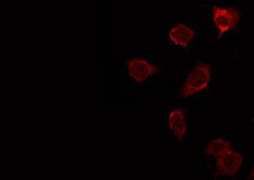
 IF (Immunofluorescence)
(AAA326401 staining NIH-3T3 by IF/ICC. The sample were fixed with PFA and permeabilized in 0.1% Triton X-100, then blocked in 10% serum for 45 minutes at 25 degree C. The primary antibody was diluted at 1/200 and incubated with the sample for 1 hour at 37 degree C. An Alexa Fluor 594 conjugated goat anti-rabbit IgG (H+L) Ab, diluted at 1/600, was used as the secondary antibody.)
IF (Immunofluorescence)
(AAA326401 staining NIH-3T3 by IF/ICC. The sample were fixed with PFA and permeabilized in 0.1% Triton X-100, then blocked in 10% serum for 45 minutes at 25 degree C. The primary antibody was diluted at 1/200 and incubated with the sample for 1 hour at 37 degree C. An Alexa Fluor 594 conjugated goat anti-rabbit IgG (H+L) Ab, diluted at 1/600, was used as the secondary antibody.)
Rabbit PSAP Polyclonal Antibody | anti-PSAP antibody
PSAP Antibody
Phosphate buffered saline, pH 7.4, 150mM NaCl, 0.02% sodium azide and 50% glycerol.
IF (Immunofluorescence)
(AAA326401 staining NIH-3T3 by IF/ICC. The sample were fixed with PFA and permeabilized in 0.1% Triton X-100, then blocked in 10% serum for 45 minutes at 25 degree C. The primary antibody was diluted at 1/200 and incubated with the sample for 1 hour at 37 degree C. An Alexa Fluor 594 conjugated goat anti-rabbit IgG (H+L) Ab, diluted at 1/600, was used as the secondary antibody.)
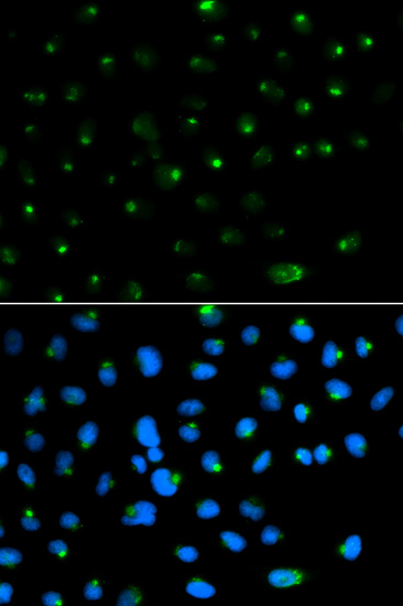
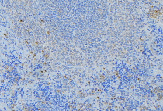
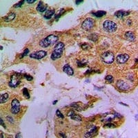
IHC (Immunohiostchemistry)
(AAA326401 at 1/100 staining Human lymph node tissue by IHC-P. The sample was formaldehyde fixed and a heat mediated antigen retrieval step in citrate buffer was performed. The sample was then blocked and incubated with the antibody for 1.5 hours at 22 degree C. An HRP conjugated goat anti-rabbit antibody was used as the secondary.)
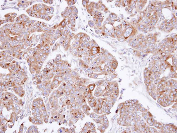
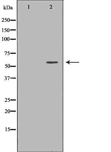
WB (Western Blot)
(Western blot analysis of extracts of HEK-293, using PSAP antibody. The lane on the left is treated with the antigen-specific peptide.)
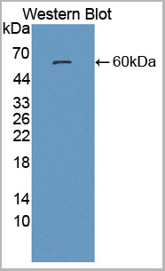
WB (Western Blot)
(Western blot analysis of extracts of HEK-293, using PSAP antibody. The lane on the left is treated with the antigen-specific peptide.)
Function: Saposin-A and saposin-C stimulate the hydrolysis of glucosylceramide by beta-glucosylceramidase (EC 3.2.1.45) and galactosylceramide by beta-galactosylceramidase (EC 3.2.1.46). Saposin-C apparently acts by combining with the enzyme and acidic lipid to form an activated complex, rather than by solubilizing the substrate.
Subunit Structure: Saposin-B is a homodimer. Prosaposin exists as a roughly half-half mixture of monomers and disulfide-linked dimers (PubMed:10406958, PubMed:12510003, PubMed:7730378, PubMed:21835174). Monomeric prosaposin interacts (via C-terminus) with sortilin/SORT1, the interaction is required for targeting to lysosomes (PubMed:14657016, PubMed:22431521).
Post-translational Modifications: The lysosomal precursor is proteolytically processed to 4 small peptides, which are similar to each other and are sphingolipid hydrolase activator proteins. N-linked glycans show a high degree of microheterogeneity. The one residue extended Saposin-B-Val is only found in 5% of the chains.
NCBI and Uniprot Product Information
Predicted: 59 kDa
Customer Reviews
Loading reviews...
Share Your Experience
Similar Products


Product Notes
The PSAP psap (Catalog #AAA326401) is an Antibody produced from Rabbit and is intended for research purposes only. The product is available for immediate purchase. The PSAP Antibody reacts with Human, Mouse, Rat and may cross-react with other species as described in the data sheet. AAA Biotech's PSAP can be used in a range of immunoassay formats including, but not limited to, ELISA, ICC (Immunocytochemistry), IF (Immunofluorescence), IHC (Immunohistochemistry), WB (Western Blot). Researchers should empirically determine the suitability of the PSAP psap for an application not listed in the data sheet. Researchers commonly develop new applications and it is an integral, important part of the investigative research process. It is sometimes possible for the material contained within the vial of "PSAP, Polyclonal Antibody" to become dispersed throughout the inside of the vial, particularly around the seal of said vial, during shipment and storage. We always suggest centrifuging these vials to consolidate all of the liquid away from the lid and to the bottom of the vial prior to opening. Please be advised that certain products may require dry ice for shipping and that, if this is the case, an additional dry ice fee may also be required.Precautions
All products in the AAA Biotech catalog are strictly for research-use only, and are absolutely not suitable for use in any sort of medical, therapeutic, prophylactic, in-vivo, or diagnostic capacity. By purchasing a product from AAA Biotech, you are explicitly certifying that said products will be properly tested and used in line with industry standard. AAA Biotech and its authorized distribution partners reserve the right to refuse to fulfill any order if we have any indication that a purchaser may be intending to use a product outside of our accepted criteria.Disclaimer
Though we do strive to guarantee the information represented in this datasheet, AAA Biotech cannot be held responsible for any oversights or imprecisions. AAA Biotech reserves the right to adjust any aspect of this datasheet at any time and without notice. It is the responsibility of the customer to inform AAA Biotech of any product performance issues observed or experienced within 30 days of receipt of said product. To see additional details on this or any of our other policies, please see our Terms & Conditions page.Item has been added to Shopping Cart
If you are ready to order, navigate to Shopping Cart and get ready to checkout.