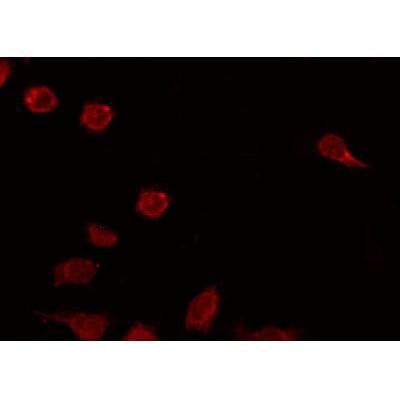
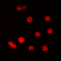
 IF (Immunofluorescence)
(Staining A549 cells by IF/ICC. The sample were fixed with PFA and permeabilized in 0.1% Triton X-100,then blocked in 10% serum for 45 minutes at 25 degree C. The primary antibody was diluted at 1/200 and incubated with the sample for 1 hour at 37 degree C. An Alexa Fluor 594 conjugated goat anti-rabbit IgG (H+L) Ab, diluted at 1/600, was used as the secondary antibody.)
IF (Immunofluorescence)
(Staining A549 cells by IF/ICC. The sample were fixed with PFA and permeabilized in 0.1% Triton X-100,then blocked in 10% serum for 45 minutes at 25 degree C. The primary antibody was diluted at 1/200 and incubated with the sample for 1 hour at 37 degree C. An Alexa Fluor 594 conjugated goat anti-rabbit IgG (H+L) Ab, diluted at 1/600, was used as the secondary antibody.)
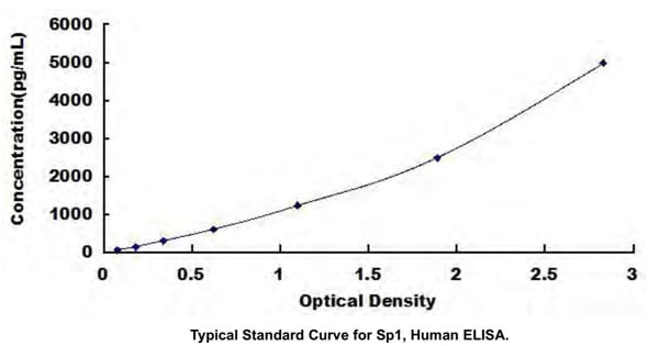
Rabbit SP1 Polyclonal Antibody | anti-SP1 antibody
Phospho-SP1 (Thr278) Antibody
Reactivity
Human, Mouse, RatPredicted Reactivity: Pig (100%), Bovine (100%), Sheep (100%), Rabbit (100%), Dog (100%)
Applications
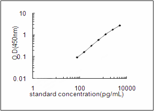
ELISA, Immunocytochemistry, Immunofluorescence, Western Blot
Purity
The antibody is from purified rabbit serum by affinity purification via sequential chromatography on phospho-peptide and non-phospho-peptide affinity columns.
Synonyms
SP1, Antibody; Phospho-SP1 (Thr278) Antibody; SP 1; SP1; Sp1 transcription factor; SP1_HUMAN; Specificity protein 1; Transcription factor Sp1; TSFP 1; TSFP1; anti-SP1 antibody
Host
Rabbit
Reactivity
Human, Mouse, Rat
Predicted Reactivity: Pig (100%), Bovine (100%), Sheep (100%), Rabbit (100%), Dog (100%)
Predicted Reactivity: Pig (100%), Bovine (100%), Sheep (100%), Rabbit (100%), Dog (100%)
Clonality
Polyclonal
Isotype
Rabbit IgG
Specificity
Phospho-SP1 (Thr278) Antibody detects endogenous levels of SP1 only when phosphorylated at Thr278.
Tissue Specificity: Up-regulated in adenocarcinomas of the stomach (at protein level). Isoform 3 is ubiquitously expressed at low levels.
Tissue Specificity: Up-regulated in adenocarcinomas of the stomach (at protein level). Isoform 3 is ubiquitously expressed at low levels.
Purity/Purification
The antibody is from purified rabbit serum by affinity purification via sequential chromatography on phospho-peptide and non-phospho-peptide affinity columns.
Form/Format
Liquid. Rabbit IgG in phosphate buffered saline, pH7.4, 150mM NaCl, 0.02% sodium azide and 50% glycerol.
Concentration
1mg/ml (varies by lot)
Applicable Applications for anti-SP1 antibody
ELISA (Peptide ELISA), ICC (Immunocytochemistry), IF (Immunofluorescence), WB (Western Blot)
Immunogen
A synthesized peptide derived from human SP1 around the phosphorylation site of Thr278.
Conjugation
Unconjugated
Fragment
Fab fragment
Post Translational Modifications
Phosphorylated on multiple serine and threonine residues. Phosphorylation is coupled to ubiquitination, sumoylation and proteolytic processing. Phosphorylation on Ser-59 enhances proteolytic cleavage. Phosphorylation on Ser-7 enhances ubiquitination and protein degradation. Hyperphosphorylation on Ser-101 in response to DNA damage has no effect on transcriptional activity. MAPK1/MAPK3-mediated phosphorylation on Thr-453 and Thr-739 enhances VEGF transcription but, represses FGF2-triggered PDGFR-alpha transcription. Also implicated in the repression of RECK by ERBB2. Hyperphosphorylated on Thr-278 and Thr-739 during mitosis by MAPK8 shielding SP1 from degradation by the ubiquitin-dependent pathway. Phosphorylated in the zinc-finger domain by calmodulin-activated PKCzeta. Phosphorylation on Ser-641 by PKCzeta is critical for TSA-activated LHR gene expression through release of its repressor, p107. Phosphorylation on Thr-668, Ser-670 and Thr-681 is stimulated by angiotensin II via the AT1 receptor inducing increased binding to the PDGF-D promoter. This phosphorylation is increased in injured artey wall. Ser-59 and Thr-681 can both be dephosphorylated by PP2A during cell-cycle interphase. Dephosphorylation on Ser-59 leads to increased chromatin association during interphase and increases the transcriptional activity. On insulin stimulation, sequentially glycosylated and phosphorylated on several C-terminal serine and threonine residues. Acetylated. Acetylation/deacetylation events affect transcriptional activity. Deacetylation leads to an increase in the expression the 12 (s)-lipooxygenase gene though recruitment of p300 to the promoter.Ubiquitinated. Ubiquitination occurs on the C-terminal proteolytically-cleaved peptide and is triggered by phosphorylation. Sumoylated with SUMO1. Sumoylation modulates proteolytic cleavage of the N-terminal repressor domain. Sumoylation levels are attenuated during tumorigenesis. Phosphorylation mediates SP1 desumoylation.Proteolytic cleavage in the N-terminal repressor domain is prevented by sumoylation. The C-terminal cleaved product is susceptible to degradation.O-glycosylated; Contains 8 N-acetylglucosamine side chains. Levels are controlled by insulin and the SP1 phosphorylation states. Insulin-mediated O-glycosylation locates SP1 to the nucleus, where it is sequentially deglycosylated and phosphorylated. O-glycosylation affects transcriptional activity through disrupting the interaction with a number of transcription factors including ELF1 and NFYA. Also inhibits interaction with the HIV1 promoter. Inhibited by peroxisomome proliferator receptor gamma (PPARgamma).
Subunit Structure
Interacts with ATF7IP, ATF7IP2, BAHD1, POGZ, HCFC1, AATF and PHC2. Interacts with HLTF; the interaction may be required for basal transcriptional activity of HLTF. Interacts (deacetylated form) with EP300; the interaction enhances gene expression. Interacts with HDAC1 and JUN. Interacts with ELF1; the interaction is inhibited by glycosylation of SP1. Interaction with NFYA; the interaction is inhibited by glycosylation of SP1. Interacts with ATF7IP and TBP. Interacts with MEIS2 isoform 4 and PBX1 isoform PBX1a. Interacts with EGR1. Interacts with SMARCA4/BRG1. Interacts with RNF112 in an oxidative stress-regulated manner (By similarity). Interacts with ZBTB7A; ZBTB7A prevents the binding to GC-rich motifs in promoters and represses the transcriptional activity of SP1. Interacts with DDX3X; this interaction potentiates SP1-induced CDKN1A/WAF1/CIP1 transcription. (Microbial infection) Interacts with varicella-zoster virus IE62 protein. (Microbial infection) Interacts with SV40 VP2/3 proteins. Interacts with SV40 major capsid protein VP1; this interaction leads to a cooperativity between the 2 proteins in DNA binding. (Microbial infection) Interacts with HIV-1 Vpr; the interaction is inhibited by SP1 O-glycosylation.
Similarity
The 9aaTAD motif is a transactivation domain present in a large number of yeast and animal transcription factors.Belongs to the Sp1 C2H2-type zinc-finger protein family.
Subcellular Location
Nucleus. Cytoplasm.
Note: Nuclear location is governed by glycosylated/phosphorylated states. Insulin promotes nuclear location, while glucagon favors cytoplasmic location.
Note: Nuclear location is governed by glycosylated/phosphorylated states. Insulin promotes nuclear location, while glucagon favors cytoplasmic location.
Preparation and Storage
Store at -20 degree C. Stable for 12 months from date of receipt.
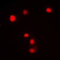
IF (Immunofluorescence)
(Staining A549 cells by IF/ICC. The sample were fixed with PFA and permeabilized in 0.1% Triton X-100,then blocked in 10% serum for 45 minutes at 25 degree C. The primary antibody was diluted at 1/200 and incubated with the sample for 1 hour at 37 degree C. An Alexa Fluor 594 conjugated goat anti-rabbit IgG (H+L) Ab, diluted at 1/600, was used as the secondary antibody.)
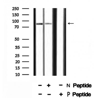
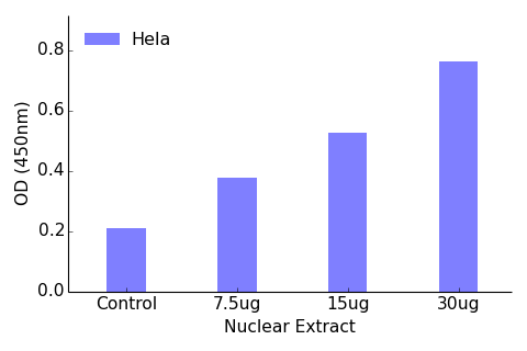
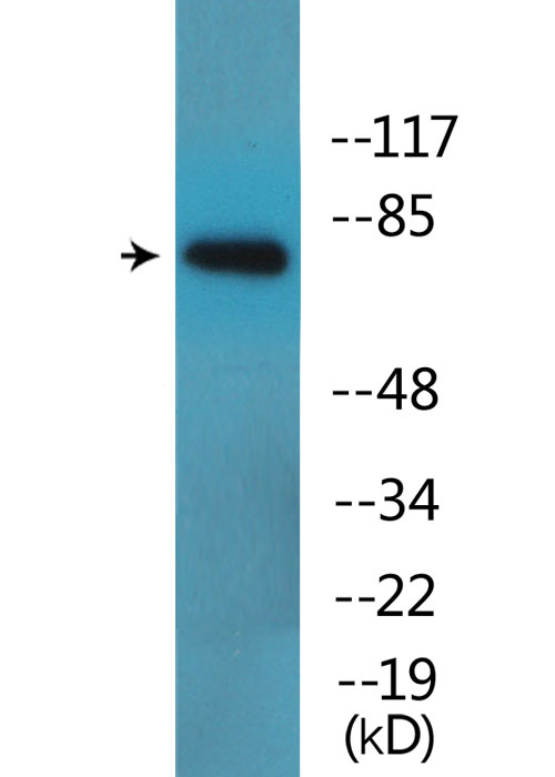
WB (Western Blot)
(Western blot analysis SP1 (Phospho-Thr278) using UV treated HeLa whole cell lysates.-/+ means absence or presence of N peptide(non-phospho peptide) and P peptide(phospho peptide).)
WB (Western Blot)
(Western blot analysis SP1 (Phospho-Thr278) using UV treated HeLa whole cell lysates.-/+ means absence or presence of N peptide(non-phospho peptide) and P peptide(phospho peptide).)
Related Product Information for anti-SP1 antibody
Transcription factor that can activate or repress transcription in response to physiological and pathological stimuli. Binds with high affinity to GC-rich motifs and regulates the expression of a large number of genes involved in a variety of processes such as cell growth, apoptosis, differentiation and immune responses. Highly regulated by post-translational modifications (phosphorylations, sumoylation, proteolytic cleavage, glycosylation and acetylation). Binds also the PDGFR-alpha G-box promoter. May have a role in modulating the cellular response to DNA damage. Implicated in chromatin remodeling. Plays an essential role in the regulation of FE65 gene expression. In complex with ATF7IP, maintains telomerase activity in cancer cells by inducing TERT and TERC gene expression. Isoform 3 is a stronger activator of transcription than isoform 1. Positively regulates the transcription of the core clock component ARNTL/BMAL1. Plays a role in the recruitment of SMARCA4/BRG1 on the c-FOS promoter. Plays a role in protecting cells against oxidative stress following brain injury by regulating the expression of RNF112 (By similarity).
NCBI and Uniprot Product Information
NCBI GI #
NCBI GeneID
NCBI Accession #
NCBI GenBank Nucleotide #
Molecular Weight
80,693 Da
NCBI Official Full Name
transcription factor Sp1 isoform c
NCBI Official Synonym Full Names
Sp1 transcription factor
NCBI Official Symbol
SP1
NCBI Protein Information
transcription factor Sp1; specificity protein 1
UniProt Protein Name
Transcription factor Sp1
UniProt Gene Name
SP1
UniProt Synonym Gene Names
TSFP1
UniProt Entry Name
SP1_HUMAN
Customer Reviews
Loading reviews...
Share Your Experience
Similar Products


Product Notes
The SP1 sp1 (Catalog #AAA329839) is an Antibody produced from Rabbit and is intended for research purposes only. The product is available for immediate purchase. The Phospho-SP1 (Thr278) Antibody reacts with Human, Mouse, Rat Predicted Reactivity: Pig (100%), Bovine (100%), Sheep (100%), Rabbit (100%), Dog (100%) and may cross-react with other species as described in the data sheet. AAA Biotech's SP1 can be used in a range of immunoassay formats including, but not limited to, ELISA (Peptide ELISA), ICC (Immunocytochemistry), IF (Immunofluorescence), WB (Western Blot). Researchers should empirically determine the suitability of the SP1 sp1 for an application not listed in the data sheet. Researchers commonly develop new applications and it is an integral, important part of the investigative research process. It is sometimes possible for the material contained within the vial of "SP1, Polyclonal Antibody" to become dispersed throughout the inside of the vial, particularly around the seal of said vial, during shipment and storage. We always suggest centrifuging these vials to consolidate all of the liquid away from the lid and to the bottom of the vial prior to opening. Please be advised that certain products may require dry ice for shipping and that, if this is the case, an additional dry ice fee may also be required.Precautions
All products in the AAA Biotech catalog are strictly for research-use only, and are absolutely not suitable for use in any sort of medical, therapeutic, prophylactic, in-vivo, or diagnostic capacity. By purchasing a product from AAA Biotech, you are explicitly certifying that said products will be properly tested and used in line with industry standard. AAA Biotech and its authorized distribution partners reserve the right to refuse to fulfill any order if we have any indication that a purchaser may be intending to use a product outside of our accepted criteria.Disclaimer
Though we do strive to guarantee the information represented in this datasheet, AAA Biotech cannot be held responsible for any oversights or imprecisions. AAA Biotech reserves the right to adjust any aspect of this datasheet at any time and without notice. It is the responsibility of the customer to inform AAA Biotech of any product performance issues observed or experienced within 30 days of receipt of said product. To see additional details on this or any of our other policies, please see our Terms & Conditions page.Item has been added to Shopping Cart
If you are ready to order, navigate to Shopping Cart and get ready to checkout.