
 IF (Immunofluorescence)
(Figure 10 Immunofluorescence Validation of TACE in Rat Brain (Pradillo et al, 2005)Cellular localization of TACE. Double immunofluorescence staining of brain sections from sham-operated (SHAM; A, C, E) and IPC-exposed animals (IPC; B, D, F) of TACE (red) and the cellular markers (green) NeuN (neurons; A, B), GFAP (astrocytes; C, D) and L. esculentum lectin (microglia and endothelium; E, F). White arrows indicate TACE-positive cells.)
IF (Immunofluorescence)
(Figure 10 Immunofluorescence Validation of TACE in Rat Brain (Pradillo et al, 2005)Cellular localization of TACE. Double immunofluorescence staining of brain sections from sham-operated (SHAM; A, C, E) and IPC-exposed animals (IPC; B, D, F) of TACE (red) and the cellular markers (green) NeuN (neurons; A, B), GFAP (astrocytes; C, D) and L. esculentum lectin (microglia and endothelium; E, F). White arrows indicate TACE-positive cells.)
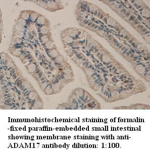
Rabbit TACE Polyclonal Antibody | anti-ADAM17 antibody
TACE Antibody
IF (Immunofluorescence)
(Figure 10 Immunofluorescence Validation of TACE in Rat Brain (Pradillo et al, 2005)Cellular localization of TACE. Double immunofluorescence staining of brain sections from sham-operated (SHAM; A, C, E) and IPC-exposed animals (IPC; B, D, F) of TACE (red) and the cellular markers (green) NeuN (neurons; A, B), GFAP (astrocytes; C, D) and L. esculentum lectin (microglia and endothelium; E, F). White arrows indicate TACE-positive cells.)
IF (Immunofluorescence)
(Figure 9 Immunofluorescence Validation of TACE in Rat Cortical Neurons (Hurtado et al., 2002)Double immunostaining of control and glutamate-exposed rat cortical cultures. (A) Control cultures show TACE immunoreactivity at the cellular membrane of some microglial cells (B) Glutamate-exposed cultures show that most microglial cells express TACE immunoreactivity.(C) Control cultures show that TACE immunostaining does not colocalize with astrocytes [glial fibrillary acidic protein (GFAP)-positive cells]. (D) Astrocyte (GFAP-positive cell) showing TACE immunoreactivity in its surface after treatment with glutamate.)
WB (Western Blot)
(Figure 8 Induced Expression Validation of TACE in Rat Cortical Neurons (Hurtado et al., 2002)Effect of oxygen–glucose deprivation(OGD) or glutamate on the levels of TACE/ADAM17 in rat cortical cultures. Western blot analysis of TACE in homogenates from control, glutamate, and OGD-exposed cultures from a representative experiment.)
WB (Western Blot)
(Figure 7 Overexpression Validation of TACE in MCF-7 Cells. (McGowan et al., 2007)ADAM-17 (TACE) protein expression, following transfection of vector and ADAM-17 cDNA, was examined by immunoblot analysis with anti-ADAM-17 (1131) antibodies in MCF-7 cells. Increased ADAM-17 was detected in ADAM-17 transfected cells.)
WB (Western Blot)
(Figure 6 KD Validation of TACE in MDA-MB-435 Cells. (McGowan et al., 2007)ADAM-17 protein expression, following transfection with ADAM-17 shRNA (two clones) or neomycin-resistant negative control vector, was examined by immunoblot analysis with anti-ADAM-17 antibodies (1131).)
WB (Western Blot)
(Figure 6 KD Validation of TACE in MDA-MB-435 Cells. (McGowan et al., 2007)ADAM-17 protein expression, following transfection with ADAM-17 shRNA (two clones) or neomycin-resistant negative control vector, was examined by immunoblot analysis with anti-ADAM-17 antibodies (1131).)
WB (Western Blot)
(Figure 5 KD Validation of TACE in Monkey COS Cells. (Wang et al., 2006)COS cells stably expressing Pref-1A were transfected with control siRNA or TACE siRNA. TACE was detected in lysates by using the anti-TACE antibody (1131). TACE expression levels were markedly reduced in TACE knockdown cell lysate.)
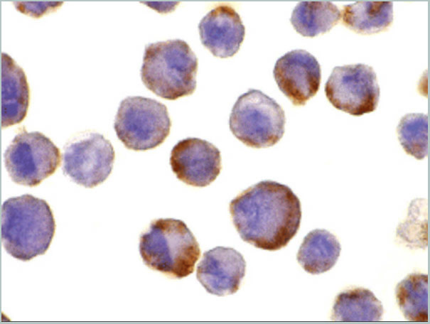
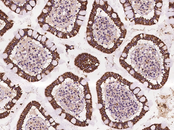
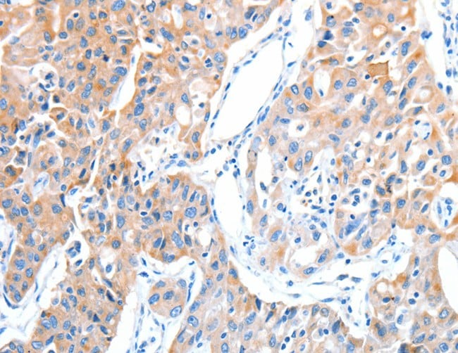
ICC (Immunocytochemistry)
(Figure 4 Immunocytochemistry Validation of TACE in HeLa CellsImmunohistochemical analysis of HeLa cells using anti-TACE antibody (1131) at 10 μg/ml. Cells was fixed with formaldehyde and blocked with 10% serum for 1 h at RT; antigen retrieval was by heat mediation with a citrate buffer (pH6). Samples were incubated with primary antibody overnight at 4˚C. A goat anti-rabbit IgG H&L (HRP) at 1/250 was used as secondary. Counter stained with Hematoxylin.)
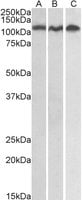
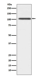
WB (Western Blot)
(strong>Figure 2 Independent Antibody Validation (IAV) via Protein Expression Profile in Cell LinesLoading: 15 μg of lysates per lane.Antibodies: TACE 1131 (0.5 μg/mL), TACE 22-001 (2 μg/mL), and GAPDH (0.02 μg/mL), 1h incubation at RT in 5% NFDM/TBST.Secondary: Goat anti-rabbit IgG HRP conjugate at 1:10000 dilution.)
NCBI and Uniprot Product Information
Customer Reviews
Loading reviews...
Share Your Experience
Similar Products


Product Notes
The ADAM17 adam17 (Catalog #AAA10912) is an Antibody produced from Rabbit and is intended for research purposes only. The product is available for immediate purchase. The TACE Antibody reacts with Human, Mouse, Rat and may cross-react with other species as described in the data sheet. AAA Biotech's TACE can be used in a range of immunoassay formats including, but not limited to, ELISA, WB (Western Blot), ICC (Immunocytochemistry), IF (Immunofluorescence), FCM/FACS (Flow Cytometry). TACE antibody can be used for detection of TACE by Western blot at 0.5 mug/mL. For immunocytochemistry use 10 mug/mL. For immunofluorescence start at 10 mug/mL. Researchers should empirically determine the suitability of the ADAM17 adam17 for an application not listed in the data sheet. Researchers commonly develop new applications and it is an integral, important part of the investigative research process. It is sometimes possible for the material contained within the vial of "TACE, Polyclonal Antibody" to become dispersed throughout the inside of the vial, particularly around the seal of said vial, during shipment and storage. We always suggest centrifuging these vials to consolidate all of the liquid away from the lid and to the bottom of the vial prior to opening. Please be advised that certain products may require dry ice for shipping and that, if this is the case, an additional dry ice fee may also be required.Precautions
All products in the AAA Biotech catalog are strictly for research-use only, and are absolutely not suitable for use in any sort of medical, therapeutic, prophylactic, in-vivo, or diagnostic capacity. By purchasing a product from AAA Biotech, you are explicitly certifying that said products will be properly tested and used in line with industry standard. AAA Biotech and its authorized distribution partners reserve the right to refuse to fulfill any order if we have any indication that a purchaser may be intending to use a product outside of our accepted criteria.Disclaimer
Though we do strive to guarantee the information represented in this datasheet, AAA Biotech cannot be held responsible for any oversights or imprecisions. AAA Biotech reserves the right to adjust any aspect of this datasheet at any time and without notice. It is the responsibility of the customer to inform AAA Biotech of any product performance issues observed or experienced within 30 days of receipt of said product. To see additional details on this or any of our other policies, please see our Terms & Conditions page.Item has been added to Shopping Cart
If you are ready to order, navigate to Shopping Cart and get ready to checkout.