 Application Data
(ACRIDINE ORANGE STAINING OF MCF-7 CELLSApoptosis was induced in human breast cancer MCF-7 cells by treating them with 0.15 uM camptothecin for 24 hours at 37°C. Cells were stained with AO in PBS for 30 minutes, then washed twice in PBS. Cells were photographed with a Nikon Microphot-FXA epifluorescence microscope at 40X using either a blue light excitation (492 nm) with a 540-550 nm emission filter (A, lysosomes appear yellowish green), or green light excitation (540 nm) with a long pass >640 nm barrier filter (B, lysosomes appear red).)
Application Data
(ACRIDINE ORANGE STAINING OF MCF-7 CELLSApoptosis was induced in human breast cancer MCF-7 cells by treating them with 0.15 uM camptothecin for 24 hours at 37°C. Cells were stained with AO in PBS for 30 minutes, then washed twice in PBS. Cells were photographed with a Nikon Microphot-FXA epifluorescence microscope at 40X using either a blue light excitation (492 nm) with a 540-550 nm emission filter (A, lysosomes appear yellowish green), or green light excitation (540 nm) with a long pass >640 nm barrier filter (B, lysosomes appear red).)
Acridine Orange Reagent
Acridine Orange
Chemical name: 3,6-acridinediamine, N, N, N’, N’-tetramethylmonohydrochloride
Molecular formula: C17H20ClN3
Appearance: Pale red/orange liquid
pH: 5.0 ± 0.5
2. Add AO to the cell sample media at 0.5 - 5 uM, equal to a final dilution of 1:2,000 - 1:200 in the cells (0.05-0.5% v/v). For example, if using AO at 1.0 uM in the final cell suspension, it must be diluted 1:1,000:
2a. First dilute it 1:100 in PBS or diH2O; e.g., put 10 uL AO into 990 uL PBS or diH2O.
2b. Pipette the diluted AO into the cell suspension at approximately 1:10; e.g., put 50 uL diluted AO into 450 uL cell suspension.
3. Incubate 15-30 minutes at 37°C.
4. Wash cells if reagent is too bright.
5. Analyze with fluorescence:
5a. Lysosomes will appear yellowish green by illuminating cells with a blue light (488 nm) excitation filter and a green light (540-550 nm) emission/barrier filter (Figures 1A and 2A).
5b. Alternatively, lysosomes will appear red when using an excitation filter of 550 nm (540-560 nm) and a long pass >610 nm emission/barrier filter (Figure 2B).
Stability: Shelf-life up to 24 months when refrigerated and protected from light.
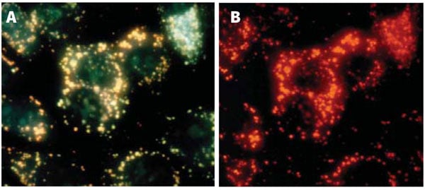
Application Data
(ACRIDINE ORANGE STAINING OF MCF-7 CELLSApoptosis was induced in human breast cancer MCF-7 cells by treating them with 0.15 uM camptothecin for 24 hours at 37°C. Cells were stained with AO in PBS for 30 minutes, then washed twice in PBS. Cells were photographed with a Nikon Microphot-FXA epifluorescence microscope at 40X using either a blue light excitation (492 nm) with a 540-550 nm emission filter (A, lysosomes appear yellowish green), or green light excitation (540 nm) with a long pass >640 nm barrier filter (B, lysosomes appear red).)
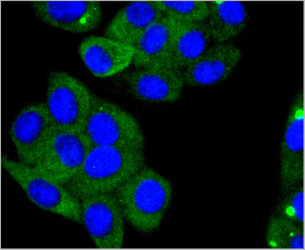
Application Data
(ACRIDINE ORANGE STAINING OF JURKAT CELLSNormal Jurkat cells stained with Acridine Orange (AO) show orange lysosomal staining. Jurkat cells were stained with 5uM AO in PBS for 60 minutes at 37°C. Photomicrographs were taken using a Nikon Eclipse E800 photomicroscope using a 460-500 nm excitation filter and a 505-560 nm emission / barrier filter set at 300X. Photo B shows the corresponding DIC image of the cells in A (AO appears faintly).)
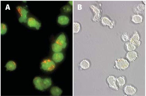
Acridine orange (AO) (MBS258066) is an orange/red fluorescent chelating dye used to reveal lysosomes and nuclei in cultured cells. Due to its metachromatic properties, AO is commonly used in fluorescence microscopy and flow cytometry analysis of cellular physiology and cell cycle status.
AO is a slightly cationic, lipophilic, weak base capable of permeating cell and organelle membrane structure3. Although quite cell permeant in the neutral form, once protonated, this dye tends to become trapped on the low pH side of the membrane barrier leading to accumulation in acidic organelle structures. Proton pump driven lysosomal acidity generates a significant pH gradient resulting in the efficient concentration of AO within the lysosome organelles6. This is sufficient to create intra-lysosomal concentrations leading to precipitation of AO into aggregated granules. These oligomeric structures exhibit a red shift (640 nm) compared to the monomeric AO that emits at 525 nm.
As AO exhibits a very broad emission range, one of several filter pairings on the fluorescence microscope can be used to view this stain. Using an excitation filter of 550 nm (540-560 nm) with a long pass >610 nm emission/barrier filter, lysosomes appear red. When illuminating with a blue light excitation filter (488 nm) and a green light emission/barrier filter (540-550 nm), lysosomes appear yellowish green instead of red. As this filter combination is very close to the maximum emission of AO, the slide may appear too bright. Excess AO may be removed by washing cells prior to viewing.
AO can be utilized in conjunction with a number of other staining techniques and fluorogenic substrates including Magic Red®-DEVDase substrate that detects caspase-3/7 activation in apoptotic cells8. Because of the overlap in emissions, be wary of dual staining with other red stains as this will yield confusing results. Red dyes should be used separately.
AO may be used neat or diluted in diH2O, PBS, or media prior to pipetting into the cell suspension. Lysosomal structures can be visualized by staining with AO at 0.5-5.0 uM. This concentration range can be obtained by diluting AO reagent stock at 1 mM) 1:2,000-1:200 (0.05-0.5% v/v) into the final cell suspension. For example, if using AO at 1.0 uM in the final cell suspension, it must be diluted 1:1,000. First dilute it 1:100 in PBS; e.g., put 10 uL AO into 990 uL PBS. Pipette the diluted AO into the cell suspension at approximately 1:10; e.g., put 50 uL diluted AO into 450 uL cell suspension. Always protect AO from bright light.
Customer Reviews
Loading reviews...
Share Your Experience
Similar Products


Product Notes
The Acridine Orange (Catalog #AAA52744) is a Reagent and is intended for research purposes only. The product is available for immediate purchase. AAA Biotech's Acridine Orange can be used in a range of immunoassay formats including, but not limited to, FM (Fluorescence Microscopy), FCM/FACS (Flow Cytometry). Researchers should empirically determine the suitability of the Acridine Orange for an application not listed in the data sheet. Researchers commonly develop new applications and it is an integral, important part of the investigative research process. It is sometimes possible for the material contained within the vial of "Acridine Orange, Reagent" to become dispersed throughout the inside of the vial, particularly around the seal of said vial, during shipment and storage. We always suggest centrifuging these vials to consolidate all of the liquid away from the lid and to the bottom of the vial prior to opening. Please be advised that certain products may require dry ice for shipping and that, if this is the case, an additional dry ice fee may also be required.Precautions
All products in the AAA Biotech catalog are strictly for research-use only, and are absolutely not suitable for use in any sort of medical, therapeutic, prophylactic, in-vivo, or diagnostic capacity. By purchasing a product from AAA Biotech, you are explicitly certifying that said products will be properly tested and used in line with industry standard. AAA Biotech and its authorized distribution partners reserve the right to refuse to fulfill any order if we have any indication that a purchaser may be intending to use a product outside of our accepted criteria.Disclaimer
Though we do strive to guarantee the information represented in this datasheet, AAA Biotech cannot be held responsible for any oversights or imprecisions. AAA Biotech reserves the right to adjust any aspect of this datasheet at any time and without notice. It is the responsibility of the customer to inform AAA Biotech of any product performance issues observed or experienced within 30 days of receipt of said product. To see additional details on this or any of our other policies, please see our Terms & Conditions page.Item has been added to Shopping Cart
If you are ready to order, navigate to Shopping Cart and get ready to checkout.