Filters
Clonality
Type
Reactivity
Gene Name
Isotype
Host
Application
Clone
1614 results for "Proteins" - showing 1350-1400


 Application Data
(Analysis of Protein Array containing more than 19,000 full-length human proteins using ERCC1 Mouse Monoclonal Antibody (ERCC1/2318). Z- and S- Score: The Z-score represents the strength of a signal that a monoclonal antibody (MAb) (in combination with a fluorescently-tagged anti-IgG secondary antibody) produces when binding to a particular protein on the HuProtTM array. Z-scores are described in units of standard deviations (SD's) above the mean value of all signals generated on that array. If targets on HuProtTM are arranged in descending order of the Z-score, the S-score is the difference (also in units of SD's) between the Z-score. S-score therefore represents the relative target specificity of a MAb to its intended target. A MAb is considered to specific to its intended target, if the MAb has an S-score of at least 2.5. For example, if a MAb binds to protein X with a Z-score of 43 and to protein Y with a Z-score of 14, then the S-score for the binding of that MAb to protein X is equal to 29.)
Application Data
(Analysis of Protein Array containing more than 19,000 full-length human proteins using ERCC1 Mouse Monoclonal Antibody (ERCC1/2318). Z- and S- Score: The Z-score represents the strength of a signal that a monoclonal antibody (MAb) (in combination with a fluorescently-tagged anti-IgG secondary antibody) produces when binding to a particular protein on the HuProtTM array. Z-scores are described in units of standard deviations (SD's) above the mean value of all signals generated on that array. If targets on HuProtTM are arranged in descending order of the Z-score, the S-score is the difference (also in units of SD's) between the Z-score. S-score therefore represents the relative target specificity of a MAb to its intended target. A MAb is considered to specific to its intended target, if the MAb has an S-score of at least 2.5. For example, if a MAb binds to protein X with a Z-score of 43 and to protein Y with a Z-score of 14, then the S-score for the binding of that MAb to protein X is equal to 29.)
ERCC1/RAD10, Monoclonal Antibody (Cat# AAA23913)
Full Name
ERCC1/RAD10 (Tumor Progression Marker)
Gene Names
ERCC1; UV20; COFS4; RAD10
Reactivity
Human
Applications
IHC
Purity
Purified Ab with BSA and Azide at 200ug/ml OR Purified Ab WITHOUT BSA and Azide at 1.0mg/ml
Pricing


 Application Data
(Analysis of Protein Array containing more than 19, 000 full-length human proteins using Mesothelin Mouse Monoclonal Antibody (MSLN/2131). Z- and S- Score: The Z-score represents the strength of a signal that a monoclonal antibody (MAb) (in combination with a fluorescently-tagged anti-IgG secondary antibody) produces when binding to a particular protein on the HuProtTM array. Z-scores are described in units of standard deviations (SD's) above the mean value of all signals generated on that array. If targets on HuProtTM are arranged in descending order of the Z-score, the S-score is the difference (also in units of SD's) between the Z-score. S-score therefore represents the relative target specificity of a MAb to its intended target. A MAb is considered to specific to its intended target, if the MAb has an S-score of at least 2.5. For example, if a MAb binds to protein X with a Z-score of 43 and to protein Y with a Z-score of 14, then the S-score for the binding of that MAb to protein X is equal to 29.)
Application Data
(Analysis of Protein Array containing more than 19, 000 full-length human proteins using Mesothelin Mouse Monoclonal Antibody (MSLN/2131). Z- and S- Score: The Z-score represents the strength of a signal that a monoclonal antibody (MAb) (in combination with a fluorescently-tagged anti-IgG secondary antibody) produces when binding to a particular protein on the HuProtTM array. Z-scores are described in units of standard deviations (SD's) above the mean value of all signals generated on that array. If targets on HuProtTM are arranged in descending order of the Z-score, the S-score is the difference (also in units of SD's) between the Z-score. S-score therefore represents the relative target specificity of a MAb to its intended target. A MAb is considered to specific to its intended target, if the MAb has an S-score of at least 2.5. For example, if a MAb binds to protein X with a Z-score of 43 and to protein Y with a Z-score of 14, then the S-score for the binding of that MAb to protein X is equal to 29.)
Mesothelin, Monoclonal Antibody (Cat# AAA23897)
Full Name
Mesothelin (Mesothelial Marker)
Gene Names
MSLN; MPF; SMRP
Reactivity
Human, Mouse, Rat. Others not known.
Applications
IHC
Pricing


 Application Data
(Analysis of Protein Array containing >19, 000 full-length human proteins using TACSTD2 Mouse Monoclonal Antibody (TACSTD2/2153) Z- and S- Score: The Z-score represents the strength of a signal that a monoclonal antibody (MAb) (in combination with a fluorescently-tagged anti-IgG secondary antibody) produces when binding to a particular protein on the HuProtTM array. Z-scores are described in units of standard deviations (SD's) above the mean value of all signals generated on that array. If targets on HuProtTM are arranged in descending order of the Z-score, the S-score is the difference (also in units of SD's) between the Z-score. S-score therefore represents the relative target specificity of a MAb to its intended target. A MAb is considered to specific to its intended target, if the MAb has an S-score of at least 2.5. For example, if a MAb binds to protein X with a Z-score of 43 and to protein Y with a Z-score of 14, then the S-score for the binding of that MAb to protein X is equal to 29.)
Application Data
(Analysis of Protein Array containing >19, 000 full-length human proteins using TACSTD2 Mouse Monoclonal Antibody (TACSTD2/2153) Z- and S- Score: The Z-score represents the strength of a signal that a monoclonal antibody (MAb) (in combination with a fluorescently-tagged anti-IgG secondary antibody) produces when binding to a particular protein on the HuProtTM array. Z-scores are described in units of standard deviations (SD's) above the mean value of all signals generated on that array. If targets on HuProtTM are arranged in descending order of the Z-score, the S-score is the difference (also in units of SD's) between the Z-score. S-score therefore represents the relative target specificity of a MAb to its intended target. A MAb is considered to specific to its intended target, if the MAb has an S-score of at least 2.5. For example, if a MAb binds to protein X with a Z-score of 43 and to protein Y with a Z-score of 14, then the S-score for the binding of that MAb to protein X is equal to 29.)
TACSTD2/TROP2, Monoclonal Antibody (Cat# AAA23900)
Full Name
TACSTD2/TROP2 (Epithelial Marker)
Gene Names
TACSTD2; EGP1; GP50; M1S1; EGP-1; TROP2; GA7331; GA733-1
Reactivity
Human. Others not known.
Applications
IHC
Pricing


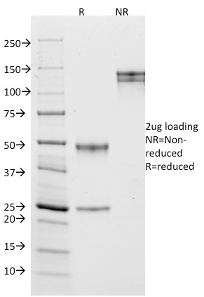
 IHC (Immunohistchemistry)
(Figure 9. IHC analysis of BAK using anti-BAK antibody (AAA11654).BAK was detected in frozen section of rat cardiac muscle tissue. Heat mediated antigen retrieval was performed in citrate buffer (pH6, epitope retrieval solution) for 20 mins. The tissue section was blocked with 10% goat serum. The tissue section was then incubated with 1ug/ml rabbit anti-BAK Antibody (AAA11654) overnight at 4 degree C. Biotinylated goat anti-rabbit IgG was used as secondary antibody and incubated for 30 minutes at 37 degree C. The tissue section was developed using Strepavidin-Biotin-Complex (SABC) with DAB as the chromogen.)
IHC (Immunohistchemistry)
(Figure 9. IHC analysis of BAK using anti-BAK antibody (AAA11654).BAK was detected in frozen section of rat cardiac muscle tissue. Heat mediated antigen retrieval was performed in citrate buffer (pH6, epitope retrieval solution) for 20 mins. The tissue section was blocked with 10% goat serum. The tissue section was then incubated with 1ug/ml rabbit anti-BAK Antibody (AAA11654) overnight at 4 degree C. Biotinylated goat anti-rabbit IgG was used as secondary antibody and incubated for 30 minutes at 37 degree C. The tissue section was developed using Strepavidin-Biotin-Complex (SABC) with DAB as the chromogen.)
BAK, Polyclonal Antibody (Cat# AAA11654)
Full Name
Anti-BAK Antibody
Gene Names
BAK1; BAK; CDN1; BCL2L7; BAK-LIKE
Reactivity
Human, Mouse, Rat
Applications
WB, IHC
Purity
Immunogen Affinity Purified
Pricing


 IHC (Immunohistochemistry)
(Figure 8. IHC analysis of RbAp48 using anti-RbAp48 antibody (AAA11671).RbAp48 was detected in frozen section of rat small intestine tissue. Heat mediated antigen retrieval was performed in citrate buffer (pH6, epitope retrieval solution) for 20 mins. The tissue section was blocked with 10% goat serum. The tissue section was then incubated with 1ug/ml rabbit anti-RbAp48 Antibody (AAA11671) overnight at 4 degree C. Biotinylated goat anti-rabbit IgG was used as secondary antibody and incubated for 30 minutes at 37 degree C. The tissue section was developed using Strepavidin-Biotin-Complex (SABC) with DAB as the chromogen.)
IHC (Immunohistochemistry)
(Figure 8. IHC analysis of RbAp48 using anti-RbAp48 antibody (AAA11671).RbAp48 was detected in frozen section of rat small intestine tissue. Heat mediated antigen retrieval was performed in citrate buffer (pH6, epitope retrieval solution) for 20 mins. The tissue section was blocked with 10% goat serum. The tissue section was then incubated with 1ug/ml rabbit anti-RbAp48 Antibody (AAA11671) overnight at 4 degree C. Biotinylated goat anti-rabbit IgG was used as secondary antibody and incubated for 30 minutes at 37 degree C. The tissue section was developed using Strepavidin-Biotin-Complex (SABC) with DAB as the chromogen.)
RbAp48, Polyclonal Antibody (Cat# AAA11671)
Full Name
Anti-RbAp48 Antibody
Gene Names
RBBP4; NURF55; RBAP48; lin-53
Reactivity
Human, Mouse, Rat
Applications
WB, IHC
Purity
Immunogen Affinity Purified
Pricing


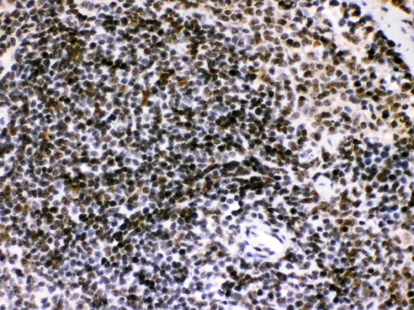
 IHC (Immunohistochemistry)
(At 1/100 staining Human gastric cancer by IHC-P. The sample was formaldehyde fixed and a heat mediated antigen retrieval step in citrate buffer was performed. The sample was then blocked and incubated with the primary antibody at 4 degree C overnight. An HRP conjugated anti-Rabbit antibody was used as the secondary antibody.)
IHC (Immunohistochemistry)
(At 1/100 staining Human gastric cancer by IHC-P. The sample was formaldehyde fixed and a heat mediated antigen retrieval step in citrate buffer was performed. The sample was then blocked and incubated with the primary antibody at 4 degree C overnight. An HRP conjugated anti-Rabbit antibody was used as the secondary antibody.)
alpha 1 Catenin, Polyclonal Antibody (Cat# AAA31379)
Full Name
alpha 1 Catenin Antibody
Gene Names
CTNNA1; CAP102
Reactivity
Human, Mouse, Rat
Applications
WB, IHC, EIA
Purity
The antiserum was purified by peptide affinity chromatography using SulfoLink Coupling Resin
Pricing


 IHC (Immunohistchemistry)
(Figure 6. IHC analysis of GLO1 using anti-GLO1 antibody (AAA19155).GLO1 was detected in paraffin-embedded section of rat spleen tissue. Heat mediated antigen retrieval was performed in citrate buffer (pH6, epitope retrieval solution) for 20 mins. The tissue section was blocked with 10% goat serum. The tissue section was then incubated with 1ug/ml rabbit anti-GLO1 Antibody (AAA19155) overnight at 4 degree C. Biotinylated goat anti-rabbit IgG was used as secondary antibody and incubated for 30 minutes at 37 degree C. The tissue section was developed using Strepavidin-Biotin-Complex (SABC) with DAB as the chromogen.)
IHC (Immunohistchemistry)
(Figure 6. IHC analysis of GLO1 using anti-GLO1 antibody (AAA19155).GLO1 was detected in paraffin-embedded section of rat spleen tissue. Heat mediated antigen retrieval was performed in citrate buffer (pH6, epitope retrieval solution) for 20 mins. The tissue section was blocked with 10% goat serum. The tissue section was then incubated with 1ug/ml rabbit anti-GLO1 Antibody (AAA19155) overnight at 4 degree C. Biotinylated goat anti-rabbit IgG was used as secondary antibody and incubated for 30 minutes at 37 degree C. The tissue section was developed using Strepavidin-Biotin-Complex (SABC) with DAB as the chromogen.)
GLO1/Glyoxalase I, Polyclonal Antibody (Cat# AAA19155)
Full Name
Anti-GLO1/Glyoxalase I Picoband antibody
Gene Names
GLO1; GLYI; GLOD1; HEL-S-74
Reactivity
Human, Mouse, Rat
No cross reactivity with other proteins.
No cross reactivity with other proteins.
Applications
EIA, IHC, WB
Pricing


 IHC (Immunohistochemistry)
(Figure 7. IHC analysis of VEGF Receptor 3 using anti-VEGF Receptor 3 antibody (AAA19147).VEGF Receptor 3 was detected in paraffin-embedded section of mouse liver tissue. Heat mediated antigen retrieval was performed in citrate buffer (pH6, epitope retrieval solution) for 20 mins. The tissue section was blocked with 10% goat serum. The tissue section was then incubated with 1ug/ml rabbit anti-VEGF Receptor 3 Antibody (AAA19147) overnight at 4 degree C. Biotinylated goat anti-rabbit IgG was used as secondary antibody and incubated for 30 minutes at 37 degree C. The tissue section was developed using Strepavidin-Biotin-Complex (SABC) with DAB as the chromogen.)
IHC (Immunohistochemistry)
(Figure 7. IHC analysis of VEGF Receptor 3 using anti-VEGF Receptor 3 antibody (AAA19147).VEGF Receptor 3 was detected in paraffin-embedded section of mouse liver tissue. Heat mediated antigen retrieval was performed in citrate buffer (pH6, epitope retrieval solution) for 20 mins. The tissue section was blocked with 10% goat serum. The tissue section was then incubated with 1ug/ml rabbit anti-VEGF Receptor 3 Antibody (AAA19147) overnight at 4 degree C. Biotinylated goat anti-rabbit IgG was used as secondary antibody and incubated for 30 minutes at 37 degree C. The tissue section was developed using Strepavidin-Biotin-Complex (SABC) with DAB as the chromogen.)
VEGF Receptor 3, Polyclonal Antibody (Cat# AAA19147)
Full Name
Anti-VEGF Receptor 3 Picoband antibody
Gene Names
FLT4; PCL; FLT-4; FLT41; LMPH1A; VEGFR3; VEGFR-3
Reactivity
Human, Mouse, Rat
Applications
EIA, IHC, WB, FC
Purity
Immunoggen affinity purified
Pricing


 IHC (Immunohistchemistry)
(Figure 6. IHC analysis of FH using anti-FH antibody (AAA19157).FH was detected in paraffin-embedded section of human mammary cancer tissue. Heat mediated antigen retrieval was performed in citrate buffer (pH6, epitope retrieval solution) for 20 mins. The tissue section was blocked with 10% goat serum. The tissue section was then incubated with 1ug/ml rabbit anti-FH Antibody (AAA19157) overnight at 4 degree C. Biotinylated goat anti-rabbit IgG was used as secondary antibody and incubated for 30 minutes at 37 degree C. The tissue section was developed using Strepavidin-Biotin-Complex (SABC) with DAB as the chromogen.)
IHC (Immunohistchemistry)
(Figure 6. IHC analysis of FH using anti-FH antibody (AAA19157).FH was detected in paraffin-embedded section of human mammary cancer tissue. Heat mediated antigen retrieval was performed in citrate buffer (pH6, epitope retrieval solution) for 20 mins. The tissue section was blocked with 10% goat serum. The tissue section was then incubated with 1ug/ml rabbit anti-FH Antibody (AAA19157) overnight at 4 degree C. Biotinylated goat anti-rabbit IgG was used as secondary antibody and incubated for 30 minutes at 37 degree C. The tissue section was developed using Strepavidin-Biotin-Complex (SABC) with DAB as the chromogen.)
FH/Fumarase, Polyclonal Antibody (Cat# AAA19157)
Full Name
Anti-FH/Fumarase Picoband Antibody
Gene Names
FH; MCL; FMRD; LRCC; HLRCC; MCUL1
Reactivity
Human, Mouse, Rat
No cross reactivity with other proteins.
No cross reactivity with other proteins.
Applications
IHC, WB
Purity
Immunogen affinity purified
Pricing


 FCM (Flow Cytometry)
(Figure 8. Flow Cytometry analysis of LLC cells using anti-Synaptotagmin 1 antibody (AAA19158).Overlay histogram showing LLC cells stained with AAA19158 (Blue line).The cells were blocked with 10% normal goat serum. And then incubated with rabbit anti-Synaptotagmin 1 Antibody (AAA19158,1ug/1x10^6 cells) for 30 min at 20 degree C. DyLight®488 conjugated goat anti-rabbit IgG (5-10ug/1x10^6 cells) was used as secondary antibody for 30 minutes at 20 degree C. Isotype control antibody (Green line) was rabbit IgG (1ug/1x106) used under the same conditions. Unlabelled sample (Red line) was also used as a control.)
FCM (Flow Cytometry)
(Figure 8. Flow Cytometry analysis of LLC cells using anti-Synaptotagmin 1 antibody (AAA19158).Overlay histogram showing LLC cells stained with AAA19158 (Blue line).The cells were blocked with 10% normal goat serum. And then incubated with rabbit anti-Synaptotagmin 1 Antibody (AAA19158,1ug/1x10^6 cells) for 30 min at 20 degree C. DyLight®488 conjugated goat anti-rabbit IgG (5-10ug/1x10^6 cells) was used as secondary antibody for 30 minutes at 20 degree C. Isotype control antibody (Green line) was rabbit IgG (1ug/1x106) used under the same conditions. Unlabelled sample (Red line) was also used as a control.)
Synaptotagmin 1, Polyclonal Antibody (Cat# AAA19158)
Full Name
Anti-Synaptotagmin 1 Picoband antibody
Gene Names
SYT1; P65; SYT; SVP65
Reactivity
Human, Mouse, Rat
No cross reactivity with other proteins.
No cross reactivity with other proteins.
Applications
IHC, WB
Pricing


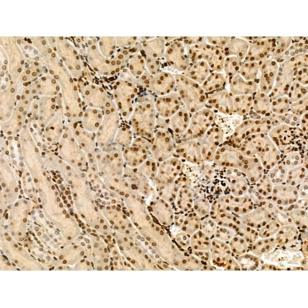
 IHC (Immunohistochemistry)
(Figure 10. IHC analysis of COX IV using anti-COX IV antibody (AAA19173).COX IV was detected in paraffin-embedded section of mouse kidney tissue. Heat mediated antigen retrieval was performed in citrate buffer (pH6, epitope retrieval solution) for 20 mins. The tissue section was blocked with 10% goat serum. The tissue section was then incubated with 2ug/ml rabbit anti-COX IV Antibody (AAA19173) overnight at 4 degree C. Biotinylated goat anti-rabbit IgG was used as secondary antibody and incubated for 30 minutes at 37 degree C. The tissue section was developed using Strepavidin-Biotin-Complex (SABC) with DAB as the chromogen.)
IHC (Immunohistochemistry)
(Figure 10. IHC analysis of COX IV using anti-COX IV antibody (AAA19173).COX IV was detected in paraffin-embedded section of mouse kidney tissue. Heat mediated antigen retrieval was performed in citrate buffer (pH6, epitope retrieval solution) for 20 mins. The tissue section was blocked with 10% goat serum. The tissue section was then incubated with 2ug/ml rabbit anti-COX IV Antibody (AAA19173) overnight at 4 degree C. Biotinylated goat anti-rabbit IgG was used as secondary antibody and incubated for 30 minutes at 37 degree C. The tissue section was developed using Strepavidin-Biotin-Complex (SABC) with DAB as the chromogen.)
COX IV, Polyclonal Antibody (Cat# AAA19173)
Full Name
Anti-COX IV Picoband Antibody
Gene Names
COX4I1; COX4; COXIV; COX4-1; COXIV-1; COX IV-1
Reactivity
Human, Mouse, Rat
No cross reactivity with other proteins.
No cross reactivity with other proteins.
Applications
EIA, IHC, WB
Pricing


 IHC (Immunohistchemistry)
(Figure 6. IHC analysis of Annexin VI using anti-Annexin VI antibody (AAA19170).Annexin VI was detected in paraffin-embedded section of rat spleen tissue. Heat mediated antigen retrieval was performed in citrate buffer (pH6, epitope retrieval solution) for 20 mins. The tissue section was blocked with 10% goat serum. The tissue section was then incubated with 2ug/ml rabbit anti-Annexin VI Antibody (AAA19170) overnight at 4 degree C. Biotinylated goat anti-rabbit IgG was used as secondary antibody and incubated for 30 minutes at 37 degree C. The tissue section was developed using Strepavidin-Biotin-Complex (SABC) with DAB as the chromogen.)
IHC (Immunohistchemistry)
(Figure 6. IHC analysis of Annexin VI using anti-Annexin VI antibody (AAA19170).Annexin VI was detected in paraffin-embedded section of rat spleen tissue. Heat mediated antigen retrieval was performed in citrate buffer (pH6, epitope retrieval solution) for 20 mins. The tissue section was blocked with 10% goat serum. The tissue section was then incubated with 2ug/ml rabbit anti-Annexin VI Antibody (AAA19170) overnight at 4 degree C. Biotinylated goat anti-rabbit IgG was used as secondary antibody and incubated for 30 minutes at 37 degree C. The tissue section was developed using Strepavidin-Biotin-Complex (SABC) with DAB as the chromogen.)
Annexin VI, Polyclonal Antibody (Cat# AAA19170)
Full Name
Anti-Annexin VI Picoband Antibody
Gene Names
ANXA6; ANX6; CBP68
Reactivity
Human, Mouse, Rat
No cross reactivity with other proteins.
No cross reactivity with other proteins.
Applications
EIA, IHC, WB
Pricing


 IHC (Immunohistchemistry)
(Figure 6. IHC analysis of TSPAN12 using anti-TSPAN12 antibody (AAA19174).TSPAN12 was detected in paraffin-embedded section of human rectal cancer tissue. Heat mediated antigen retrieval was performed in citrate buffer (pH6, epitope retrieval solution) for 20 mins. The tissue section was blocked with 10% goat serum. The tissue section was then incubated with 1ug/ml rabbit anti-TSPAN12 Antibody (AAA19174) overnight at 4 degree C. Biotinylated goat anti-rabbit IgG was used as secondary antibody and incubated for 30 minutes at 37 degree C. The tissue section was developed using Strepavidin-Biotin-Complex (SABC) with DAB as the chromogen.)
IHC (Immunohistchemistry)
(Figure 6. IHC analysis of TSPAN12 using anti-TSPAN12 antibody (AAA19174).TSPAN12 was detected in paraffin-embedded section of human rectal cancer tissue. Heat mediated antigen retrieval was performed in citrate buffer (pH6, epitope retrieval solution) for 20 mins. The tissue section was blocked with 10% goat serum. The tissue section was then incubated with 1ug/ml rabbit anti-TSPAN12 Antibody (AAA19174) overnight at 4 degree C. Biotinylated goat anti-rabbit IgG was used as secondary antibody and incubated for 30 minutes at 37 degree C. The tissue section was developed using Strepavidin-Biotin-Complex (SABC) with DAB as the chromogen.)
TSPAN12, Polyclonal Antibody (Cat# AAA19174)
Full Name
Anti-TSPAN12 Picoband antibody
Gene Names
TSPAN12; EVR5; NET2; NET-2; TM4SF12
Reactivity
Human, Mouse, Rat
No cross reactivity with other proteins.
No cross reactivity with other proteins.
Applications
EIA, IHC, WB
Pricing


 IHC (Immunohistchemistry)
(Figure 6. IHC analysis of Musashi 1/Msi1 using anti- Musashi 1/Msi1 antibody (AAA19172).Musashi 1/Msi1 was detected in paraffin-embedded section of human mammary cancer tissues. Heat mediated antigen retrieval was performed in citrate buffer (pH6, epitope retrieval solution) for 20 mins. The tissue section was blocked with 10% goat serum. The tissue section was then incubated with 1ug/ml rabbit anti- Musashi 1/Msi1 Antibody (AAA19172) overnight at 4 degree C. Biotinylated goat anti-rabbit IgG was used as secondary antibody and incubated for 30 minutes at 37 degree C. The tissue section was developed using Strepavidin-Biotin-Complex (SABC) with DAB as the chromogen.)
IHC (Immunohistchemistry)
(Figure 6. IHC analysis of Musashi 1/Msi1 using anti- Musashi 1/Msi1 antibody (AAA19172).Musashi 1/Msi1 was detected in paraffin-embedded section of human mammary cancer tissues. Heat mediated antigen retrieval was performed in citrate buffer (pH6, epitope retrieval solution) for 20 mins. The tissue section was blocked with 10% goat serum. The tissue section was then incubated with 1ug/ml rabbit anti- Musashi 1/Msi1 Antibody (AAA19172) overnight at 4 degree C. Biotinylated goat anti-rabbit IgG was used as secondary antibody and incubated for 30 minutes at 37 degree C. The tissue section was developed using Strepavidin-Biotin-Complex (SABC) with DAB as the chromogen.)
Musashi 1/Msi1, Polyclonal Antibody (Cat# AAA19172)
Full Name
Anti-Musashi 1/Msi1 Picoband Antibody
Reactivity
Human, Mouse, Rat
No cross reactivity with other proteins
No cross reactivity with other proteins
Applications
IHC, WB
Purity
Immunogen affinity purified
Pricing


 IHC (Immunohistochemistry)
(Figure 7. IHC analysis of MED4 using anti-MED4 antibody (AAA19178).MED4 was detected in paraffin-embedded section of human rectal cancer tissue. Heat mediated antigen retrieval was performed in citrate buffer (pH6, epitope retrieval solution) for 20 mins. The tissue section was blocked with 10% goat serum. The tissue section was then incubated with 1ug/ml rabbit anti-MED4 Antibody (AAA19178) overnight at 4 degree C. Biotinylated goat anti-rabbit IgG was used as secondary antibody and incubated for 30 minutes at 37 degree C. The tissue section was developed using Strepavidin-Biotin-Complex (SABC) with DAB as the chromogen.)
IHC (Immunohistochemistry)
(Figure 7. IHC analysis of MED4 using anti-MED4 antibody (AAA19178).MED4 was detected in paraffin-embedded section of human rectal cancer tissue. Heat mediated antigen retrieval was performed in citrate buffer (pH6, epitope retrieval solution) for 20 mins. The tissue section was blocked with 10% goat serum. The tissue section was then incubated with 1ug/ml rabbit anti-MED4 Antibody (AAA19178) overnight at 4 degree C. Biotinylated goat anti-rabbit IgG was used as secondary antibody and incubated for 30 minutes at 37 degree C. The tissue section was developed using Strepavidin-Biotin-Complex (SABC) with DAB as the chromogen.)
MED4, Polyclonal Antibody (Cat# AAA19178)
Full Name
Anti-MED4 Picoband Antibody
Gene Names
MED4; ARC36; VDRIP; DRIP36; TRAP36; HSPC126
Reactivity
Human, Mouse, Rat
No cross reactivity with other proteins.
No cross reactivity with other proteins.
Applications
EIA, IHC, WB
Purity
Immunogen affinity purified
Pricing


 IF (Immunofluorescence)
(Immunofluorescent analysis of 4% paraformaldehyde-fixed, 0.1% Triton X-100 permeabilized MCF-7 (human breast cancer cell line) cells labeling Pdx1 with at 1:25 dilution, followed by DyLight 488-conjugated IgG goat anti-rabbit secondary antibody at 1:200 dilution (green). Immunofluorescence image showing cytoplasm staining on MCF-7 cell line. Cytoplasmic actin is detected with DyLight 554 Phalloidin (PD18466410) at 1:100 dilution (red). The nuclear counter stain is DAPI (blue).)
IF (Immunofluorescence)
(Immunofluorescent analysis of 4% paraformaldehyde-fixed, 0.1% Triton X-100 permeabilized MCF-7 (human breast cancer cell line) cells labeling Pdx1 with at 1:25 dilution, followed by DyLight 488-conjugated IgG goat anti-rabbit secondary antibody at 1:200 dilution (green). Immunofluorescence image showing cytoplasm staining on MCF-7 cell line. Cytoplasmic actin is detected with DyLight 554 Phalloidin (PD18466410) at 1:100 dilution (red). The nuclear counter stain is DAPI (blue).)
OPN-a/b, Polyclonal Antibody (Cat# AAA26858)
Full Name
OPN-a/b, NT (SPP1, BNSP, OPN, Osteopontin, Bone sialoprotein 1, Nephropontin, Secreted phosphoprotein 1, Urinary stone protein, Uropontin) (Biotin)
Gene Names
SPP1; OPN; BNSP; BSPI; ETA-1
Reactivity
Human
Applications
WB, IHC, IF, EIA
Purity
Purified by Protein A and Peptide Affinity Chromatography.
Pricing
 FCM (Flow Cytometry)
(Figure 2. Flow Cytometry analysis of A549 cells using anti-WEE1 antibody (AAA19240).Overlay histogram showing A549 cells stained with AAA19240 (Blue line). The cells were blocked with 10% normal goat serum. And then incubated with rabbit anti-WEE1 Antibody (AAA19240, 1μg/1x106 cells) for 30 min at 20 degree C. DyLight®488 conjugated goat anti-rabbit IgG (5-10μg/1x106 cells) was used as secondary antibody for 30 minutes at 20 degree C. Isotype control antibody (Green line) was rabbit IgG (1μg/1x106) used under the same conditions. Unlabelled sample (Red line) was also used as a control.)
FCM (Flow Cytometry)
(Figure 2. Flow Cytometry analysis of A549 cells using anti-WEE1 antibody (AAA19240).Overlay histogram showing A549 cells stained with AAA19240 (Blue line). The cells were blocked with 10% normal goat serum. And then incubated with rabbit anti-WEE1 Antibody (AAA19240, 1μg/1x106 cells) for 30 min at 20 degree C. DyLight®488 conjugated goat anti-rabbit IgG (5-10μg/1x106 cells) was used as secondary antibody for 30 minutes at 20 degree C. Isotype control antibody (Green line) was rabbit IgG (1μg/1x106) used under the same conditions. Unlabelled sample (Red line) was also used as a control.)
WEE1, Polyclonal Antibody (Cat# AAA19240)
Full Name
Anti-WEE1 Antibody
Gene Names
WEE1; WEE1A; WEE1hu
Reactivity
Human, Mouse
Applications
WB, FC, EIA
Purity
Immunogen affinity purified.
Pricing


 WB (Western Blot)
(WB Suggested Anti-GSR Antibody Titration: 0.2-1 ug/mlELISA Titer: 1:312500Positive Control: Hela cell lysate)
WB (Western Blot)
(WB Suggested Anti-GSR Antibody Titration: 0.2-1 ug/mlELISA Titer: 1:312500Positive Control: Hela cell lysate)
GSR, Polyclonal Antibody (Cat# AAA23553)
Full Name
GSR antibody - N-terminal region
Gene Names
GSR; GR; HEL-75; HEL-S-122m
Reactivity
Tested Reactivity: Human, Mouse, Zebrafish
Predicted Reactivity: Cow, Dog, Guinea Pig, Horse, Human, Mouse, Rabbit, Rat
Predicted Reactivity: Cow, Dog, Guinea Pig, Horse, Human, Mouse, Rabbit, Rat
Applications
IHC, WB
Purity
Affinity Purified
Pricing


 WB (Western Blot)
(WB Suggested Anti-SHH Antibody Titration: 0.2-1 ug/mlPositive Control: HepG2 cell lysate)
WB (Western Blot)
(WB Suggested Anti-SHH Antibody Titration: 0.2-1 ug/mlPositive Control: HepG2 cell lysate)
SHH, Polyclonal Antibody (Cat# AAA23516)
Full Name
SHH antibody - N-terminal region
Gene Names
SHH; TPT; HHG1; HLP3; HPE3; SMMCI; ShhNC; TPTPS; MCOPCB5
Reactivity
Predicted Reactivity: Cow, Dog, Goat, Guinea Pig, Horse, Human, Mouse, Rabbit, Rat, Zebrafish, Chicken (Tested Reactivity: Human, Mouse, Chicken)
Applications
IHC, WB
Purity
Affinity Purified
Pricing


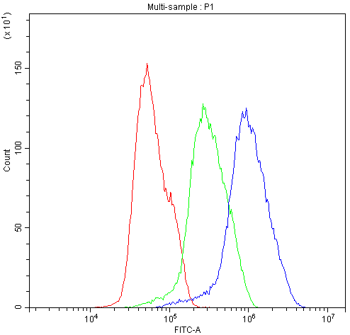
 FCM (Flow Cytometry)
(Figure 7. Flow Cytometry analysis of SiHa cells using anti-POR antibody (AAA11661).Overlay histogram showing SiHa cells stained with AAA11661 (Blue line).The cells were blocked with 10% normal goat serum. And then incubated with rabbit anti-POR Antibody (AAA11661,1ug/1x10^6 cells) for 30 min at 20 degree C. DyLight®488 conjugated goat anti-rabbit IgG (5-10ug/1x10^6 cells) was used as secondary antibody for 30 minutes at 20 degree C. Isotype control antibody (Green line) was rabbit IgG (1ug/1x106) used under the same conditions. Unlabelled sample (Red line) was also used as a control.)
FCM (Flow Cytometry)
(Figure 7. Flow Cytometry analysis of SiHa cells using anti-POR antibody (AAA11661).Overlay histogram showing SiHa cells stained with AAA11661 (Blue line).The cells were blocked with 10% normal goat serum. And then incubated with rabbit anti-POR Antibody (AAA11661,1ug/1x10^6 cells) for 30 min at 20 degree C. DyLight®488 conjugated goat anti-rabbit IgG (5-10ug/1x10^6 cells) was used as secondary antibody for 30 minutes at 20 degree C. Isotype control antibody (Green line) was rabbit IgG (1ug/1x106) used under the same conditions. Unlabelled sample (Red line) was also used as a control.)
POR, Polyclonal Antibody (Cat# AAA11661)
Full Name
Anti-POR Antibody
Gene Names
POR; CPR; CYPOR; P450R
Reactivity
Human, Mouse, Rat
Applications
WB, IHC
Purity
Immunogen Affinity Purified
Pricing


 Application Data
(Analysis of Protein Array containing more than 19, 000 full-length human proteins using PU.1 Mouse Monoclonal Antibody (PU1/2446). Z- and S- Score: The Z-score represents the strength of a signal that a monoclonal antibody (MAb) (in combination with a fluorescently-tagged anti-IgG secondary antibody) produces when binding to a particular protein on the HuProtTM array. Z-scores are described in units of standard deviations (SD's) above the mean value of all signals generated on that array. If targets on HuProtTM are arranged in descending order of the Z-score, the S-score is the difference (also in units of SD's) between the Z-score. S-score therefore represents the relative target specificity of a MAb to its intended target. A MAb is considered to specific to its intended target, if the MAb has an S-score of at least 2.5. For example, if a MAb binds to protein X with a Z-score of 43 and to protein Y with a Z-score of 14, then the S-score for the binding of that MAb to protein X is equal to 29.)
Application Data
(Analysis of Protein Array containing more than 19, 000 full-length human proteins using PU.1 Mouse Monoclonal Antibody (PU1/2446). Z- and S- Score: The Z-score represents the strength of a signal that a monoclonal antibody (MAb) (in combination with a fluorescently-tagged anti-IgG secondary antibody) produces when binding to a particular protein on the HuProtTM array. Z-scores are described in units of standard deviations (SD's) above the mean value of all signals generated on that array. If targets on HuProtTM are arranged in descending order of the Z-score, the S-score is the difference (also in units of SD's) between the Z-score. S-score therefore represents the relative target specificity of a MAb to its intended target. A MAb is considered to specific to its intended target, if the MAb has an S-score of at least 2.5. For example, if a MAb binds to protein X with a Z-score of 43 and to protein Y with a Z-score of 14, then the S-score for the binding of that MAb to protein X is equal to 29.)
PU.1 (SPI-1), Monoclonal Antibody (Cat# AAA23903)
Full Name
PU.1 (SPI-1) (B-Cell Marker)
Gene Names
SPI1; OF; PU.1; SFPI1; SPI-1; SPI-A
Reactivity
Human. Others not known.
Applications
WB, IHC
Pricing


 Application Data
(Analysis of Protein Array containing more than 19, 000 full-length human proteins using CD10 Mouse Monoclonal Antibody (MME/1870)Z- and S- Score: The Z-score represents the strength of a signal that a monoclonal antibody (MAb) (in combination with a fluorescently-tagged anti-IgG secondary antibody) produces when binding to a particular protein on the HuProtTM array. Z-scores are described in units of standard deviations (SD's) above the mean value of all signals generated on that array. If targets on HuProtTM are arranged in descending order of the Z-score, the S-score is the difference (also in units of SD's) between the Z-score. S-score therefore represents the relative target specificity of a MAb to its intended target. A MAb is considered to specific to its intended target, if the MAb has an S-score of at least 2.5. For example, if a MAb binds to protein X with a Z-score of 43 and to protein Y with a Z-score of 14, then the S-score for the binding of that MAb to protein X is equal to 29.)
Application Data
(Analysis of Protein Array containing more than 19, 000 full-length human proteins using CD10 Mouse Monoclonal Antibody (MME/1870)Z- and S- Score: The Z-score represents the strength of a signal that a monoclonal antibody (MAb) (in combination with a fluorescently-tagged anti-IgG secondary antibody) produces when binding to a particular protein on the HuProtTM array. Z-scores are described in units of standard deviations (SD's) above the mean value of all signals generated on that array. If targets on HuProtTM are arranged in descending order of the Z-score, the S-score is the difference (also in units of SD's) between the Z-score. S-score therefore represents the relative target specificity of a MAb to its intended target. A MAb is considered to specific to its intended target, if the MAb has an S-score of at least 2.5. For example, if a MAb binds to protein X with a Z-score of 43 and to protein Y with a Z-score of 14, then the S-score for the binding of that MAb to protein X is equal to 29.)
CD10, Monoclonal Antibody (Cat# AAA23901)
Full Name
CD10 (Membrane Metalloendopeptidase)
Gene Names
MME; NEP; SFE; CD10; CALLA; CMT2T; SCA43
Reactivity
Human. Others not tested.
Applications
IHC
Pricing


 Application Data
(Analysis of Protein Array containing more than 19,000 full-length human proteins using HER-2 Mouse Monoclonal Antibody (ERBB2/3080). Z- and S- Score: The Z-score represents the strength of a signal that a monoclonal antibody (MAb) (in combination with a fluorescently-tagged anti-IgG secondary antibody) produces when binding to a particular protein on the HuProtTM array. Z-scores are described in units of standard deviations (SD's) above the mean value of all signals generated on that array. If targets on HuProtTM are arranged in descending order of the Z-score, the S-score is the difference (also in units of SD's) between the Z-score. S-score therefore represents the relative target specificity of a MAb to its intended target. A MAb is considered to specific to its intended target, if the MAb has an S-score of at least 2.5. For example, if a MAb binds to protein X with a Z-score of 43 and to protein Y with a Z-score of 14, then the S-score for the binding of that MAb to protein X is equal to 29.)
Application Data
(Analysis of Protein Array containing more than 19,000 full-length human proteins using HER-2 Mouse Monoclonal Antibody (ERBB2/3080). Z- and S- Score: The Z-score represents the strength of a signal that a monoclonal antibody (MAb) (in combination with a fluorescently-tagged anti-IgG secondary antibody) produces when binding to a particular protein on the HuProtTM array. Z-scores are described in units of standard deviations (SD's) above the mean value of all signals generated on that array. If targets on HuProtTM are arranged in descending order of the Z-score, the S-score is the difference (also in units of SD's) between the Z-score. S-score therefore represents the relative target specificity of a MAb to its intended target. A MAb is considered to specific to its intended target, if the MAb has an S-score of at least 2.5. For example, if a MAb binds to protein X with a Z-score of 43 and to protein Y with a Z-score of 14, then the S-score for the binding of that MAb to protein X is equal to 29.)
HER-2/c-erbB-2/neu/CD340, Monoclonal Antibody (Cat# AAA23912)
Full Name
HER-2/c-erbB-2/neu/CD340
Gene Names
ERBB2; NEU; NGL; HER2; TKR1; CD340; HER-2; MLN 19; HER-2/neu
Reactivity
Human
Applications
IHC
Purity
Purified Ab with BSA and Azide at 200ug/ml OR Purified Ab WITHOUT BSA and Azide at 1.0mg/ml
Pricing


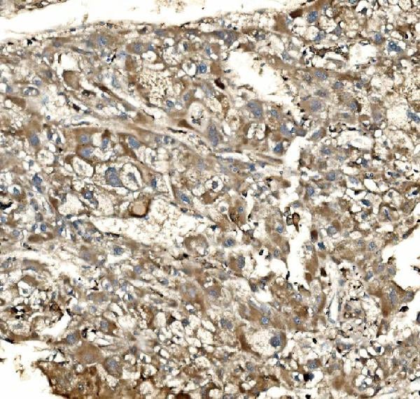
 IHC (Immunohistochemistry)
(At 1/100 staining Human colorectal cancer and adjacent normal tissues by IHC-P. The sample was formaldehyde fixed and a heat mediated antigen retrieval step in citrate buffer was performed. The sample was then blocked and incubated with the primary antibody at 4 degree C overnight. An HRP conjugated anti-Rabbit antibody was used as the secondary antibody.)
IHC (Immunohistochemistry)
(At 1/100 staining Human colorectal cancer and adjacent normal tissues by IHC-P. The sample was formaldehyde fixed and a heat mediated antigen retrieval step in citrate buffer was performed. The sample was then blocked and incubated with the primary antibody at 4 degree C overnight. An HRP conjugated anti-Rabbit antibody was used as the secondary antibody.)
HP1 alpha, Polyclonal Antibody (Cat# AAA31332)
Full Name
Phospho-HP1 alpha (Ser92) Antibody
Gene Names
CBX5; HP1; HP1A
Reactivity
Human, Mouse, Rat
Applications
IHC, EIA
Purity
The antibody is from purified rabbit serum by affinity purification via sequential chromatography on phospho-peptide and non-phospho-peptide affinity columns.
Pricing


 FCM (Flow Cytometry)
(Figure 7. Flow Cytometry analysis of A431 cells using anti-BIK antibody (AAA11666).Overlay histogram showing A431 cells stained with AAA11666 (Blue line).The cells were blocked with 10% normal goat serum. And then incubated with rabbit anti-BIK Antibody (AAA11666,1ug/1x10^6 cells) for 30 min at 20 degree C. DyLight®488 conjugated goat anti-rabbit IgG (5-10ug/1x10^6 cells) was used as secondary antibody for 30 minutes at 20 degree C. Isotype control antibody (Green line) was rabbit IgG (1ug/1x106) used under the same conditions. Unlabelled sample (Red line) was also used as a control.)
FCM (Flow Cytometry)
(Figure 7. Flow Cytometry analysis of A431 cells using anti-BIK antibody (AAA11666).Overlay histogram showing A431 cells stained with AAA11666 (Blue line).The cells were blocked with 10% normal goat serum. And then incubated with rabbit anti-BIK Antibody (AAA11666,1ug/1x10^6 cells) for 30 min at 20 degree C. DyLight®488 conjugated goat anti-rabbit IgG (5-10ug/1x10^6 cells) was used as secondary antibody for 30 minutes at 20 degree C. Isotype control antibody (Green line) was rabbit IgG (1ug/1x106) used under the same conditions. Unlabelled sample (Red line) was also used as a control.)
Bik, Polyclonal Antibody (Cat# AAA11666)
Full Name
Anti-Bik Antibody
Gene Names
BIK; BP4; NBK; BIP1
Reactivity
Human, Mouse, Rat
Applications
WB, IHC
Purity
Immunogen Affinity Purified
Pricing


 Application Data
(Analysis of Protein Array containing more than 19,000 full-length human proteins using CAIX-Monospecific Mouse Monoclonal Antibody (CA9/3406). Z- and S- Score: The Z-score represents the strength of a signal that a monoclonal antibody (MAb) (in combination with a fluorescently-tagged anti-IgG secondary antibody) produces when binding to a particular protein on the HuProtTM array. Z-scores are described in units of standard deviations (SD's) above the mean value of all signals generated on that array. If targets on HuProtTM are arranged in descending order of the Z-score, the S-score is the difference (also in units of SD's) between the Z-score. S-score therefore represents the relative target specificity of a MAb to its intended target. A MAb is considered to specific to its intended target, if the MAb has an S-score of at least 2.5. For example, if a MAb binds to protein X with a Z-score of 43 and to protein Y with a Z-score of 14, then the S-score for the binding of that MAb to protein X is equal to 29.)
Application Data
(Analysis of Protein Array containing more than 19,000 full-length human proteins using CAIX-Monospecific Mouse Monoclonal Antibody (CA9/3406). Z- and S- Score: The Z-score represents the strength of a signal that a monoclonal antibody (MAb) (in combination with a fluorescently-tagged anti-IgG secondary antibody) produces when binding to a particular protein on the HuProtTM array. Z-scores are described in units of standard deviations (SD's) above the mean value of all signals generated on that array. If targets on HuProtTM are arranged in descending order of the Z-score, the S-score is the difference (also in units of SD's) between the Z-score. S-score therefore represents the relative target specificity of a MAb to its intended target. A MAb is considered to specific to its intended target, if the MAb has an S-score of at least 2.5. For example, if a MAb binds to protein X with a Z-score of 43 and to protein Y with a Z-score of 14, then the S-score for the binding of that MAb to protein X is equal to 29.)
Renal Cell Carcinoma, Monoclonal Antibody (Cat# AAA23952)
Full Name
Renal Cell Carcinoma (Carbonic Anhydrase IX)
Gene Names
CA9; MN; CAIX
Reactivity
Human
Applications
FC/FACS, IHC, WB
Purity
Purified Ab with BSA and Azide at 200ug/ml or Purified Ab with BSA and Azide at 200ug/ml or Purified Ab WITHOUT BSA and Azide at 1.0mg/ml
Pricing


 FCM (Flow Cytometry)
(Figure 8. Flow Cytometry analysis of U20S cells using anti-Dynamin 1 antibody (AAA19160).Overlay histogram showing U20S cells stained with AAA19160 (Blue line).The cells were blocked with 10% normal goat serum. And then incubated with rabbit anti-DNM1 Antibody (AAA19160,1ug/1x10^6 cells) for 30 min at 20 degree C. DyLight®488 conjugated goat anti-rabbit IgG (5-10ug/1x10^6 cells) was used as secondary antibody for 30 minutes at 20 degree C. Isotype control antibody (Green line) was rabbit IgG (1ug/1x106) used under the same conditions. Unlabelled sample (Red line) was also used as a control.)
FCM (Flow Cytometry)
(Figure 8. Flow Cytometry analysis of U20S cells using anti-Dynamin 1 antibody (AAA19160).Overlay histogram showing U20S cells stained with AAA19160 (Blue line).The cells were blocked with 10% normal goat serum. And then incubated with rabbit anti-DNM1 Antibody (AAA19160,1ug/1x10^6 cells) for 30 min at 20 degree C. DyLight®488 conjugated goat anti-rabbit IgG (5-10ug/1x10^6 cells) was used as secondary antibody for 30 minutes at 20 degree C. Isotype control antibody (Green line) was rabbit IgG (1ug/1x106) used under the same conditions. Unlabelled sample (Red line) was also used as a control.)
Dynamin 1, Polyclonal Antibody (Cat# AAA19160)
Full Name
Anti-Dynamin 1 Picoband antibody
Gene Names
DNM1; DNM; EIEE31
Reactivity
Human, Mouse, Rat
No cross reactivity with other proteins.
No cross reactivity with other proteins.
Applications
EIA, FC/FACS, IHC, ICC, WB
Pricing


 IF (Immunofluorescence)
(Immunofluorescent analysis of 4% paraformaldehyde-fixed, 0.1% Triton X-100 permeabilized U-2 OS (human osteosarcoma cell line) cells labeling DLL3 with (1:25), followed by Dylight 488- conjugated goat anti- rabbit IgG secondary antibody at (1:200) (green). Immunofluorescence image showing nucleus and weak cytoplasm staining on U-2 OScell line. Cytoplasmic actin is detected with Dylight 554 Phalloidin (1:100) (red).)
IF (Immunofluorescence)
(Immunofluorescent analysis of 4% paraformaldehyde-fixed, 0.1% Triton X-100 permeabilized U-2 OS (human osteosarcoma cell line) cells labeling DLL3 with (1:25), followed by Dylight 488- conjugated goat anti- rabbit IgG secondary antibody at (1:200) (green). Immunofluorescence image showing nucleus and weak cytoplasm staining on U-2 OScell line. Cytoplasmic actin is detected with Dylight 554 Phalloidin (1:100) (red).)
DLL3, Polyclonal Antibody (Cat# AAA26850)
Full Name
DLL3, CT (DLL3, Delta-like protein 3, Drosophila Delta homolog 3) (APC)
Gene Names
DLL3; SCDO1
Reactivity
Human
Applications
IF, IHC, WB
Purity
Purified by Protein A Affinity Chromatography.
Pricing


 FCM (Flow Cytometry)
(Figure 8. Flow Cytometry analysis of U20S cells using anti-SV2A antibody (AAA19279).Overlay histogram showing U20S cells stained with AAA19279 (Blue line). The cells were blocked with 10% normal goat serum. And then incubated with rabbit anti-SV2A Antibody (AAA19279, 1μg/1x106 cells) for 30 min at 20 degree C. DyLight®488 conjugated goat anti-rabbit IgG (5-10μg/1x106 cells) was used as secondary antibody for 30 minutes at 20 degree C. Isotype control antibody (Green line) was rabbit IgG (1μg/1x106) used under the same conditions. Unlabelled sample (Red line) was also used as a control.)
FCM (Flow Cytometry)
(Figure 8. Flow Cytometry analysis of U20S cells using anti-SV2A antibody (AAA19279).Overlay histogram showing U20S cells stained with AAA19279 (Blue line). The cells were blocked with 10% normal goat serum. And then incubated with rabbit anti-SV2A Antibody (AAA19279, 1μg/1x106 cells) for 30 min at 20 degree C. DyLight®488 conjugated goat anti-rabbit IgG (5-10μg/1x106 cells) was used as secondary antibody for 30 minutes at 20 degree C. Isotype control antibody (Green line) was rabbit IgG (1μg/1x106) used under the same conditions. Unlabelled sample (Red line) was also used as a control.)
SV2A, Polyclonal Antibody (Cat# AAA19279)
Full Name
Anti-SV2A Antibody
Gene Names
SV2A; SV2
Reactivity
Human, Mouse, Rat
Applications
WB, IHC-P, FC/FACS/FCM
Purity
Immunogen affinity purified.
Pricing


 IF (Immunofluorescence)
(Figure 6. IF analysis of EEF2 using anti- EEF2 antibody (AAA19228).EEF2 was detected in immunocytochemical section of MCF-7 cells. Enzyme antigen retrieval was performed using IHC enzyme antigen retrieval reagent for 15 mins. The cells were blocked with 10% goat serum. And then incubated with 5μg/mL rabbit anti-EEF2 Antibody (AAA19228) overnight at 4 degree C. DyLight®488 Conjugated Goat Anti-Rabbit IgG was used as secondary antibody at 1:100 dilution and incubated for 30 minutes at 37 degree C. The section was counterstained with DAPI. Visualize using a fluorescence microscope and filter sets appropriate for the label used.)
IF (Immunofluorescence)
(Figure 6. IF analysis of EEF2 using anti- EEF2 antibody (AAA19228).EEF2 was detected in immunocytochemical section of MCF-7 cells. Enzyme antigen retrieval was performed using IHC enzyme antigen retrieval reagent for 15 mins. The cells were blocked with 10% goat serum. And then incubated with 5μg/mL rabbit anti-EEF2 Antibody (AAA19228) overnight at 4 degree C. DyLight®488 Conjugated Goat Anti-Rabbit IgG was used as secondary antibody at 1:100 dilution and incubated for 30 minutes at 37 degree C. The section was counterstained with DAPI. Visualize using a fluorescence microscope and filter sets appropriate for the label used.)
EEF2, Polyclonal Antibody (Cat# AAA19228)
Full Name
Anti-EEF2 Antibody
Gene Names
EEF2; EF2; EF-2; EEF-2
Reactivity
Human
Applications
WB, IHC-P, ICC, IF, EIA
Purity
Immunogen affinity purified.
Pricing


 FCM (Flow Cytometry)
(Figure 6. Flow Cytometry analysis of Hela cells using anti-CD147/Emmprin antibody (AAA11678).Overlay histogram showing Hela cells stained with AAA11678 (Blue line).The cells were blocked with 10% normal goat serum. And then incubated with rabbit anti-CD147/Emmprin Antibody (AAA11678,1ug/1x10^6 cells) for 30 min at 20 degree C. DyLight 488 conjugated goat anti-rabbit IgG (5-10ug/1x10^6 cells) was used as secondary antibody for 30 minutes at 20 degree C. Isotype control antibody (Green line) was rabbit IgG (1ug/1x106) used under the same conditions. Unlabelled sample (Red line) was also used as a control.)
FCM (Flow Cytometry)
(Figure 6. Flow Cytometry analysis of Hela cells using anti-CD147/Emmprin antibody (AAA11678).Overlay histogram showing Hela cells stained with AAA11678 (Blue line).The cells were blocked with 10% normal goat serum. And then incubated with rabbit anti-CD147/Emmprin Antibody (AAA11678,1ug/1x10^6 cells) for 30 min at 20 degree C. DyLight 488 conjugated goat anti-rabbit IgG (5-10ug/1x10^6 cells) was used as secondary antibody for 30 minutes at 20 degree C. Isotype control antibody (Green line) was rabbit IgG (1ug/1x106) used under the same conditions. Unlabelled sample (Red line) was also used as a control.)
CD147/Emmprin, Polyclonal Antibody (Cat# AAA11678)
Full Name
Anti-CD147/Emmprin Antibody
Gene Names
BSG; OK; 5F7; TCSF; CD147; EMMPRIN
Reactivity
Human, Mouse, Rat
Applications
WB, IHC
Purity
Immunogen affinity purified.
Pricing


 FCM (Flow Cytometry)
(Figure 8. Flow Cytometry analysis of U20S cells using anti-NFIA antibody (AAA11691).Overlay histogram showing U20S cells stained with AAA11691 (Blue line).The cells were blocked with 10% normal goat serum. And then incubated with rabbit anti-NFIA Antibody (AAA11691,1ug/1x10^6 cells) for 30 min at 20 degree C. DyLight®488 conjugated goat anti-rabbit IgG (5-10ug/1x10^6 cells) was used as secondary antibody for 30 minutes at 20 degree C. Isotype control antibody (Green line) was rabbit IgG (1ug/1x106) used under the same conditions. Unlabelled sample (Red line) was also used as a control.)
FCM (Flow Cytometry)
(Figure 8. Flow Cytometry analysis of U20S cells using anti-NFIA antibody (AAA11691).Overlay histogram showing U20S cells stained with AAA11691 (Blue line).The cells were blocked with 10% normal goat serum. And then incubated with rabbit anti-NFIA Antibody (AAA11691,1ug/1x10^6 cells) for 30 min at 20 degree C. DyLight®488 conjugated goat anti-rabbit IgG (5-10ug/1x10^6 cells) was used as secondary antibody for 30 minutes at 20 degree C. Isotype control antibody (Green line) was rabbit IgG (1ug/1x106) used under the same conditions. Unlabelled sample (Red line) was also used as a control.)
NFIA, Polyclonal Antibody (Cat# AAA11691)
Full Name
Anti-NFIA Antibody
Gene Names
NFIA; CTF; NF1-A; NFI-A; NFI-L; NF-I/A
Reactivity
Human, Mouse, Rat
Applications
WB, IHC
Purity
Immunogen affinity purified.
Pricing


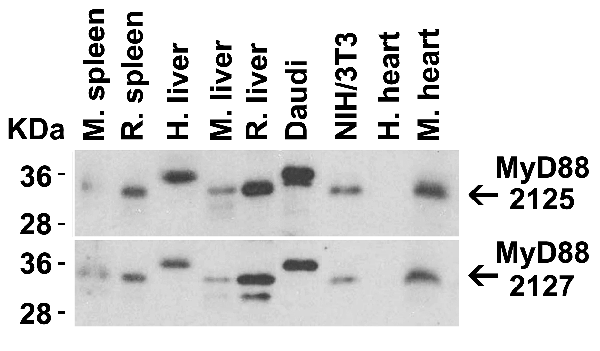


 IP (Immunoprecipitation)
(Figure 13 Immunoprecipitation Validation in HEK293 cells (Kawai et al., 2004)HEK293 cells were transiently transfected with DYKDDDDK-IRF7. Ccell lysates were immunoprecipitated with control rabbit anti-mouse immunoglobulin serum (IgG) or anti-MyD88 (Ab1 and Ab2), followed by immunoblotting with anti-DYKDDDDK.)
IP (Immunoprecipitation)
(Figure 13 Immunoprecipitation Validation in HEK293 cells (Kawai et al., 2004)HEK293 cells were transiently transfected with DYKDDDDK-IRF7. Ccell lysates were immunoprecipitated with control rabbit anti-mouse immunoglobulin serum (IgG) or anti-MyD88 (Ab1 and Ab2), followed by immunoblotting with anti-DYKDDDDK.)
MYD88, Polyclonal Antibody (Cat# AAA10930)
Full Name
MYD88 Antibody
Gene Names
MYD88; MYD88D
Reactivity
Human, Mouse
Applications
EIA, WB, IHC, IF
Purity
MYD88 Antibody is affinity chromatography purified via peptide column.
Pricing


 FCM (Flow Cytometry)
(Figure 6. Flow Cytometry analysis of PC-3 cells using anti-ADK antibody (AAA11690).Overlay histogram showing PC-3 cells stained with AAA11690 (Blue line).The cells were blocked with 10% normal goat serum. And then incubated with rabbit anti-ADK Antibody (AAA11690,1ug/1x10^6 cells) for 30 min at 20 degree C. DyLight®488 conjugated goat anti-rabbit IgG (5-10ug/1x10^6 cells) was used as secondary antibody for 30 minutes at 20 degree C. Isotype control antibody (Green line) was rabbit IgG (1ug/1x106) used under the same conditions. Unlabelled sample (Red line) was also used as a control.)
FCM (Flow Cytometry)
(Figure 6. Flow Cytometry analysis of PC-3 cells using anti-ADK antibody (AAA11690).Overlay histogram showing PC-3 cells stained with AAA11690 (Blue line).The cells were blocked with 10% normal goat serum. And then incubated with rabbit anti-ADK Antibody (AAA11690,1ug/1x10^6 cells) for 30 min at 20 degree C. DyLight®488 conjugated goat anti-rabbit IgG (5-10ug/1x10^6 cells) was used as secondary antibody for 30 minutes at 20 degree C. Isotype control antibody (Green line) was rabbit IgG (1ug/1x106) used under the same conditions. Unlabelled sample (Red line) was also used as a control.)
ADK, Polyclonal Antibody (Cat# AAA11690)
Full Name
Anti-ADK Antibody
Gene Names
ADK; AK
Reactivity
Human, Mouse, Rat
Applications
WB, IHC
Purity
Immunogen affinity purified.
Pricing


 IF (Immunofluorescence)
(Figure 10 Immunofluorescence images of PUMA in the P14 rat retina (Wakabayashi et al., 2012)PUMA expression in the rat retina detected by anti-PUMA antibodies (3043). The specimens were counterstained with Hoechst 33258 to visualize nuclei (+DNA). GCL, ganglion cell layer; INL, inner nuclear layer; IPL, inner plexiform layer; ONL, outer nuclear layer; OPL, outer plexiform layer; P, postnatal day.)
IF (Immunofluorescence)
(Figure 10 Immunofluorescence images of PUMA in the P14 rat retina (Wakabayashi et al., 2012)PUMA expression in the rat retina detected by anti-PUMA antibodies (3043). The specimens were counterstained with Hoechst 33258 to visualize nuclei (+DNA). GCL, ganglion cell layer; INL, inner nuclear layer; IPL, inner plexiform layer; ONL, outer nuclear layer; OPL, outer plexiform layer; P, postnatal day.)
PUMA, Polyclonal Antibody (Cat# AAA10934)
Full Name
PUMA Antibody
Gene Names
BBC3; JFY1; PUMA; JFY-1
Reactivity
Human
Applications
EIA, WB, IHC, IF
Purity
PUMA Antibody is affinity chromatography purified via peptide column.
Pricing


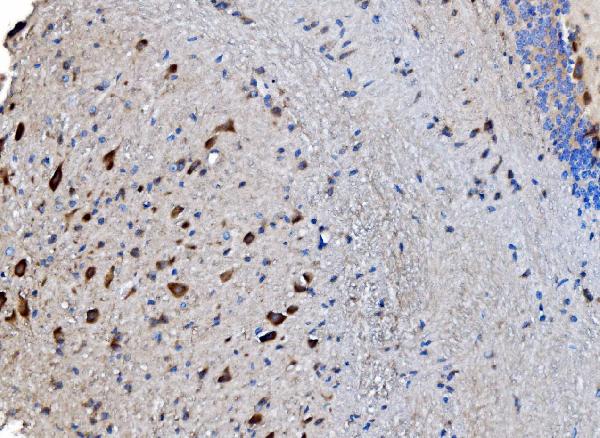
 IHC (Immunohistochemistry)
(Immunohistochemical analysis of paraffin-embedded Human brain section using Pink 1 (AAA28759). AAA28759 was diluted at 1:1000 dilution. A undiluted binotinylated goat polyvalent antibody was used as the secondary, followed by DAB staining)
IHC (Immunohistochemistry)
(Immunohistochemical analysis of paraffin-embedded Human brain section using Pink 1 (AAA28759). AAA28759 was diluted at 1:1000 dilution. A undiluted binotinylated goat polyvalent antibody was used as the secondary, followed by DAB staining)
S100B, Polyclonal Antibody (Cat# AAA28759)
Full Name
S100B Antibody
Gene Names
S100B; NEF; S100; S100-B; S100beta
Reactivity
Human, Mouse, Rat (Predicted: Rabbit)
Applications
WB, IHC-P-Leica, FC/FACS, IHC, EIA
Purity
Peptide Affinity Purified Rabbit Polyclonal Antibody (Pab)
Pricing


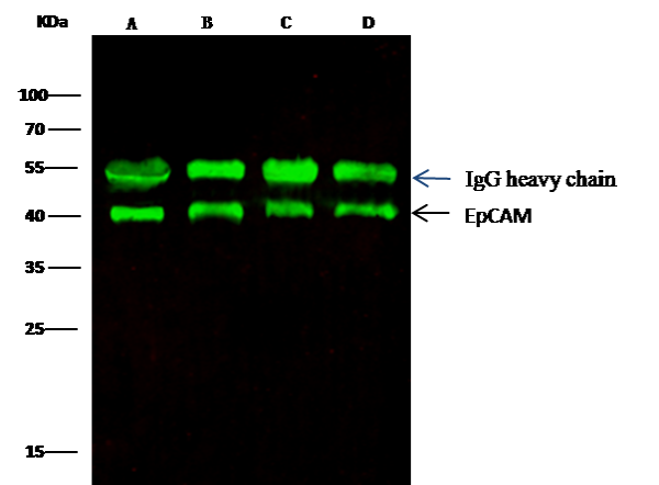 IP (Immunoprecipitation)
(EpCAM was immunoprecipitated using:Lane A:0.5 mg MCF-7 Whole Cell LysateLane B:0.5 mg A431 Whole Cell LysateLane C:0.5 mg HepG2 Whole Cell LysateLane D:0.5 mg Caco-2 Whole Cell Lysate0.5 uL anti-EpCAM rabbit monoclonal antibody and 15 ul of 50 % Protein G agarose.Primary antibody:Anti-EpCAM rabbit monoclonal antibody,at 1:5000 dilution Secondary antibody:Dylight 800-labeled antibody to rabbit IgG (H+L), at 1:5000 dilution Developed using the odssey technique.Performed under reducing conditions.Predicted band size: 40 kDaObserved band size: 40 kDa)
IP (Immunoprecipitation)
(EpCAM was immunoprecipitated using:Lane A:0.5 mg MCF-7 Whole Cell LysateLane B:0.5 mg A431 Whole Cell LysateLane C:0.5 mg HepG2 Whole Cell LysateLane D:0.5 mg Caco-2 Whole Cell Lysate0.5 uL anti-EpCAM rabbit monoclonal antibody and 15 ul of 50 % Protein G agarose.Primary antibody:Anti-EpCAM rabbit monoclonal antibody,at 1:5000 dilution Secondary antibody:Dylight 800-labeled antibody to rabbit IgG (H+L), at 1:5000 dilution Developed using the odssey technique.Performed under reducing conditions.Predicted band size: 40 kDaObserved band size: 40 kDa)
EpCAM, Monoclonal Antibody (Cat# AAA27736)
Full Name
Recombinant Anti-EpCAM Antibody, Rabbit Monoclonal
Gene Names
EPCAM; ESA; KSA; M4S1; MK-1; DIAR5; EGP-2; EGP40; KS1/4; MIC18; TROP1; EGP314; HNPCC8; TACSTD1
Reactivity
Human
Applications
WB, EIA, EIA, Det, IHC-P, FC/FACS/FCM, ICC, IF, IP
Purity
Protein A
Pricing


 Application Data
(Analysis of Protein Array containing more than 19,000 full-length human proteins using AMACR/p504S Mouse Monoclonal Antibody (AMACR/1864). Z- and S- Score: The Z-score represents the strength of a signal that a monoclonal antibody (MAb) (in combination with a fluorescently-tagged anti-IgG secondary antibody) produces when binding to a particular protein on the HuProtTM array. Z-scores are described in units of standard deviations (SD's) above the mean value of all signals generated on that array. If targets on HuProtTM are arranged in descending order of the Z-score, the S-score is the difference (also in units of SD's) between the Z-score. S-score therefore represents the relative target specificity of a MAb to its intended target. A MAb is considered to specific to its intended target, if the MAb has an S-score of at least 2.5. For example, if a MAb binds to protein X with a Z-score of 43 and to protein Y with a Z-score of 14, then the S-score for the binding of that MAb to protein X is equal to 29.)
Application Data
(Analysis of Protein Array containing more than 19,000 full-length human proteins using AMACR/p504S Mouse Monoclonal Antibody (AMACR/1864). Z- and S- Score: The Z-score represents the strength of a signal that a monoclonal antibody (MAb) (in combination with a fluorescently-tagged anti-IgG secondary antibody) produces when binding to a particular protein on the HuProtTM array. Z-scores are described in units of standard deviations (SD's) above the mean value of all signals generated on that array. If targets on HuProtTM are arranged in descending order of the Z-score, the S-score is the difference (also in units of SD's) between the Z-score. S-score therefore represents the relative target specificity of a MAb to its intended target. A MAb is considered to specific to its intended target, if the MAb has an S-score of at least 2.5. For example, if a MAb binds to protein X with a Z-score of 43 and to protein Y with a Z-score of 14, then the S-score for the binding of that MAb to protein X is equal to 29.)
AMACR/p504S, Monoclonal Antibody (Cat# AAA23906)
Full Name
AMACR/p504S (Prostate Cancer Marker)
Gene Names
AMACR; RM; RACE; CBAS4; P504S; AMACRD
Reactivity
Human. Others not known.
Applications
WB, Formalin-Fixed
Purity
Purified Ab with BSA and Azide at 200ug/ml OR Purified Ab WITHOUT BSA and Azide at 1.0mg/ml
Pricing


 WB (Western Blot)
(Figure 1. Western blot analysis of Lyn using anti-Lyn antibody (AAA19150). Electrophoresis was performed on a 5-20% SDS-PAGE gel at 70V (Stacking gel) / 90V (Resolving gel) for 2-3 hours. The sample well of each lane was loaded with 50ug of sample under reducing conditions. Lane 1: human 293T whole cell lysate. After Electrophoresis, proteins were transferred to a Nitrocellulose membrane at 150mA for 50-90 minutes. Blocked the membrane with 5% Non-fat Milk/ TBS for 1.5 hour at RT. The membrane was incubated with rabbit anti-Lyn antigen affinity purified polyclonal antibody at 0.5ug/mL overnight at 4 degree C, then washed with TBS-0.1%Tween 3 times with 5 minutes each and probed with a goat anti-rabbit IgG-HRP secondary antibody at a dilution of 1:10000 for 1.5 hour at RT. The signal is developed using an Enhanced Chemiluminescent detection (ECL) kit with Tanon 5200 system. A specific band was detected for Lyn at approximately 59KD. The expected band size for Lyn is at 59KD.)
WB (Western Blot)
(Figure 1. Western blot analysis of Lyn using anti-Lyn antibody (AAA19150). Electrophoresis was performed on a 5-20% SDS-PAGE gel at 70V (Stacking gel) / 90V (Resolving gel) for 2-3 hours. The sample well of each lane was loaded with 50ug of sample under reducing conditions. Lane 1: human 293T whole cell lysate. After Electrophoresis, proteins were transferred to a Nitrocellulose membrane at 150mA for 50-90 minutes. Blocked the membrane with 5% Non-fat Milk/ TBS for 1.5 hour at RT. The membrane was incubated with rabbit anti-Lyn antigen affinity purified polyclonal antibody at 0.5ug/mL overnight at 4 degree C, then washed with TBS-0.1%Tween 3 times with 5 minutes each and probed with a goat anti-rabbit IgG-HRP secondary antibody at a dilution of 1:10000 for 1.5 hour at RT. The signal is developed using an Enhanced Chemiluminescent detection (ECL) kit with Tanon 5200 system. A specific band was detected for Lyn at approximately 59KD. The expected band size for Lyn is at 59KD.)
Lyn, Polyclonal Antibody (Cat# AAA19150)
Full Name
Anti-Lyn Picoband Antibody
Gene Names
LYN; JTK8; p53Lyn; p56Lyn
Reactivity
Human, Mouse, Rat
No cross reactivity with other proteins.
No cross reactivity with other proteins.
Applications
IHC, WB
Purity
Immunogen affinity purified
Pricing


 FCM (Flow Cytometry)
(Figure 6. Flow Cytometry analysis of U20S cells using anti-PPID antibody (AAA19159).Overlay histogram showing U20S cells stained with AAA19159 (Blue line).The cells were blocked with 10% normal goat serum. And then incubated with rabbit anti-PPID Antibody (AAA19159,1ug/1x10^6 cells) for 30 min at 20 degree C. DyLight®488 conjugated goat anti-rabbit IgG (5-10ug/1x10^6 cells) was used as secondary antibody for 30 minutes at 20 degree C. Isotype control antibody (Green line) was rabbit IgG (1ug/1x106) used under the same conditions. Unlabelled sample (Red line) was also used as a control.)
FCM (Flow Cytometry)
(Figure 6. Flow Cytometry analysis of U20S cells using anti-PPID antibody (AAA19159).Overlay histogram showing U20S cells stained with AAA19159 (Blue line).The cells were blocked with 10% normal goat serum. And then incubated with rabbit anti-PPID Antibody (AAA19159,1ug/1x10^6 cells) for 30 min at 20 degree C. DyLight®488 conjugated goat anti-rabbit IgG (5-10ug/1x10^6 cells) was used as secondary antibody for 30 minutes at 20 degree C. Isotype control antibody (Green line) was rabbit IgG (1ug/1x106) used under the same conditions. Unlabelled sample (Red line) was also used as a control.)
PPID/Cyclophilin 40, Polyclonal Antibody (Cat# AAA19159)
Full Name
Anti-PPID/Cyclophilin 40 Picoband antibody
Gene Names
PPID; CYPD; CYP-40
Reactivity
Human, Mouse, Rat
No cross reactivity with other proteins.
No cross reactivity with other proteins.
Applications
EIA, FC/FACS, IHC, ICC, WB
Pricing


 IHC (Immunohistchemistry)
(Figure 6. IHC analysis of MED15 using anti-MED15 antibody (AAA19169).MED15 was detected in paraffin-embedded section of rat brain tissue. Heat mediated antigen retrieval was performed in citrate buffer (pH6, epitope retrieval solution) for 20 mins. The tissue section was blocked with 10% goat serum. The tissue section was then incubated with 1ug/ml rabbit anti-MED15 Antibody (AAA19169) overnight at 4 degree C. Biotinylated goat anti-rabbit IgG was used as secondary antibody and incubated for 30 minutes at 37 degree C. The tissue section was developed using Strepavidin-Biotin-Complex (SABC) with DAB as the chromogen.)
IHC (Immunohistchemistry)
(Figure 6. IHC analysis of MED15 using anti-MED15 antibody (AAA19169).MED15 was detected in paraffin-embedded section of rat brain tissue. Heat mediated antigen retrieval was performed in citrate buffer (pH6, epitope retrieval solution) for 20 mins. The tissue section was blocked with 10% goat serum. The tissue section was then incubated with 1ug/ml rabbit anti-MED15 Antibody (AAA19169) overnight at 4 degree C. Biotinylated goat anti-rabbit IgG was used as secondary antibody and incubated for 30 minutes at 37 degree C. The tissue section was developed using Strepavidin-Biotin-Complex (SABC) with DAB as the chromogen.)
MED15/Pcqap, Polyclonal Antibody (Cat# AAA19169)
Full Name
Anti-MED15/Pcqap Picoband Antibody
Gene Names
MED15; TIG1; CAG7A; CTG7A; PCQAP; TIG-1; TNRC7; ARC105
Reactivity
Human, Mouse, Rat
No cross reactivity with other proteins.
No cross reactivity with other proteins.
Applications
EIA, IHC, WB
Purity
Immunogen affinity purified
Pricing


 FCM (Flow Cytometry)
(Figure 6. Flow Cytometry analysis of MCF-7 cells using anti-Caspase-7/CASP7 antibody (AAA19232).Overlay histogram showing MCF-7 cells stained with AAA19232 (Blue line). The cells were blocked with 10% normal goat serum. And then incubated with rabbit anti-Caspase-7/CASP7 Antibody (AAA19232, 1μg/1x106 cells) for 30 min at 20 degree C. DyLight®488 conjugated goat anti-rabbit IgG (5-10μg/1x106 cells) was used as secondary antibody for 30 minutes at 20 degree C. Isotype control antibody (Green line) was rabbit IgG (1μg/1x106) used under the same conditions. Unlabelled sample (Red line) was also used as a control.)
FCM (Flow Cytometry)
(Figure 6. Flow Cytometry analysis of MCF-7 cells using anti-Caspase-7/CASP7 antibody (AAA19232).Overlay histogram showing MCF-7 cells stained with AAA19232 (Blue line). The cells were blocked with 10% normal goat serum. And then incubated with rabbit anti-Caspase-7/CASP7 Antibody (AAA19232, 1μg/1x106 cells) for 30 min at 20 degree C. DyLight®488 conjugated goat anti-rabbit IgG (5-10μg/1x106 cells) was used as secondary antibody for 30 minutes at 20 degree C. Isotype control antibody (Green line) was rabbit IgG (1μg/1x106) used under the same conditions. Unlabelled sample (Red line) was also used as a control.)
Caspase-7/CASP7, Polyclonal Antibody (Cat# AAA19232)
Full Name
Anti-Caspase-7/CASP7 Antibody
Gene Names
CASP7; MCH3; CMH-1; LICE2; CASP-7; ICE-LAP3
Reactivity
Human, Rat
Applications
WB, IHC-P, ICC, IF, FC/FACS/FCM, EIA
Purity
Immunogen affinity purified.
Pricing


 FCM (Flow Cytometry)
(Figure 10. Flow Cytometry analysis of RH35 cells using anti-Prohibitin/PHB antibody (AAA19234).Overlay histogram showing RH35 cells stained with AAA19234 (Blue line). The cells were blocked with 10% normal goat serum. And then incubated with rabbit anti-Prohibitin/PHB Antibody (AAA19234, 1μg/1x106 cells) for 30 min at 20 degree C. DyLight®488 conjugated goat anti-rabbit IgG (5-10μg/1x106 cells) was used as secondary antibody for 30 minutes at 20 degree C. Isotype control antibody (Green line) was rabbit IgG (1μg/1x106) used under the same conditions. Unlabelled sample (Red line) was also used as a control.)
FCM (Flow Cytometry)
(Figure 10. Flow Cytometry analysis of RH35 cells using anti-Prohibitin/PHB antibody (AAA19234).Overlay histogram showing RH35 cells stained with AAA19234 (Blue line). The cells were blocked with 10% normal goat serum. And then incubated with rabbit anti-Prohibitin/PHB Antibody (AAA19234, 1μg/1x106 cells) for 30 min at 20 degree C. DyLight®488 conjugated goat anti-rabbit IgG (5-10μg/1x106 cells) was used as secondary antibody for 30 minutes at 20 degree C. Isotype control antibody (Green line) was rabbit IgG (1μg/1x106) used under the same conditions. Unlabelled sample (Red line) was also used as a control.)
Prohibitin/PHB, Polyclonal Antibody (Cat# AAA19234)
Full Name
Anti-Prohibitin/PHB Antibody
Gene Names
PHB; PHB1
Reactivity
Human, Mouse, Rat
Applications
WB, IHC-P, FC/FACS/FCM, EIA
Purity
Immunogen affinity purified.
Pricing


 FCM (Flow Cytometry)
(Figure 8. Flow Cytometry analysis of U251 cells using anti-GABARAP antibody (AAA19250).Overlay histogram showing U251 cells stained with AAA19250 (Blue line). The cells were blocked with 10% normal goat serum. And then incubated with rabbit anti-GABARAP Antibody (AAA19250, 1μg/1x106 cells) for 30 min at 20 degree C. DyLight®488 conjugated goat anti-rabbit IgG (5-10μg/1x106 cells) was used as secondary antibody for 30 minutes at 20 degree C. Isotype control antibody (Green line) was rabbit IgG (1μg/1x106) used under the same conditions. Unlabelled sample (Red line) was also used as a control.)
FCM (Flow Cytometry)
(Figure 8. Flow Cytometry analysis of U251 cells using anti-GABARAP antibody (AAA19250).Overlay histogram showing U251 cells stained with AAA19250 (Blue line). The cells were blocked with 10% normal goat serum. And then incubated with rabbit anti-GABARAP Antibody (AAA19250, 1μg/1x106 cells) for 30 min at 20 degree C. DyLight®488 conjugated goat anti-rabbit IgG (5-10μg/1x106 cells) was used as secondary antibody for 30 minutes at 20 degree C. Isotype control antibody (Green line) was rabbit IgG (1μg/1x106) used under the same conditions. Unlabelled sample (Red line) was also used as a control.)
GABARAP, Polyclonal Antibody (Cat# AAA19250)
Full Name
Anti-GABARAP Antibody
Gene Names
GABARAP; MM46; ATG8A; GABARAP-a
Reactivity
Human, Mouse, Rat
Applications
WB, IHC-P, FC/FACS/FCM
Purity
Immunogen affinity purified.
Pricing


 FCM (Flow Cytometry)
(Figure 10. Flow Cytometry analysis of MCF-7 cells using anti-DDX1 antibody (AAA19378).Overlay histogram showing MCF-7 cells stained with AAA19378 (Blue line). The cells were blocked with 10% normal goat serum. And then incubated with mouse anti- DDX1 Antibody (AAA19378, 1μg/1x106 cells) for 30 min at 20 degree C. DyLight®488 conjugated goat anti-mouse IgG (BA1126, 5-10μg/1x106 cells) was used as secondary antibody for 30 minutes at 20 degree C. Isotype control antibody (Green line) was mouse IgG (1μg/1x106) used under the same conditions. Unlabelled sample (Red line) was also used as a control.)
FCM (Flow Cytometry)
(Figure 10. Flow Cytometry analysis of MCF-7 cells using anti-DDX1 antibody (AAA19378).Overlay histogram showing MCF-7 cells stained with AAA19378 (Blue line). The cells were blocked with 10% normal goat serum. And then incubated with mouse anti- DDX1 Antibody (AAA19378, 1μg/1x106 cells) for 30 min at 20 degree C. DyLight®488 conjugated goat anti-mouse IgG (BA1126, 5-10μg/1x106 cells) was used as secondary antibody for 30 minutes at 20 degree C. Isotype control antibody (Green line) was mouse IgG (1μg/1x106) used under the same conditions. Unlabelled sample (Red line) was also used as a control.)
DDX1, Monoclonal Antibody (Cat# AAA19378)
Full Name
Anti-DDX1 Antibody (monoclonal, 3I10)
Gene Names
DDX1; DBP-RB; UKVH5d
Reactivity
Human, Mouse, Rat
Applications
WB, IHC-P, FC/FACS/FCM
Purity
Immunogen affinity purified.
Pricing


 FCM (Flow Cytometry)
(Figure 6. Flow Cytometry analysis of MCF-7 cells using anti- PCK2 antibody (AAA19384).Overlay histogram showing MCF-7 cells stained with AAA19384 (Blue line). The cells were blocked with 10% normal goat serum. And then incubated with mouse anti-PCK2 Antibody (AAA19384, 1μg/1x106 cells) for 30 min at 20 degree C. DyLight®488 conjugated goat anti-mouse IgG (BA1126, 5-10μg/1x106 cells) was used as secondary antibody for 30 minutes at 20 degree C. Isotype control antibody (Green line) was mouse IgG (1μg/1x106) used under the same conditions. Unlabelled sample (Red line) was also used as a control.)
FCM (Flow Cytometry)
(Figure 6. Flow Cytometry analysis of MCF-7 cells using anti- PCK2 antibody (AAA19384).Overlay histogram showing MCF-7 cells stained with AAA19384 (Blue line). The cells were blocked with 10% normal goat serum. And then incubated with mouse anti-PCK2 Antibody (AAA19384, 1μg/1x106 cells) for 30 min at 20 degree C. DyLight®488 conjugated goat anti-mouse IgG (BA1126, 5-10μg/1x106 cells) was used as secondary antibody for 30 minutes at 20 degree C. Isotype control antibody (Green line) was mouse IgG (1μg/1x106) used under the same conditions. Unlabelled sample (Red line) was also used as a control.)
PCK2, Monoclonal Antibody (Cat# AAA19384)
Full Name
Anti-PCK2 Antibody (monoclonal, 3F7)
Gene Names
PCK2; PEPCK; PEPCK2; PEPCK-M
Reactivity
Human, Mouse, Rat, Monkey
Applications
WB, IHC-P, ICC, IF, FC/FACS/FCM
Purity
Immunogen affinity purified.
Pricing


 FCM (Flow Cytometry)
(Figure 6. Flow Cytometry analysis of SiHa cells using anti- Aldolase/ALDOA antibody (AAA19385).Overlay histogram showing SiHa cells stained with AAA19385 (Blue line). The cells were blocked with 10% normal goat serum. And then incubated with mouse anti-Aldolase/ALDOA Antibody (AAA19385, 1μg/1x106 cells) for 30 min at 20 degree C. DyLight®488 conjugated goat anti-mouse IgG (BA1126, 5-10μg/1x106 cells) was used as secondary antibody for 30 minutes at 20 degree C. Isotype control antibody (Green line) was mouse IgG (1μg/1x106) used under the same conditions. Unlabelled sample (Red line) was also used as a control.)
FCM (Flow Cytometry)
(Figure 6. Flow Cytometry analysis of SiHa cells using anti- Aldolase/ALDOA antibody (AAA19385).Overlay histogram showing SiHa cells stained with AAA19385 (Blue line). The cells were blocked with 10% normal goat serum. And then incubated with mouse anti-Aldolase/ALDOA Antibody (AAA19385, 1μg/1x106 cells) for 30 min at 20 degree C. DyLight®488 conjugated goat anti-mouse IgG (BA1126, 5-10μg/1x106 cells) was used as secondary antibody for 30 minutes at 20 degree C. Isotype control antibody (Green line) was mouse IgG (1μg/1x106) used under the same conditions. Unlabelled sample (Red line) was also used as a control.)
Aldolase/ALDOA, Monoclonal Antibody (Cat# AAA19385)
Full Name
Anti-Aldolase/ALDOA Antibody (monoclonal, 6H8)
Gene Names
ALDOA; ALDA; GSD12
Reactivity
Human
Applications
WB, IHC-P, ICC, IF, FC/FACS/FCM
Purity
Immunogen affinity purified.
Pricing


 FCM (Flow Cytometry)
(Figure 6. Flow Cytometry analysis of A549 cells using anti-Ch TOG/CKAP5 antibody (AAA19386).Overlay histogram showing A549 cells stained with AAA19386 (Blue line). The cells were blocked with 10% normal goat serum. And then incubated with mouse anti- Ch TOG/CKAP5 Antibody (AAA19386, 1μg/1x106 cells) for 30 min at 20 degree C. DyLight®488 conjugated goat anti-mouse IgG (BA1126, 5-10μg/1x106 cells) was used as secondary antibody for 30 minutes at 20 degree C. Isotype control antibody (Green line) was mouse IgG (1μg/1x106) used under the same conditions. Unlabelled sample (Red line) was also used as a control.)
FCM (Flow Cytometry)
(Figure 6. Flow Cytometry analysis of A549 cells using anti-Ch TOG/CKAP5 antibody (AAA19386).Overlay histogram showing A549 cells stained with AAA19386 (Blue line). The cells were blocked with 10% normal goat serum. And then incubated with mouse anti- Ch TOG/CKAP5 Antibody (AAA19386, 1μg/1x106 cells) for 30 min at 20 degree C. DyLight®488 conjugated goat anti-mouse IgG (BA1126, 5-10μg/1x106 cells) was used as secondary antibody for 30 minutes at 20 degree C. Isotype control antibody (Green line) was mouse IgG (1μg/1x106) used under the same conditions. Unlabelled sample (Red line) was also used as a control.)
ch TOG/CKAP5, Monoclonal Antibody (Cat# AAA19386)
Full Name
Anti-ch TOG/CKAP5 Antibody (monoclonal, 3C13)
Gene Names
CKAP5; TOG; MSPS; TOGp; CHTOG; ch-TOG
Reactivity
Human, Mouse, Rat
Applications
WB, IHC-P, ICC, IF, FC/FACS/FCM
Purity
Immunogen affinity purified.
Pricing


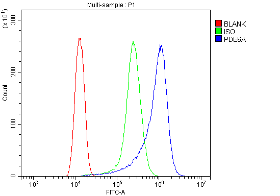 FCM (Flow Cytometry)
(Figure 6. Flow Cytometry analysis of HELA cells using anti-PDE6 alpha/PDE6A antibody (AAA19318).Overlay histogram showing HELA cells stained with AAA19318 (Blue line). The cells were blocked with 10% normal goat serum. And then incubated with rabbit anti-PDE6 alpha/PDE6A Antibody (AAA19318, 1μg/1x106 cells) for 30 min at 20 degree C. DyLight®488 conjugated goat anti-rabbit IgG (5-10μg/1x106 cells) was used as secondary antibody for 30 minutes at 20 degree C. Isotype control antibody (Green line) was rabbit IgG (1μg/1x106) used under the same conditions. Unlabelled sample (Red line) was also used as a control.)
FCM (Flow Cytometry)
(Figure 6. Flow Cytometry analysis of HELA cells using anti-PDE6 alpha/PDE6A antibody (AAA19318).Overlay histogram showing HELA cells stained with AAA19318 (Blue line). The cells were blocked with 10% normal goat serum. And then incubated with rabbit anti-PDE6 alpha/PDE6A Antibody (AAA19318, 1μg/1x106 cells) for 30 min at 20 degree C. DyLight®488 conjugated goat anti-rabbit IgG (5-10μg/1x106 cells) was used as secondary antibody for 30 minutes at 20 degree C. Isotype control antibody (Green line) was rabbit IgG (1μg/1x106) used under the same conditions. Unlabelled sample (Red line) was also used as a control.)
PDE6 alpha/PDE6A, Polyclonal Antibody (Cat# AAA19318)
Full Name
Anti-PDE6 alpha/PDE6A Antibody
Gene Names
PDE6A; PDEA; RP43; CGPR-A; PDE6A
Reactivity
Human, Mouse, Rat
Applications
WB, IHC-P, ICC, IF, FC/FACS/FCM, EIA
Purity
Immunogen affinity purified.
Pricing
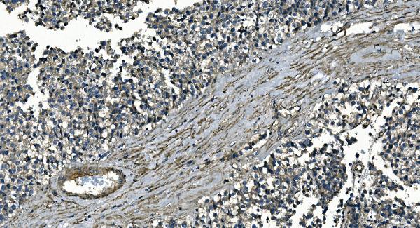


 FCM (Flow Cytometry)
(Figure 9. Flow Cytometry analysis of U937 cells using anti-GNG7 antibody (AAA19335).Overlay histogram showing U937 cells stained with AAA19335 (Blue line). The cells were blocked with 10% normal goat serum. And then incubated with rabbit anti-GNG7 Antibody (AAA19335, 1μg/1x106 cells) for 30 min at 20 degree C. DyLight®488 conjugated goat anti-rabbit IgG (5-10μg/1x106 cells) was used as secondary antibody for 30 minutes at 20 degree C. Isotype control antibody (Green line) was rabbit IgG (1μg/1x106) used under the same conditions. Unlabelled sample (Red line) was also used as a control.)
FCM (Flow Cytometry)
(Figure 9. Flow Cytometry analysis of U937 cells using anti-GNG7 antibody (AAA19335).Overlay histogram showing U937 cells stained with AAA19335 (Blue line). The cells were blocked with 10% normal goat serum. And then incubated with rabbit anti-GNG7 Antibody (AAA19335, 1μg/1x106 cells) for 30 min at 20 degree C. DyLight®488 conjugated goat anti-rabbit IgG (5-10μg/1x106 cells) was used as secondary antibody for 30 minutes at 20 degree C. Isotype control antibody (Green line) was rabbit IgG (1μg/1x106) used under the same conditions. Unlabelled sample (Red line) was also used as a control.)
GNG7, Polyclonal Antibody (Cat# AAA19335)
Full Name
Anti-GNG7 Antibody
Reactivity
Human, Mouse, Rat
Applications
WB, IHC-P, FC/FACS/FCM, EIA
Purity
Immunogen affinity purified.
Pricing