Rabbit anti-Human STAT3-Y705 Monoclonal Antibody | anti-STAT3 antibody
Phospho-STAT3-Y705 Rabbit mAb
IHC-P: 1:50-1:200
IF/ICC: 1:50-1:200
ELISA: Recommended starting concentration is 1ug/mL.
Please optimize the concentration based on your specific assay requirements.
ICC (Immunocytochemistry)
(Confocal imaging of paraffin-embedded Mouse colon tissue using Phospho-STAT3-Y705 Rabbit mAb (AAA28610, dilution 1:200) followed by a further incubation with Cy3 Goat Anti-Rabbit IgG (H+L) (AS007, dilution 1:500) (Red). DAPI was used for nuclear staining (Blue). High pressure antigen retrieval performed with 0.01M Citrate Buffer (pH 6.0) prior to IF staining. Objective: 40x.)
IHC (Immunohistchemistry)
(Immunohistochemistry analysis of paraffin-embedded Mouse kidney using Phospho-STAT3-Y705 Rabbit mAb (AAA28610) at dilution of 1:400 (40x lens). High pressure antigen retrieval performed with 0.01M Citrate Bufferr (pH 6.0) prior to IHC staining.)
IHC (Immunohistochemistry)
(Immunohistochemistry analysis of paraffin-embedded Mouse colon using Phospho-STAT3-Y705 Rabbit mAb (AAA28610) at dilution of 1:400 (40x lens). High pressure antigen retrieval performed with 0.01M Citrate Bufferr (pH 6.0) prior to IHC staining.)
IHC (Immunohistochemistry)
(Immunohistochemistry analysis of paraffin-embedded Rat kidney using Phospho-STAT3-Y705 Rabbit mAb (AAA28610) at dilution of 1:100 (40x lens). High pressure antigen retrieval performed with 0.01M Citrate Bufferr (pH 6.0) prior to IHC staining.)
IHC (Immunohistochemistry)
(Immunohistochemistry analysis of paraffin-embedded Mouse kidney using Phospho-STAT3-Y705 Rabbit mAb (AAA28610) at dilution of 1:100 (40x lens). High pressure antigen retrieval performed with 0.01M Citrate Bufferr (pH 6.0) prior to IHC staining.)
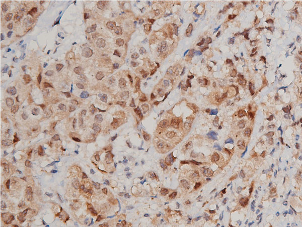
IHC (Immunohistochemistry)
(Immunohistochemistry analysis of paraffin-embedded Human colon carcinoma using Phospho-STAT3-Y705 Rabbit mAb (AAA28610) at dilution of 1:100 (40x lens). High pressure antigen retrieval performed with 0.01M Citrate Bufferr (pH 6.0) prior to IHC staining.)
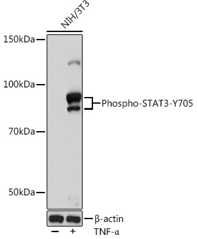
WB (Western Blot)
(Western blot analysis of lysates from NIH/3T3 cells, using Phospho-STAT3-Y705 Rabbit mAb (AAA28610) at 1:1000 dilution. NIH/3T3 cells were treated by TNF-? (20 ng/mL) at 37? for 30 minutes.Secondary antibody: HRP-conjugated Goat anti-Rabbit IgG (H+L) (AS014) at 1:10000 dilution.Lysates/proteins: 25ug per lane.Blocking buffer: 3% nonfat dry milk in TBST.Detection: ECL Enhanced Kit (RM00021).Exposure time: 180s.)
NCBI and Uniprot Product Information
Observed MW: 79kDa/88kDa