Loading...

Skip to main content

Call us on + 1 (800) 604-9114 for more information about our products

Have a question or looking for a specific datasheet Manual/COA/MSDS?
Submit a Question

Interested to get a quote about our products?
Request a Quote

IHC (Immunohistochemistry) (At 1/200 staining Human tonsil tissue sections by IHC-P. The tissue was formaldehyde fixed and a heat mediated antigen retrieval step in citrate buffer was performed. The tissue was then blocked and incubated with the antibody for 1.5 hours at 22 degree C. An HRP conjugated goat anti-rabbit antibody was used as the secondary antibody.)

Rabbit TBK1 Polyclonal Antibody | anti-TBK1 antibody

Phospho-TBK1 (Ser172) Antibody

Gene Names
TBK1; NAK; T2K
Reactivity
Human, Mouse, Rat
Predicted Reactivity: Pig (100%), Zebrafish (100%), Bovine (100%), Horse (100%), Sheep (100%), Rabbit (100%), Dog (100%), Chicken (100%), Xenopus (100%)
Applications
Western Blot, Immunohistochemistry, ELISA
Purity
The antibody is from purified rabbit serum by affinity purification via sequential chromatography on phospho-peptide and non-phospho-peptide affinity columns.
Synonyms
TBK1; Polyclonal Antibody; Phospho-TBK1 (Ser172) Antibody; EC 2.7.11.1; FLJ11330; FTDALS4; NAK; NF kappa B activating kinase; NF kB activating kinase; NF-kappa-B-activating kinase; Serine/threonine protein kinase TBK 1; Serine/threonine protein kinase TBK1; Serine/threonine-protein kinase TBK1; T2K; TANK binding kinase 1; TANK-binding kinase 1; TBK 1; Tbk1; TBK1_HUMAN; anti-TBK1 antibody
Ordering
For Research Use Only!
Host
Rabbit
Reactivity
Human, Mouse, Rat
Predicted Reactivity: Pig (100%), Zebrafish (100%), Bovine (100%), Horse (100%), Sheep (100%), Rabbit (100%), Dog (100%), Chicken (100%), Xenopus (100%)
Clonality
Polyclonal
Isotype
Rabbit IgG
Specificity
Phospho-TBK1 (Ser172) Antibody detects endogenous levels of TBK1 only when phosphorylated at Ser172.
Tissue Specificity: Ubiquitous with higher expression in testis. Expressed in the ganglion cells, nerve fiber layer and microvasculature of the retina.
Purity/Purification
The antibody is from purified rabbit serum by affinity purification via sequential chromatography on phospho-peptide and non-phospho-peptide affinity columns.
Form/Format
Liquid. Rabbit IgG in phosphate buffered saline, pH7.4, 150mM NaCl, 0.02% sodium azide and 50% glycerol.
Concentration
1mg/ml (varies by lot)
Applicable Applications for anti-TBK1 antibody
Western Blot (WB), Immunohistochemistry (IHC), Peptide ELISA (EIA)
Application Notes
WB: 1:1000-3000
IHC: 1:50-1:200
Peptide ELISA: 1:20,000-1:40,000
Immunogen
A synthesized peptide derived from human TBK1 around the phosphorylation site of Ser172.
Conjugation
Unconjugated
Fragment
Fab fragment
Post Translational Modifications
Autophosphorylation at Ser-172 activates the kinase, and is an essential step for virus-triggered signaling. Phosphorylated by IKBKB/IKKB at Ser-172. Phosphorylation requires homodimerization and ubiquitination at Lys-30 and Lys-401. Dephosphorylated at Ser-172 by PPM1B and this negatively regulates its role in mediating antiviral response.'Lys-63'-linked polyubiquitination by MIB1 after RNA virus infection, or by NRDP1 after LPS stimulation at Lys-30 and Lys-401, participates in kinase activation. 'Lys-48'-linked polyubiquitination at Lys-670 by DTX4 leads to proteasomal degradation. 'Lys-48'-linked polyubiquitination by TRAIP also leads to proteasomal degradation. 'Lys-63'-linked polyubiquitination by RNF128 at Lys-30 and Lys-401 leads to the activation of antiviral responses.
Subunit Structure
Homodimer. Interacts with DDX3X, TIRAP and TRAF2. Part of a ternary complex consisting of TANK, TRAF2 and TBK1. Interacts with AZI2, TANK and TBKBP1; these interactions are mutually exclusive and mediate TBK1 activation. Interacts with GSK3B; this interaction promotes TBK1 self-association and autophosphorylation. Interacts with SIKE1; SIKE1 is associated with TBK1 under physiological condition and dissociated from TBK1 upon viral infection or TLR3 stimulation. Interacts with IRF3, leading to IRF3 phosphorylation. Interacts with DDX58/RIG-I. Interacts with CYLD. Interacts with OPTN and TRAF3. Interacts with SRC. Interacts with the exocyst complex subunit SEC5/EXOC2; this interaction is sufficient to trigger TBK1 activity. Interacts with STING1, leading to STING1 phosphorylation. Interacts with IFIT3 (via N-terminus). Interacts with MAVS; interaction only takes place in the presence of IFIT3 and leads to MAVS phosphorylation. Interacts (via protein kinase domain) with TTLL12 (via TTL domain); the interaction prevents MAVS binding to TBK1. Interacts with TICAM1; this interaction is enhanced in the presence of WDFY1 and leads to TICAM1 phosphorylation. Interacts with TRIM26. Interacts with TRIM23. Interacts with TTC4 and IKBKE. Interacts with HNRNPA2B1. (Microbial infection) Interacts with Borna disease virus (BDV) P protein leading to its phosphorylation. (Microbial infection) Interacts with Ebola virus protein VP35. (Microbial infection) Interacts with HCV NS3; this interaction leads to inhibition of cellular antiviral response by blocking necessary interactions between the TBK1 and its substrates IRF3 and IRF7. (Microbial infection) Interacts with herpes simplex virus 1 protein ICP34.5.
Similarity
Comprises A N-terminal kinase domain, a ubiquitin-like domain and a C-terminal coiled-coil region mediating homodimerization.Belongs to the protein kinase superfamily. Ser/Thr protein kinase family. I-kappa-B kinase subfamily.
Subcellular Location
Cytoplasm.
Note: Upon mitogen stimulation or triggering of the immune system, TBK1 is recruited to the exocyst by EXOC2.
Preparation and Storage
Store at -20 degree C. Stable for 12 months from date of receipt.

IHC (Immunohistochemistry)

(At 1/200 staining Human tonsil tissue sections by IHC-P. The tissue was formaldehyde fixed and a heat mediated antigen retrieval step in citrate buffer was performed. The tissue was then blocked and incubated with the antibody for 1.5 hours at 22 degree C. An HRP conjugated goat anti-rabbit antibody was used as the secondary antibody.)

IHC (Immunohistochemistry) (At 1/200 staining Human tonsil tissue sections by IHC-P. The tissue was formaldehyde fixed and a heat mediated antigen retrieval step in citrate buffer was performed. The tissue was then blocked and incubated with the antibody for 1.5 hours at 22 degree C. An HRP conjugated goat anti-rabbit antibody was used as the secondary antibody.)

IHC (Immunohistochemistry)

(At 1/200 staining Mouse kidney tissue sections by IHC-P. The tissue was formaldehyde fixed and a heat mediated antigen retrieval step in citrate buffer was performed. The tissue was then blocked and incubated with the antibody for 1.5 hours at 22 degree C. An HRP conjugated goat anti-rabbit antibody was used as the secondary antibody.)

IHC (Immunohistochemistry) (At 1/200 staining Mouse kidney tissue sections by IHC-P. The tissue was formaldehyde fixed and a heat mediated antigen retrieval step in citrate buffer was performed. The tissue was then blocked and incubated with the antibody for 1.5 hours at 22 degree C. An HRP conjugated goat anti-rabbit antibody was used as the secondary antibody.)

IHC (Immunohistochemistry)

(At 1/200 staining Rat lung tissue sections by IHC-P. The tissue was formaldehyde fixed and a heat mediated antigen retrieval step in citrate buffer was performed. The tissue was then blocked and incubated with the antibody for 1.5 hours at 22 degree C. An HRP conjugated goat anti-rabbit antibody was used as the secondary antibody.)

IHC (Immunohistochemistry) (At 1/200 staining Rat lung tissue sections by IHC-P. The tissue was formaldehyde fixed and a heat mediated antigen retrieval step in citrate buffer was performed. The tissue was then blocked and incubated with the antibody for 1.5 hours at 22 degree C. An HRP conjugated goat anti-rabbit antibody was used as the secondary antibody.)

IHC (Immunohistochemistry)

(At 1/100 staining Rat brain tissue by IHC-P. The sample was formaldehyde fixed and a heat mediated antigen retrieval step in citrate buffer was performed. The sample was then blocked and incubated with the primary antibody at 4 degree C overnight. An HRP conjugated anti-Rabbit antibody was used as the secondary antibody.)

IHC (Immunohistochemistry) (At 1/100 staining Rat brain tissue by IHC-P. The sample was formaldehyde fixed and a heat mediated antigen retrieval step in citrate buffer was performed. The sample was then blocked and incubated with the primary antibody at 4 degree C overnight. An HRP conjugated anti-Rabbit antibody was used as the secondary antibody.)

IHC (Immunohistochemistry)

(At 1/100 staining Mouse spleen tissue by IHC-P. The sample was formaldehyde fixed and a heat mediated antigen retrieval step in citrate buffer was performed. The sample was then blocked and incubated with the primary antibody at 4 degree C overnight. An HRP conjugated anti-Rabbit antibody was used as the secondary antibody.)

IHC (Immunohistochemistry) (At 1/100 staining Mouse spleen tissue by IHC-P. The sample was formaldehyde fixed and a heat mediated antigen retrieval step in citrate buffer was performed. The sample was then blocked and incubated with the primary antibody at 4 degree C overnight. An HRP conjugated anti-Rabbit antibody was used as the secondary antibody.)

IHC (Immunohistochemistry)

(At 1/100 staining Mouse kidney tissue by IHC-P. The sample was formaldehyde fixed and a heat mediated antigen retrieval step in citrate buffer was performed. The sample was then blocked and incubated with the primary antibody at 4 degree C overnight. An HRP conjugated anti-Rabbit antibody was used as the secondary antibody.)

IHC (Immunohistochemistry) (At 1/100 staining Mouse kidney tissue by IHC-P. The sample was formaldehyde fixed and a heat mediated antigen retrieval step in citrate buffer was performed. The sample was then blocked and incubated with the primary antibody at 4 degree C overnight. An HRP conjugated anti-Rabbit antibody was used as the secondary antibody.)

IHC (Immunohistochemistry)

(At 1/100 staining Mouse ovary tissue by IHC-P. The sample was formaldehyde fixed and a heat mediated antigen retrieval step in citrate buffer was performed. The sample was then blocked and incubated with the primary antibody at 4 degree C overnight. An HRP conjugated anti-Rabbit antibody was used as the secondary antibody.)

IHC (Immunohistochemistry) (At 1/100 staining Mouse ovary tissue by IHC-P. The sample was formaldehyde fixed and a heat mediated antigen retrieval step in citrate buffer was performed. The sample was then blocked and incubated with the primary antibody at 4 degree C overnight. An HRP conjugated anti-Rabbit antibody was used as the secondary antibody.)

IHC (Immunohistochemistry)

(At 1/100 staining Mouse stomach tissue by IHC-P. The sample was formaldehyde fixed and a heat mediated antigen retrieval step in citrate buffer was performed. The sample was then blocked and incubated with the primary antibody at 4 degree C overnight. An HRP conjugated anti-Rabbit antibody was used as the secondary antibody.)

IHC (Immunohistochemistry) (At 1/100 staining Mouse stomach tissue by IHC-P. The sample was formaldehyde fixed and a heat mediated antigen retrieval step in citrate buffer was performed. The sample was then blocked and incubated with the primary antibody at 4 degree C overnight. An HRP conjugated anti-Rabbit antibody was used as the secondary antibody.)

IHC (Immunohistchemistry)

(At 1/100 staining Mouse testis tissue by IHC-P. The sample was formaldehyde fixed and a heat mediated antigen retrieval step in citrate buffer was performed. The sample was then blocked and incubated with the primary antibody at 4 degree C overnight. An HRP conjugated anti-Rabbit antibody was used as the secondary antibody.)

IHC (Immunohistchemistry) (At 1/100 staining Mouse testis tissue by IHC-P. The sample was formaldehyde fixed and a heat mediated antigen retrieval step in citrate buffer was performed. The sample was then blocked and incubated with the primary antibody at 4 degree C overnight. An HRP conjugated anti-Rabbit antibody was used as the secondary antibody.)

IHC (Immunohistochemistry)

(At 1/100 staining Human mammary cancer by IHC-P. The sample was formaldehyde fixed and a heat mediated antigen retrieval step in citrate buffer was performed. The sample was then blocked and incubated with the primary antibody at 4 degree C overnight. An HRP conjugated anti-Rabbit antibody was used as the secondary antibody.)

IHC (Immunohistochemistry) (At 1/100 staining Human mammary cancer by IHC-P. The sample was formaldehyde fixed and a heat mediated antigen retrieval step in citrate buffer was performed. The sample was then blocked and incubated with the primary antibody at 4 degree C overnight. An HRP conjugated anti-Rabbit antibody was used as the secondary antibody.)

IHC (Immunohistchemistry)

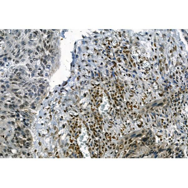
(At 1/100 staining Human lung cancer by IHC-P. The sample was formaldehyde fixed and a heat mediated antigen retrieval step in citrate buffer was performed. The sample was then blocked and incubated with the primary antibody at 4 degree C overnight. An HRP conjugated anti-Rabbit antibody was used as the secondary antibody.)

IHC (Immunohistchemistry) (At 1/100 staining Human lung cancer by IHC-P. The sample was formaldehyde fixed and a heat mediated antigen retrieval step in citrate buffer was performed. The sample was then blocked and incubated with the primary antibody at 4 degree C overnight. An HRP conjugated anti-Rabbit antibody was used as the secondary antibody.)

IHC (Immunohistochemistry)

(At 1/100 staining Human lung cancer by IHC-P. The sample was formaldehyde fixed and a heat mediated antigen retrieval step in citrate buffer was performed. The sample was then blocked and incubated with the primary antibody at 4 degree C overnight. An HRP conjugated anti-Rabbit antibody was used as the secondary antibody.)

IHC (Immunohistochemistry) (At 1/100 staining Human lung cancer by IHC-P. The sample was formaldehyde fixed and a heat mediated antigen retrieval step in citrate buffer was performed. The sample was then blocked and incubated with the primary antibody at 4 degree C overnight. An HRP conjugated anti-Rabbit antibody was used as the secondary antibody.)

IHC (Immunohistochemistry)

(At 1/100 staining Rat stomach tissue by IHC-P. The sample was formaldehyde fixed and a heat mediated antigen retrieval step in citrate buffer was performed. The sample was then blocked and incubated with the primary antibody at 4 degree C overnight. An HRP conjugated anti-Rabbit antibody was used as the secondary antibody.)

IHC (Immunohistochemistry) (At 1/100 staining Rat stomach tissue by IHC-P. The sample was formaldehyde fixed and a heat mediated antigen retrieval step in citrate buffer was performed. The sample was then blocked and incubated with the primary antibody at 4 degree C overnight. An HRP conjugated anti-Rabbit antibody was used as the secondary antibody.)

IHC (Immunohistochemistry)

(At 1/100 staining Rat testis tissue by IHC-P. The sample was formaldehyde fixed and a heat mediated antigen retrieval step in citrate buffer was performed. The sample was then blocked and incubated with the primary antibody at 4 degree C overnight. An HRP conjugated anti-Rabbit antibody was used as the secondary antibody.)

IHC (Immunohistochemistry) (At 1/100 staining Rat testis tissue by IHC-P. The sample was formaldehyde fixed and a heat mediated antigen retrieval step in citrate buffer was performed. The sample was then blocked and incubated with the primary antibody at 4 degree C overnight. An HRP conjugated anti-Rabbit antibody was used as the secondary antibody.)

IHC (Immunohistochemistry)

(Staining Rat colon tissue by IHC-P. The sample was formaldehyde fixed and a heat mediated antigen retrieval step in citrate buffer was performed. The sample was then blocked and incubated with the primary antibody at 4 degree C overnight. An HRP conjugated anti-Rabbit antibody was used as the secondary antibody.)

IHC (Immunohistochemistry) (Staining Rat colon tissue by IHC-P. The sample was formaldehyde fixed and a heat mediated antigen retrieval step in citrate buffer was performed. The sample was then blocked and incubated with the primary antibody at 4 degree C overnight. An HRP conjugated anti-Rabbit antibody was used as the secondary antibody.)

WB (Western Blot)

(Western blot analysis of TBK1 (Phospho-Ser172) using PMA treated HeLa whole cell lysates.-/+ means absence or presence of N peptide(non-phospho peptide) and P peptide(phospho peptide).)

WB (Western Blot) (Western blot analysis of TBK1 (Phospho-Ser172) using PMA treated HeLa whole cell lysates.-/+ means absence or presence of N peptide(non-phospho peptide) and P peptide(phospho peptide).)
Related Product Information for anti-TBK1 antibody
Serine/threonine kinase that plays an essential role in regulating inflammatory responses to foreign agents. Following activation of toll-like receptors by viral or bacterial components, associates with TRAF3 and TANK and phosphorylates interferon regulatory factors (IRFs) IRF3 and IRF7 as well as DDX3X. This activity allows subsequent homodimerization and nuclear translocation of the IRFs leading to transcriptional activation of pro-inflammatory and antiviral genes including IFNA and IFNB. In order to establish such an antiviral state, TBK1 form several different complexes whose composition depends on the type of cell and cellular stimuli. Plays a key role in IRF3 activation: acts by first phosphorylating innate adapter proteins MAVS, STING1 and TICAM1 on their pLxIS motif, leading to recruitment of IRF3, thereby licensing IRF3 for phosphorylation by TBK1. Phosphorylated IRF3 dissociates from the adapter proteins, dimerizes, and then enters the nucleus to induce expression of interferons. Thus, several scaffolding molecules including FADD, TRADD, MAVS, AZI2, TANK or TBKBP1/SINTBAD can be recruited to the TBK1-containing-complexes. Under particular conditions, functions as a NF-kappa-B effector by phosphorylating NF-kappa-B inhibitor alpha/NFKBIA, IKBKB or RELA to translocate NF-Kappa-B to the nucleus. Restricts bacterial proliferation by phosphorylating the autophagy receptor OPTN/Optineurin on 'Ser-177', thus enhancing LC3 binding affinity and antibacterial autophagy. Phosphorylates SMCR8 component of the C9orf72-SMCR8 complex, promoting autophagosome maturation. Phosphorylates and activates AKT1. Seems to play a role in energy balance regulation by sustaining a state of chronic, low-grade inflammation in obesity, wich leads to a negative impact on insulin sensitivity (By similarity). Attenuates retroviral budding by phosphorylating the endosomal sorting complex required for transport-I (ESCRT-I) subunit VPS37C. Phosphorylates Borna disease virus (BDV) P protein. Plays an essential role in the TLR3- and IFN-dependent control of herpes virus HSV-1 and HSV-2 infections in the central nervous system.

NCBI and Uniprot Product Information

NCBI GI #
NCBI GeneID
NCBI Accession #
NCBI GenBank Nucleotide #
UniProt Accession #
Molecular Weight
84kDa
NCBI Official Full Name
serine/threonine-protein kinase TBK1
NCBI Official Synonym Full Names
TANK-binding kinase 1
NCBI Official Symbol
TBK1
NCBI Official Synonym Symbols
NAK; T2K
NCBI Protein Information
serine/threonine-protein kinase TBK1; NF-kB-activating kinase; NF-kappa-B-activating kinase
UniProt Protein Name
Serine/threonine-protein kinase TBK1
UniProt Gene Name
TBK1
UniProt Synonym Gene Names
NAK
UniProt Entry Name
TBK1_HUMAN

Similar Products

Product Notes

The TBK1 tbk1 (Catalog #AAA31411) is an Antibody produced from Rabbit and is intended for research purposes only. The product is available for immediate purchase. The Phospho-TBK1 (Ser172) Antibody reacts with Human, Mouse, Rat Predicted Reactivity: Pig (100%), Zebrafish (100%), Bovine (100%), Horse (100%), Sheep (100%), Rabbit (100%), Dog (100%), Chicken (100%), Xenopus (100%) and may cross-react with other species as described in the data sheet. AAA Biotech's TBK1 can be used in a range of immunoassay formats including, but not limited to, Western Blot (WB), Immunohistochemistry (IHC), Peptide ELISA (EIA). WB: 1:1000-3000 IHC: 1:50-1:200 Peptide ELISA: 1:20,000-1:40,000. Researchers should empirically determine the suitability of the TBK1 tbk1 for an application not listed in the data sheet. Researchers commonly develop new applications and it is an integral, important part of the investigative research process. It is sometimes possible for the material contained within the vial of "TBK1, Polyclonal Antibody" to become dispersed throughout the inside of the vial, particularly around the seal of said vial, during shipment and storage. We always suggest centrifuging these vials to consolidate all of the liquid away from the lid and to the bottom of the vial prior to opening. Please be advised that certain products may require dry ice for shipping and that, if this is the case, an additional dry ice fee may also be required.

Precautions

All products in the AAA Biotech catalog are strictly for research-use only, and are absolutely not suitable for use in any sort of medical, therapeutic, prophylactic, in-vivo, or diagnostic capacity. By purchasing a product from AAA Biotech, you are explicitly certifying that said products will be properly tested and used in line with industry standard. AAA Biotech and its authorized distribution partners reserve the right to refuse to fulfill any order if we have any indication that a purchaser may be intending to use a product outside of our accepted criteria.

Disclaimer

Though we do strive to guarantee the information represented in this datasheet, AAA Biotech cannot be held responsible for any oversights or imprecisions. AAA Biotech reserves the right to adjust any aspect of this datasheet at any time and without notice. It is the responsibility of the customer to inform AAA Biotech of any product performance issues observed or experienced within 30 days of receipt of said product. To see additional details on this or any of our other policies, please see our Terms & Conditions page.

Item has been added to Shopping Cart

If you are ready to order, navigate to Shopping Cart and get ready to checkout.

Submit a Question

Please complete the form below and a representative will contact you as soon as possible.

Request more Information

Please complete the form below and a representative will contact you as soon as possible.

Request a Manual

Please complete the form below and a representative will contact you as soon as possible.

Request a Quote

Please complete the form below and a representative will contact you as soon as possible.