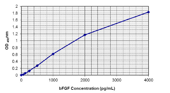
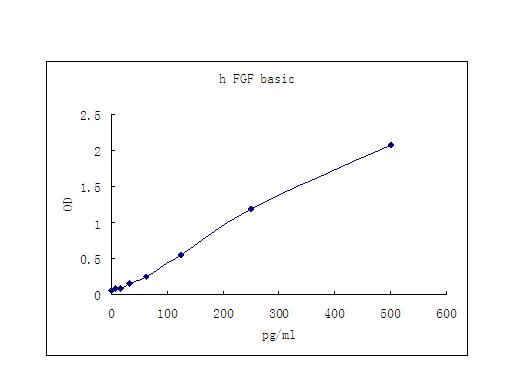
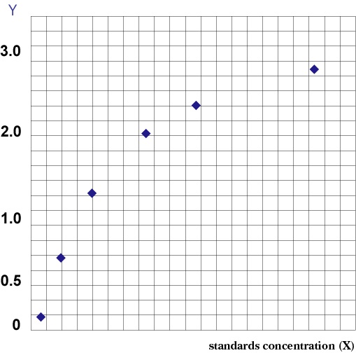
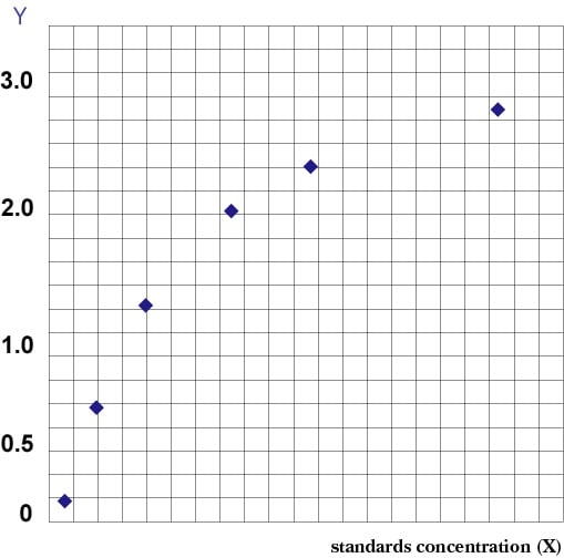
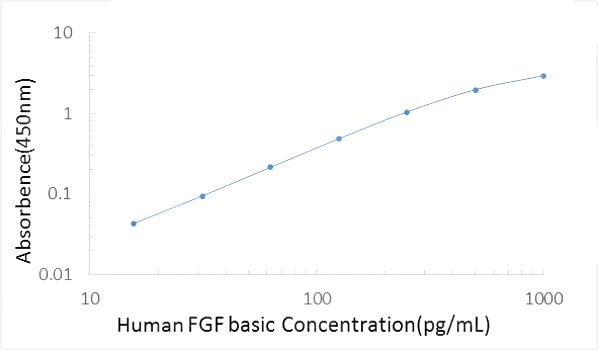
 Standard Curve (Sample)
Basic Fibroblast Growth Factor ELISA Kit | b-FGF elisa kit
Basic Fibroblast Growth Factor (b-FGF)
Gene Names
FGF2; BFGF; FGFB; FGF-2; HBGF-2
Synonyms
Basic Fibroblast Growth Factor; N/A; Basic Fibroblast Growth Factor (b-FGF); b-FGF elisa kit
Specificity
This sandwich ELISA recognises both natural and recombinant human bFGF. This kit exhibits no detectable cross-reactivity with human; TGF-beta, GM-CSF, MCP-3, INF-r, EGF, IL-1alpha, IL-8, IL-1beta, TNF-alpha, IL-16, MCSF, IL-5, and IL-16.
Sequence Length
114
Samples
Serum, plasma, cell culture supernatant, urine, and other biological fluids
Assay Type
Quantitative Sandwich
Intra-assay Precision
To determine within-run precision, three different samples of known concentration were assayed by using 10 replicates in 1 assay.
Inter-assay Precision
To determine between-run precision, three different samples of known concentration were assayed by using replicates on different assays.
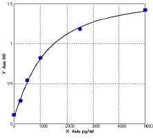
Standard Curve (Sample)
Standard Curve (Sample)
Related Product Information for b-FGF elisa kit
Intended Uses: This Human bFGF ELISA kit is to be used for the in vitro quantitative determination of human basic fibroblast growth factor (bFGF) concentrations in serum, plasma, cell culture supernatant, urine, and other biological fluids. This kit is intended FOR LABORATORY RESEARCH USE ONLY.
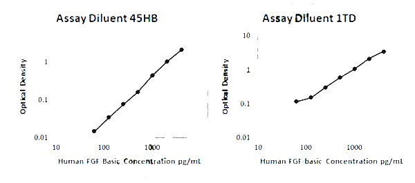
Principle of the Assay: This bFGF enzyme-linked immunosorbent assay (ELISA) applies a technique called a quantitative sandwich immunoassay. The microtiter plate provided in this kit has been pre-coated with a monoclonal specific for bFGF. Standards or samples are then added to the appropriate microtiter plate wells and incubated. bFGF, if present, will bind and become immobilized by the antibody pre-coated on the wells. The microtiter plate wells are thoroughly washed to remove unbound bFGF and other components of sample. In order to quantitative the amount of bFGF present in the sample, a standardized preparation of horseradish peroxidase (HRP)-conjugated antibody specific for bFGF is added to each well to "sandwich" the bFGF immobilised during the first incubation. The microtiter plate then undergoes a second incubation. The wells are thoroughly washed to remove all unbound HRP-conjugated antibodies and a TMB (3,3'5,5' tetramethyl-benzidine) substrate solution is added to each well. The enzyme (HRP) and substrate are allowed to react over a short incubation period. Only those wells that contain bFGF and enzyme-conjugated antibody will exhibit a change in colour. The enzyme-substrate reaction is terminated by the addition of a sulphuric acid solution and the colour change is measured by spectrophotometer at a wavelength of 450 nm +/- 2 nm. In order to measure the concentration of bFGF in the samples, this kit contains two calibration diluents (Calibrator Diluent I for serum/plasma testing and Calibrator Diluent II for cell culture supernatant/ urine testing). According to the testing system, the provided standard is diluted (2-fold) with the appropriate Calibrator Diluent and assayed at the same time as the samples. This allows the operator to produce a standard curve of Optical Density (O.D.) versus bFGF concentration (pg/mL). The concentration of bFGF in the samples is then determined by comparing the O.D. of the samples to the standard curve.
Background/Introduction: When proteins synthesized by one cell can diffuse over small distances to induce changes in neighboring cells, the event is called a paracrine interaction, and the diffusible proteins are called paracrine factors or growth and differentiation factors (GDFs). Many of these paracrine factors can be grouped into four major families on the basis of their structures. These families are the fibroblast growth factor (FGF) family, the Hedgehog family, the Wingless (Wnt) family, and the TGF-beta superfamily. The fibroblast growth factor (FGF) family currently has over a dozen structurally related members. FGF-1 is also known as acidic FGF; FGF-2 is called basic FGF(bFGF); and FGF7 sometimes goes by the name of keratinocyte growth factor. Over a dozen distinct FGF genes are known in vertebrates, and they can generate hundreds of protein isoforms by varying their RNA splicing or initiation codons in different tissues. FGFs can activate a set of receptor tyrosine kinases called the fibroblast growth factor receptors (FGFRs). The receptor tyrosine kinases are proteins that extend through the cell membrane. On the extracellular side is the portion of the protein that binds the paracrine factor. On the intracellular side is a dormant tyrosine kinase (i.e., a protein that can phosphorylate another protein by splitting ATP). When the FGF receptor binds an FGF (and only when it binds an FGF), the dormant kinase is activated, and it phosphorylates certain proteins within the responding cell. The proteins are now activated and can perform new functions. FGFs are associated with several developmental functions, including angiogenesis (blood vessel formation), mesoderm formation, and axon extension. While FGFs can often substitute for one another, their expression patterns give them separate functions. Basic FGF especially important in angiogenesis, and FGF8 is important for the development of the midbrain and limbs. The FGFs have been showed 30-50% overall sequence homology at the amino acid level. They are present at significantly higher concentrations than the neurotrophins; FGF-1 and bFGF concentrations, respectively, are approximately 500-fold and 50-fold greater than that of NGF. The major FGF translation products do not possess a signal peptide sequence and are found principally within the cytoplasm of cells in which they are expressed. It is suspected that other members of the FGF family influence development. For example, there is evidence that FGF-8 is involved in axial specification and patterning of limb development. FGFs stimulate the proliferation of neurons in the developing nervous system and glial cells throughout life. bFGF stimulates the proliferation of multipotential stem cells, which subsequently give rise to neurons of the cortex.The FGFs also exhibit trophic activity toward mature neurons, promoting the survival of these cells without stimulating DNA synthesis. There is some evidence that the FGFs may play a critical role in facilitating axonal regeneration in the PNS and provide trophic support to neurons following trauma or injury.The complexity of FGF action is compounded by the existence of at least four receptors for FGF (FGFR1-4). Three FGFRs are expressed in the CNS, where they exist as multiple alternatively spliced products. All of the FGFRs are ligand-activated tyrosine kinases and comprise a distinct subfamily of the receptor tyrosine kinases. The interaction of the various FGFs with the four FGFRs and their multiple mRNA splice products is bewilderingly complex and incompletely understood. FGFR1 appears to be expressed exclusively in neurons, while principally glial cells express bFGFR and FGFR3. Interestingly, neurons of the substantia nigra and some motor neurons appear to express both FGF-1 and its receptor, FGFR1, suggesting that FGF-1 may act in an autocrine fashion to support these cells. Both receptors are expressed prior to the appearance of their ligands, FGF-1 and bFGF. These data support the view that other FGF family members are more functionally relevant species during embryogenesis. FGFR1 is expressed in the primitive neuroepithelium. A novel aspect of FGF biology is the ability of these growth factors to bind to cell surface proteoglycans, specifically heparan sulfate proteoglycans. Indeed, it appears that these proteoglycans can act as low-affinity receptors for the FGFs. It is thought that FGFs bind to the proteoglycans, which effectively immobilize them and induce or stabilize an active conformation, facilitating binding to the FGFR. bFGF is the most extensively studied member in FGF family, and it is found in almost all tissues of mesodermal and neuroectodermal origin and also in tumors related to these tissues. Endothelial cells produce large amounts of this factor. Some bFGF is associated with the extracellular matrix of the subendothelial cells. bFGF is an 18 kDa protein with a length of 155 amino acids and an isoelectric point of 9.6. It does not contain disulfide bonds and is not glycosylated. bFGF stimulates the growth of fibroblasts, myoblasts, osteoblasts, neuronal cells, endothelial cells, keratinocytes, chondrocytes, and many other cell types. bFGF has been shown to be a promoting or inhibitory modulator of cellular differentiation. bFGF is not only a mitogen for chondrocytes but also inhibits their terminal differentiation. It was stated that the highest levels of bFGF was connected with the advancement of neoplastic process, especially when metastases coexisted at the same time. It was also demonstrated that the higher levels of bFGF gave worse prognosis as far as survival was concerned. These observations suggest the bFGF and its family may play an active role in variety of pathological condition. This human bFGF test kit is for quantitative measurement of human bFGF in serum and cell culture supernatants in vitro.
Product Categories/Family for b-FGF elisa kit
NCBI and Uniprot Product Information
NCBI GI #
NCBI GeneID
Molecular Weight
21,203 Da
NCBI Official Full Name
basic fibroblast growth factor, partial
NCBI Official Synonym Full Names
fibroblast growth factor 2 (basic)
NCBI Official Symbol
FGF2
NCBI Official Synonym Symbols
BFGF; FGFB; FGF-2; HBGF-2
NCBI Protein Information
fibroblast growth factor 2; basic fibroblast growth factor bFGF; heparin-binding growth factor 2; prostatropin
UniProt Protein Name
Fibroblast growth factor 2
UniProt Gene Name
FGF2
UniProt Entry Name
FGF2_HUMAN
Customer Reviews
Loading reviews...
Share Your Experience
Similar Products


Product Notes
The b-FGF fgf2 (Catalog #AAA77841) is an ELISA Kit and is intended for research purposes only. The product is available for immediate purchase. It is sometimes possible for the material contained within the vial of "Basic Fibroblast Growth Factor, ELISA Kit" to become dispersed throughout the inside of the vial, particularly around the seal of said vial, during shipment and storage. We always suggest centrifuging these vials to consolidate all of the liquid away from the lid and to the bottom of the vial prior to opening. Please be advised that certain products may require dry ice for shipping and that, if this is the case, an additional dry ice fee may also be required.Precautions
All products in the AAA Biotech catalog are strictly for research-use only, and are absolutely not suitable for use in any sort of medical, therapeutic, prophylactic, in-vivo, or diagnostic capacity. By purchasing a product from AAA Biotech, you are explicitly certifying that said products will be properly tested and used in line with industry standard. AAA Biotech and its authorized distribution partners reserve the right to refuse to fulfill any order if we have any indication that a purchaser may be intending to use a product outside of our accepted criteria.Disclaimer
Though we do strive to guarantee the information represented in this datasheet, AAA Biotech cannot be held responsible for any oversights or imprecisions. AAA Biotech reserves the right to adjust any aspect of this datasheet at any time and without notice. It is the responsibility of the customer to inform AAA Biotech of any product performance issues observed or experienced within 30 days of receipt of said product. To see additional details on this or any of our other policies, please see our Terms & Conditions page.Item has been added to Shopping Cart
If you are ready to order, navigate to Shopping Cart and get ready to checkout.