Colorimetric vs Fluorometric ELISA: Which Detection Method is Better?
 Cynthia
6 min read
Cynthia
6 min read
In this Article
All of the products listed in AAA Biotech’s catalog are strictly for research-use only (RUO).

The detection method is a major choice when it comes to designing an ELISA. Two commonly used methods are colorimetric and fluorometric. While both methods rely on an enzyme-linked reaction, the signals produced are different.
Since each detection method has its advantages and ideal applications, it can be tricky to determine which one may be best suited for your needs.
To help you choose the right one, this blog will help explain what each method is, address common questions, and go over some important differences.
Key Takeaways
- The colorimetric test is primarily determined by observing the visible color change.
- Fluorometric ELISA is more sensitive, but also more expensive.
- Colorimetric reactions can typically be stopped and are more stable.
- Fluorescent signals are susceptible to photobleaching and often require more precise timing for reading.
What Does It Mean for ELISA to Be a Colorimetric Test?
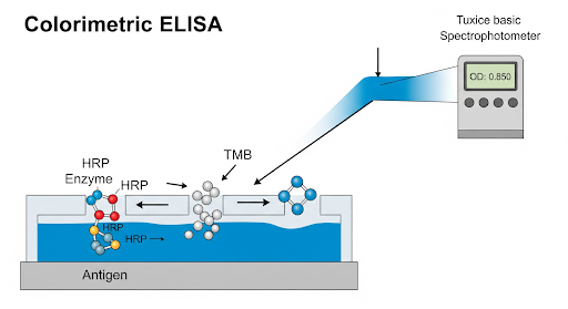
When an ELISA is described as a colorimetric test, it means the assay’s readout is based on a visible color change. In a typical colorimetric ELISA, the detection antibody is linked to an enzyme (commonly HRP – horseradish peroxidase, or AP – alkaline phosphatase) that reacts with a chromogenic substrate to produce a colored product.
For instance, HRP can convert the substrate TMB (3,3',5,5'-tetramethylbenzidine) into a solution that turns deep blue and then yellow upon addition of the “stop solution”. The intensity of the color development is then measured by a spectrophotometer (microplate reader) as an optical density (OD) value at a specific wavelength, typically 450 nm for TMB after addition og the stop solution. In sandwich ELISA, the higher the OD value, the more target analyte is present in the sample.
As for practicality, a colorimetric ELISA is straightforward, as a darker color indicates a positive result, and you can see the difference in wells with the naked eye. This is also the reason why it is the most commonly used ELISA detection method.
Calling an ELISA “colorimetric” simply emphasizes that a color-producing reaction is the detection mechanism.
NOTE: Unfamiliar with ELISA? Check our beginner’s guide to ELISA.
Can ELISA Be Fluorometric?
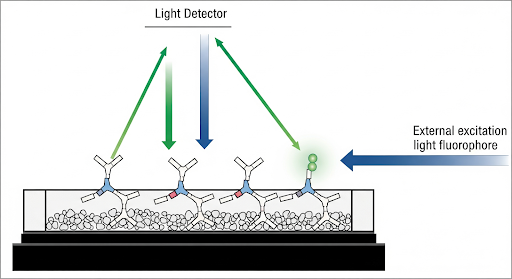
Yes, ELISAs can also be performed with a fluorescent readout. While there’s no color change for obvious indication, the principle is the same, i.e., an enzyme or detection reagent generates a signal in proportion to the target abundance, but that signal is “fluorescence” rather than regularly visible color.
There are two approaches for using this detection method.
1. Using an enzyme (HRP or AP) with a fluorogenic substrate: Using an alkaline phosphatase can convert 4-MUP (4-methylumbelliferyl phosphate) into a fluorescent product (4-MU) that emits light when excited at the right wavelength.
2. Direct fluorescent labeling: Another approach is to directly tag the detecting antibody with a fluorescent dye.
In either case, the outcome is that wells with more target produce a stronger fluorescent signal instead of a deeper color.
However, it should be noted that implementing a fluorescent ELISA requires appropriate equipment and setup (i.e., instead of a regular absorbance spectrophotometer, you need a fluorometer or a multi-mode plate reader capable of fluorescence detection). The instrument will shine an excitation light of a specific wavelength onto each well and then measure the emitted fluorescence at a higher wavelength. Because fluorescence can potentially shine through or scatter between wells, these assays are usually run in opaque black-walled plates (as opposed to clear plates) to minimize any crosstalk and background interference. You won’t see fluorescence with the naked eye in most cases (the signals are typically in UV or low visible ranges), so the readout is entirely instrument-dependent, reported in relative fluorescence units (RFU).
Fluorescent ELISA is a powerful detection method because it often provides greater analytical sensitivity. In fact, fluorescence-based detection offers enhanced sensitivity and a broader dynamic range compared to traditional colorimetric ELISA. It is ideal for applications requiring low detection limits where a colorimetric ELISA might not be sensitive enough.
Colorimetric vs Fluorescent ELISA: Key Differences
Below are some key differences between colorimetric and fluorometric ELISA detection:
| Aspect | Colorimetric | Fluorescent |
|---|---|---|
| Detection Principle | Absorbance (measures reduction in transmitted light). The colored product absorbs light at a specific wavelength. | Light Emission (measures light given off by a glowing solution). A fluorophore emits light at one wavelength after excitation. |
| Sensitivity & Dynamic Range | Generally less sensitive; detects higher analyte amounts. Narrower dynamic range (e.g., OD 0 to 2) | More sensitive. Can detect lower analyte amounts. Broader dynamic range and better signal-to-noise ratio |
| Instrumentation Requirements | Standard microplate reader/Spectrophotometer | Fluorometer with correct excitation and emission filters/optics |
| Microplate Type | Clear, transparent plates | Opaque black plates |
| Signal Stability | More stable | Less stable |
| Background and Interference | Affected by colored compounds or turbidity in the sample | Low inherent background but susceptible to autofluorescence from samples, plastics, or reagents |
| Ease of Use and Cost | Easier to use and inexpensive | Requires more method optimization and expensive |
Fluorometer vs Colorimeter: Instrument Differences
A common question is how a fluorometer differs from a colorimeter (or standard spectrophotometric microplate reader) used in ELISA. In essence, a colorimeter is an instrument that measures the intensity of color by detecting how much light a sample absorbs, whereas a fluorometer measures the intensity of emitted light from a sample that has been excited by a light source.
In the context of ELISA:
A colorimeter (often the term is used interchangeably with “microplate absorbance reader” or a simple “spectrophotometer”) passes a beam of light at a specific wavelength through the ELISA well and records how much of that light is absorbed by the sample in the well. The result is given as an absorbance value or “optical density” (OD). It’s essentially measuring how much the colored product in the ELISA blocks/absorbs the light. The more color, the higher the absorbance and the less light that reaches the detector.
A fluorometer, by contrast, has both a light source and an emission detection system. It shines an excitation light of a defined wavelength onto the well and then detects emission light at a different wavelength coming from the well. The key is that the fluorometer is measuring light that the sample produces (via fluorescence) rather than measuring how much light the sample absorbed. The detected signal is usually reported in arbitrary Relative Fluorescence Units (RFU).
NOTE: Many modern instruments are actually multi-mode microplate readers that can function as both a colorimeter and a fluorometer (and sometimes a luminometer) in one.
Which Detection Method is Best?
Neither method is universally “better” in all aspects; each has its pros and cons.
Where colorimetric ELISAs are easy to use and perfectly adequate for many purposes, fluorescent ELISAs offer superior sensitivity and flexibility for demanding applications.
By understanding the differences (absorbance vs. fluorescence, equipment needs, sensitivity trade-offs), you can choose the ELISA detection method that best fits your experiment’s requirements.
Faq's
What is the fundamental difference in the signal measured?
Colorimetric ELISA measures absorbed light (Optical Density) due to a color change. Fluorometric ELISA measures emitted light (Relative Fluorescence Units, RFU) from a glowing product.
Which method offers better sensitivity?
Fluorometric ELISA is generally more sensitive and offers a wider dynamic range, making it better for detecting low-abundance targets.
Are the signals stable?
Colorimetric signals (e.g., stopped TMB reaction) are generally more stable over time. Fluorescent signals can photobleach (fade) with light exposure and often require immediate reading.
Why are different microplates used for each detection method?
Clear/transparent plates are used for colorimetric assays so light can pass through for absorbance measurement. Opaque black plates are used for fluorescent assays to minimize light crosstalk and background interference.
Which detection method is cheaper?
Colorimetric ELISA is generally considered easier to perform and more cost-effective due to cheaper substrates and standard equipment.
Lead Clinical Research Coordinator (LCRC)
Cynthia Lee is the President of AAA Biotech and specializes in understanding highly validated and characterized monoclonal/polyclonal antibodies, recombinant proteins, and ELISA kits.