Galisepticum (Negative)
M. synoviae (Negative)
M. melearidis (Negative)
Application Data
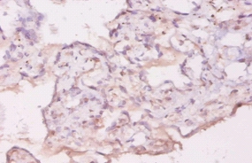
(Ref. 3: Isolation, culture and immunocytochemical characterization of HUCPC. The dissection under sterile condition of foetal and full-term cords was performed to expose the WJ, the vein and arteries (A). After the in vitro expansion of foetal HUCPC (B) the cells ..)
Application Data
(Ref. 2:Immunostaining of cells with anti-NeuN and anti-GFAP showing the neural-specific differentiation of MSCs. Anti-NeuN and anti-GFAP stain nuclei and cytoplasm, respectively. While many cells cultured under three-dimensional conditions (collagen (b), collagen–FN:)
Application Data
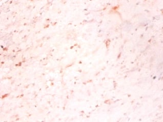
(Ref. 1: Nascent C2C12 skeletal myotubes on collagen patterns. (A) Patterns of collagen-coated rectangles, 20 μm wide and 200-1000 μm long, surrounded by interpenetrating polymer network (IPN). Even after 14 days of incubation with fluorescent collagen, the IPN shows no significant adsorption based on intensity. In addition, when measured by AFM, the apparent elastic modulus of the IPN is approximately half that of the collagen-coated glass. Scale bar, 200 μm. (B) Myoblasts plated onto the patterned coverslips were either stained for vinculin (and imaged by TIRF for clarity) or triple-stained for actin (red) myosin (green) and nuclei (blue). The cells invariably fuse to form multinucleated myotubes. The arrow points to the vinculin clustered at the edges of the patterned myotube, indicative of classical focal adhesion structures. The actin and myosin images show no striations and are of the same nascent myotube at day 6. Bars: in A 200 μm; in B 10 μm.)
Manufactured at an IS09000 facility. Animals are sourced from a specific USDA-Registered facility using a veterinary-inspected flock. Chick Embryo Extract, Ultrafiltrate is protein free. It does not support the growth of viruses.
Similar Products
Product Notes
The Chick Embryo Extract, Ultrafiltrate (Catalog #AAA14749) is a Reagent produced from Chicken and is intended for research purposes only. The product is available for immediate purchase. Recommended Dilution: 1ml of extract/100ml of media. Researchers should empirically determine the suitability of the Chick Embryo Extract, Ultrafiltrate for an application not listed in the data sheet. Researchers commonly develop new applications and it is an integral, important part of the investigative research process. It is sometimes possible for the material contained within the vial of "Chick Embryo Extract, Ultrafiltrate, Reagent" to become dispersed throughout the inside of the vial, particularly around the seal of said vial, during shipment and storage. We always suggest centrifuging these vials to consolidate all of the liquid away from the lid and to the bottom of the vial prior to opening. Please be advised that certain products may require dry ice for shipping and that, if this is the case, an additional dry ice fee may also be required.Precautions
All products in the AAA Biotech catalog are strictly for research-use only, and are absolutely not suitable for use in any sort of medical, therapeutic, prophylactic, in-vivo, or diagnostic capacity. By purchasing a product from AAA Biotech, you are explicitly certifying that said products will be properly tested and used in line with industry standard. AAA Biotech and its authorized distribution partners reserve the right to refuse to fulfill any order if we have any indication that a purchaser may be intending to use a product outside of our accepted criteria.Disclaimer
Though we do strive to guarantee the information represented in this datasheet, AAA Biotech cannot be held responsible for any oversights or imprecisions. AAA Biotech reserves the right to adjust any aspect of this datasheet at any time and without notice. It is the responsibility of the customer to inform AAA Biotech of any product performance issues observed or experienced within 30 days of receipt of said product. To see additional details on this or any of our other policies, please see our Terms & Conditions page.Item has been added to Shopping Cart
If you are ready to order, navigate to Shopping Cart and get ready to checkout.


