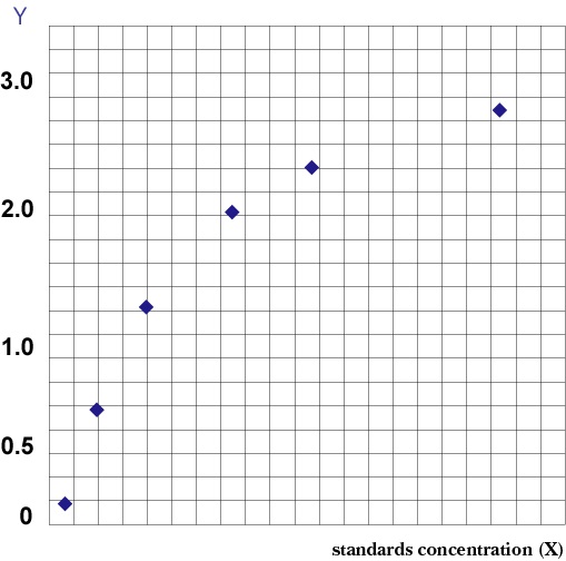
 Application Data
(TGF-? is a negative regulator of NK cell generation CD11cdnR and wild-type mice were injected (i.p.)Four days later, mice were sacrificed and HSC-enriched bone marrow cells were cultured in the presence of IL-7, SCF, and Flt3L.)
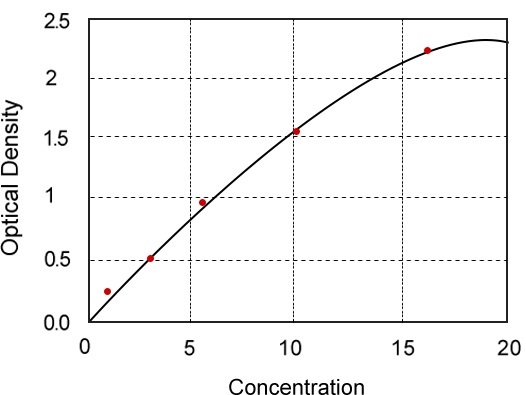
Application Data
(TGF-? is a negative regulator of NK cell generation CD11cdnR and wild-type mice were injected (i.p.)Four days later, mice were sacrificed and HSC-enriched bone marrow cells were cultured in the presence of IL-7, SCF, and Flt3L.)
Interleukin-7 Active Protein | Il7 active protein
Mouse Interleukin-7 Recombinant
Initially, experiments with non-irradiated or irradiated pLN2 were performed with no difference in the outcome (data not shown).
Ex vivo stromal cells were isolated as described above and non-adherent cells washed away after overnight culture (density comparable with lines); they were non-proliferative and therefore not irradiated.
BMDC: They were activated with 0.5 ug/ml LPS for 6 h at 37 degree C.
2 h after LPS addition 1 uM SIINFEKL peptide was added.
1×10⁴ BM-DC were added per 24-well.
T cells: They were obtained ex vivo from spleen and pLN dissected from CO2-killed WT B6 and OT-I transgenic mice and suspended by meshing.
Erythrocytes were removed using red blood cell lysis buffer (Tris-Ammonium chloride based).
CD8 cells were enriched by panning using antibodies to B220 (RA3-6B2), CD4 (H129.19.6), CD11b (M1/70) and CD11c (N418).OT-I cells were labeled with 2 uM CFSE.
Only 50% of WT B6 T cells were CFSE-labeled to identify the peak of undivided cells, with the 50% unlabeled T cells showing the background fluorescence of T cells.
Per 24-well 0.04×10⁶ CFSE+ OT-I cells together with 0.98×10⁶ unlabeled B6 WT cells and 0.98×10⁶ CFSE+ B6 WT cells were added.
The assay was performed in complete RPMI enriched with 1×MEM, 3 ng/ml murine IL-7 and 10 U/ml human IL-2.
After 2-4 days, cells were harvested, counted and analyzed by flow cytometry.
Transwell assays used were 0.4 um transwell chambers (HTS 24-well).
Blocking experiments used 10 uM indomethacin, 1 uM 1400W (?=?dihydrochloride), 10 uM 1-Methyl-L-tryptophan (1-MT), 200 uM (S)-(2-Boronoethyl)-L-cysteine, 10 ug/ml anti-PD-L1 (MIH5, eBioscience), 20 ug/ml anti-IL-10 (kind gift from,,) or 30 ug/ml anti-TGFbeta (clone 1D11.16.8).
To better compare the decrease of T cell proliferation between experiments the attenuating effect by the stromal cells was defined as percent inhibition in divided OT-I T cell numbers relative to the 'no stroma' control: {1-(number of proliferated cells)stroma/(number of proliferated cells)no stroma}*100).
The 'no stroma' control was considered as 0% inhibition; absence of proliferating OT-I cells was considered as 100% inhibition.Functional assaysBone marrow was harvested from C57BL/6 mice and plated in a six-well dish at 1?×?106 cells/ml in 2?ml of lymphocyte media (RPMI 1640 supplemented with 10% FCS, 5?mM l-Alanyl-l-Glutamine, 100?IU/ml penicillin-streptomycin and 2?mM beta-mercaptoethanol with 5?ng/ml of Daedalus IL-7 or commercial IL-7.
Media with IL-7 was replaced at Day 3 and the resulting cell populations were evaluated by fluorescence-activated cell sorting (FACS) at Day 5.
Cells were stained with fluorescein isothiocyanate (FITC) anti-mouse IgM and PE anti-mouse B220 (BD) in FACS staining buffer (PBS with 2.5% FCS) and analyzed on an LSRII flow cytometer (BD) and using FlowJo software.
Recombinant human LIF was tested for its ability to maintain pluripotency of murine ES cells.
Briefly, murine ES cells (AB1 ES cells kindly provided by Steve Jones, University of Massachusetts) were plated on irradiated feeder cells with either commercial human LIF or Daedalus recombinant human LIF at 10?ng/ml.
Cells were split every 2 days with fresh media and stained with APC anti-SSEA1 (BD) for analysis by flow cytometry.Differentiation of CD34+ cellsErythroid differentiation was carried out on MS-5 stromal cells as previously described 4 cells were plated in 2 mL medium'>serum-free expansion medium containing 100 ng/ml SCF, 5 ng/ml IL-3, 3 U/ml erythropoietin (EPO), and 1 mM hydrocortisone and cultured in 6-wells plates at 37 degree C and 5% CO2 for 4 days.
The cells were then diluted into 6 ml of the same medium in a 25 cm2 flask and cultured for an additional 4 days.
Expanded cells were washed with basal medium and resuspended in 25 ml of basal medium with 3 U/ml EPO and plated onto a newly confluent 75 cm2 flask of MS-5 feeder cells and cultured for 3 days.
For final maturation, erythroid cells were co-cultured on feeder cells for 10 days using basal medium without cytokines with one medium change after 5 days at which time 2×10⁶ cells were harvested for nucleic acid purification.
T-cell differentiation was carried on OP9 stromal cells as previously described 4 nucleofected cells were resuspended in alphaMEM with 20% FBS, 5 ng/ml IL-7, and 5 ng/ml Flt3L (both,,) onto sub-confluent OP9-DLL1 cells.
Fresh media with cytokines was replaced every 3 to 4 days.
B-cell differentiation was carried out on MS-5 stromal cells as reported 5 nucleofected CD34+ cells were resuspended in 15 ml of alphaMEM with 10% FBS, 10 ng/ml SCF, and 10 ng/ml G-CSF and seeded onto a confluent MS-5 layer in a 75 cm2 flask.
Ten milliliters of fresh medium was added after 7 days and cells were maintained with twice weekly half-media changes.
Myeloid and granulocyte differentiations were performed on MS-5 stromal cells as described 5 nucleofected CD34+ cells were seeded in 1 ml IMDM with 10% FBS, 2 mM L-alanyl-glutamine, 50 ng/ml SCF, and 50 ng/ml IL-3 in six-well plates.
After 3 days in culture, cells were washed and seeded into 25 cm2 flasks in basal medium plus 50 ng/ml SCF, 50 ng/ml IL-3, and 10 ng/ml G-CSF.
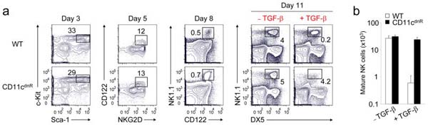
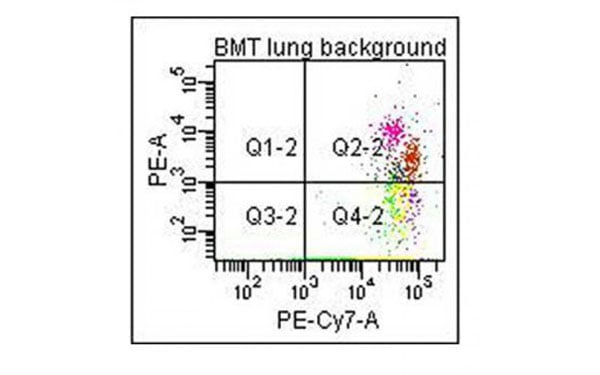
Cells were maintained at 2×10⁵ to 8×10⁵ cells per ml in complete medium with cytokines for 10 days until harvest.In vitro Generation of NK cellsThe generation of NK cells from stem cell precursors was conducted in vitro, based on protocol previously described.Briefly, CD11cdnR and wild-type mice were first treated (i.p.) with 5-FU (5-Fluorouracil) provided at 150 mg/kg body weight.
Four days later, mice were sacrificed and HSC-enriched bone marrow cells were cultured at 10,000 cells/well in complete RPMI supplemented with 0.5 ng/ml murine IL-7, 30 ng/ml mouse SCF, and 100 U/ml murine Flt3L (eBiosciences).
Cells were re-fed with the same media on day 3.
On day 5, cultures were harvested and re-plated at 15,000/well on a confluent monolayer of OP9 stromal cells in complete RPMI containing 30 ng/ml murine IL-15.
On day 8, cultures were re-fed with the same media with or without 5 ng/ml human TGF-beta.
On day 11, cells were harvested and analyzed by flow cytometry.
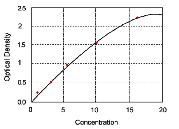
Application Data
(TGF-? is a negative regulator of NK cell generation CD11cdnR and wild-type mice were injected (i.p.)Four days later, mice were sacrificed and HSC-enriched bone marrow cells were cultured in the presence of IL-7, SCF, and Flt3L.)
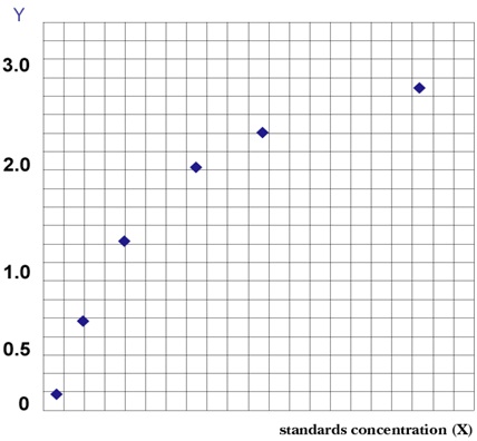
Application Data
(TGF-? is a negative regulator of NK cell generation CD11cdnR and wild-type mice were injected (i.p.)Four days later, mice were sacrificed and HSC-enriched bone marrow cells were cultured in the presence of IL-7, SCF, and Flt3L.)
Application Data
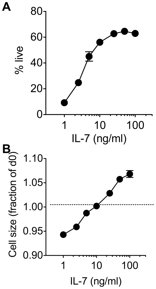
(IL-7 mediated growth and survival is dose dependent in vitro.CD8 T cells were enriched from C57Bl6/J donors and cultured with a range of IL-7 concentrations.)
Application Data
(IL-7 mediated growth and survival is dose dependent in vitro.CD8 T cells were enriched from C57Bl6/J donors and cultured with a range of IL-7 concentrations.)
Application Data
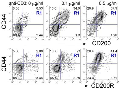
(T cells co-express CD200 and CD200R following TCR activation.C. Naïve LN cells were stimulated with anti-CD3 (1 ug/ml) + anti-CD28 (2 ug/ml) either transiently on Ab-coated wells for the first 2d then removed to fresh media (blue lines) or chronically with Ab-coated aAPC present throughout the culture (red lines) in Th1 (top row), Th2 (middle row) or non-polarising (IL-7, bottom row) conditions.)
Application Data
(T cells co-express CD200 and CD200R following TCR activation.C. Naïve LN cells were stimulated with anti-CD3 (1 ug/ml) + anti-CD28 (2 ug/ml) either transiently on Ab-coated wells for the first 2d then removed to fresh media (blue lines) or chronically with Ab-coated aAPC present throughout the culture (red lines) in Th1 (top row), Th2 (middle row) or non-polarising (IL-7, bottom row) conditions.)
NCBI and Uniprot Product Information
Customer Reviews
Loading reviews...
Share Your Experience
Similar Products


Product Notes
The Il7 il7 (Catalog #AAA76391) is an Active Protein produced from E coli (Note: Host protein expression may be lot-specific and can be subject to change during production, including bulk orders.) and is intended for research purposes only. The product is available for immediate purchase. The amino acid sequence is listed below: MFHVSFRYIF GIPPLILVLL PVTSSECHIK DKEGKAYESV LMISIDELDK MTGTDSNCPN NEPNFFRKHV CDDTKEAAFL NRAARKLKQF LKMNISEEFN VHLLTVSQGT QTLVNCTSKE EKNVKEQKKN DACFLKRLLR EIKTCWNKIL KGSI. It is sometimes possible for the material contained within the vial of "Interleukin-7, Active Protein" to become dispersed throughout the inside of the vial, particularly around the seal of said vial, during shipment and storage. We always suggest centrifuging these vials to consolidate all of the liquid away from the lid and to the bottom of the vial prior to opening. Please be advised that certain products may require dry ice for shipping and that, if this is the case, an additional dry ice fee may also be required.Precautions
All products in the AAA Biotech catalog are strictly for research-use only, and are absolutely not suitable for use in any sort of medical, therapeutic, prophylactic, in-vivo, or diagnostic capacity. By purchasing a product from AAA Biotech, you are explicitly certifying that said products will be properly tested and used in line with industry standard. AAA Biotech and its authorized distribution partners reserve the right to refuse to fulfill any order if we have any indication that a purchaser may be intending to use a product outside of our accepted criteria.Disclaimer
Though we do strive to guarantee the information represented in this datasheet, AAA Biotech cannot be held responsible for any oversights or imprecisions. AAA Biotech reserves the right to adjust any aspect of this datasheet at any time and without notice. It is the responsibility of the customer to inform AAA Biotech of any product performance issues observed or experienced within 30 days of receipt of said product. To see additional details on this or any of our other policies, please see our Terms & Conditions page.Item has been added to Shopping Cart
If you are ready to order, navigate to Shopping Cart and get ready to checkout.