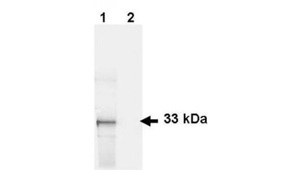

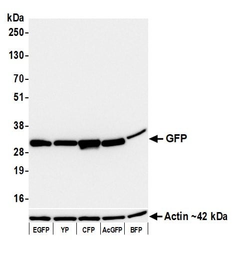
 WB (Western Blot)
(Western Blot of GFP antibody. Lane 1: HeLa cells. Lane 2: mock transfected HeLa cell lysate. Load: 35 ug per lane. GFP antibody at 1 ug/ml for 1 h at room temperature. IRDye 800 conjugated Donkey-a-Goat IgG [H&L] MX7 (605-732-125) secondary antibody at 1:2,500 for 45 min at RT. Block: 5% Blocking buffer overnight at 4 deg C. Size: 27 kDa, 33 kDa for GFP..)
WB (Western Blot)
(Western Blot of GFP antibody. Lane 1: HeLa cells. Lane 2: mock transfected HeLa cell lysate. Load: 35 ug per lane. GFP antibody at 1 ug/ml for 1 h at room temperature. IRDye 800 conjugated Donkey-a-Goat IgG [H&L] MX7 (605-732-125) secondary antibody at 1:2,500 for 45 min at RT. Block: 5% Blocking buffer overnight at 4 deg C. Size: 27 kDa, 33 kDa for GFP..)
GFP Antibody | anti-GFP antibody
GFP antibody (biotin)
WB (Western Blot)
(Western Blot of GFP antibody. Lane 1: HeLa cells. Lane 2: mock transfected HeLa cell lysate. Load: 35 ug per lane. GFP antibody at 1 ug/ml for 1 h at room temperature. IRDye 800 conjugated Donkey-a-Goat IgG [H&L] MX7 (605-732-125) secondary antibody at 1:2,500 for 45 min at RT. Block: 5% Blocking buffer overnight at 4 deg C. Size: 27 kDa, 33 kDa for GFP..)
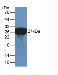
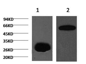
WB (Western Blot)
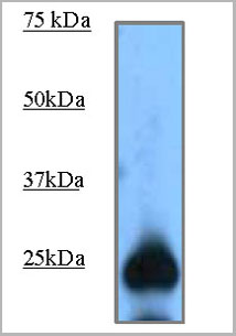
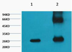
(Western blot of Biotin GFP antibody. Lane 1: GFP. Lane 2: None. Load: 50 ng per lane. GFP antibody Biotin conjugated at 1:1,000 for 60 min at RT. Peroxidase streptavidin secondary antibody at 1:40,000 for 30 min at RT. Blocking: Blocking buffer for 30 min at RT. Size: 28 kDa, 28 kDa for GFP..)
WB (Western Blot)
(Western blot of Biotin GFP antibody. Lane 1: GFP. Lane 2: None. Load: 50 ng per lane. GFP antibody Biotin conjugated at 1:1,000 for 60 min at RT. Peroxidase streptavidin secondary antibody at 1:40,000 for 30 min at RT. Blocking: Blocking buffer for 30 min at RT. Size: 28 kDa, 28 kDa for GFP..)
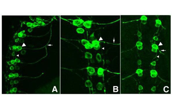
IF (Immunofluorescence)
(Tissue: Drosophila melanogaster late stage embryonic central nervous system. Fixation: 0.5% PFA. Antigen retrieval: not required. Anti-GFP antibody at a 1:1,000 for 1 h at RT. AlexaFluor 488 conjugated anti-Goat antibody at 1:300 for 45 min at RT. Panel A: shows a lateral view (ventral left). Panels B and C: shows ventral views of whole mount embryos at 63x magnification (plus 2x digital zoom). In all panels, anterior is up. Staining: tau-GFP cell bodies (large arrowhead) and axons of motorneurons (arrow) and interneurons (small arrowhead) as green fluorescent signal.)
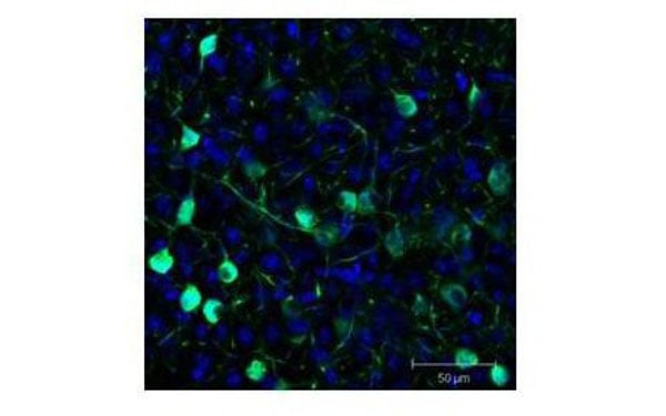
IF (Immunofluorescence)
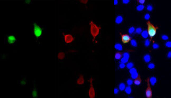
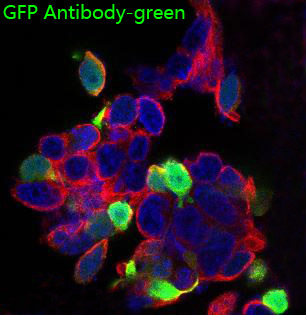
(issue: Sf-1:Cre mice crossed to the Z/EG reporter line. Mouse brain (coronal view, 20X magnification). Fixation: 4%PFA/PBS with o/n fixation, and subsequently transferred to a 30% sucrose solution. Antigen retrieval: frozen in OCT freezing medium (Sakura) and cryostat sectioned at 40 microns. Goat anti-GFP was used at 1:500 dilution in free floating imunnohistochemistry to detect GFP. Fluorchrome conjugated Anti-goat IgG secondary antibody was used for detection at 1:500 at 1:10,000 for 45 min at RT. Localization: Sf-1+ neurons and their processes of the ventromedial nucleus of the hypothalamus. Staining: eGFP as green fluorescent signal and sections were counterstained with DAPI.)
IF (Immunofluorescence)
(issue: Sf-1:Cre mice crossed to the Z/EG reporter line. Mouse brain (coronal view, 20X magnification). Fixation: 4%PFA/PBS with o/n fixation, and subsequently transferred to a 30% sucrose solution. Antigen retrieval: frozen in OCT freezing medium (Sakura) and cryostat sectioned at 40 microns. Goat anti-GFP was used at 1:500 dilution in free floating imunnohistochemistry to detect GFP. Fluorchrome conjugated Anti-goat IgG secondary antibody was used for detection at 1:500 at 1:10,000 for 45 min at RT. Localization: Sf-1+ neurons and their processes of the ventromedial nucleus of the hypothalamus. Staining: eGFP as green fluorescent signal and sections were counterstained with DAPI.)
NCBI and Uniprot Product Information
Customer Reviews
Loading reviews...
Share Your Experience
Similar Products

Product Notes
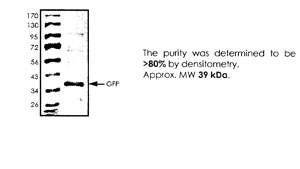
The GFP gfp (Catalog #AAA75019) is an Antibody and is intended for research purposes only. The product is available for immediate purchase. The GFP antibody (biotin) reacts with Assay by immunoelectrophoresis resulted in a single precipitation arc against anti-goat serum, anti-biotin and purified and partially purified green fluorescent protein (Aequorea victoria) Serum. no reaction was observed against Human, Mouse, and Rat ser and may cross-react with other species as described in the data sheet. It is sometimes possible for the material contained within the vial of "GFP, Antibody" to become dispersed throughout the inside of the vial, particularly around the seal of said vial, during shipment and storage. We always suggest centrifuging these vials to consolidate all of the liquid away from the lid and to the bottom of the vial prior to opening. Please be advised that certain products may require dry ice for shipping and that, if this is the case, an additional dry ice fee may also be required.Precautions
All products in the AAA Biotech catalog are strictly for research-use only, and are absolutely not suitable for use in any sort of medical, therapeutic, prophylactic, in-vivo, or diagnostic capacity. By purchasing a product from AAA Biotech, you are explicitly certifying that said products will be properly tested and used in line with industry standard. AAA Biotech and its authorized distribution partners reserve the right to refuse to fulfill any order if we have any indication that a purchaser may be intending to use a product outside of our accepted criteria.Disclaimer
Though we do strive to guarantee the information represented in this datasheet, AAA Biotech cannot be held responsible for any oversights or imprecisions. AAA Biotech reserves the right to adjust any aspect of this datasheet at any time and without notice. It is the responsibility of the customer to inform AAA Biotech of any product performance issues observed or experienced within 30 days of receipt of said product. To see additional details on this or any of our other policies, please see our Terms & Conditions page.Item has been added to Shopping Cart
If you are ready to order, navigate to Shopping Cart and get ready to checkout.