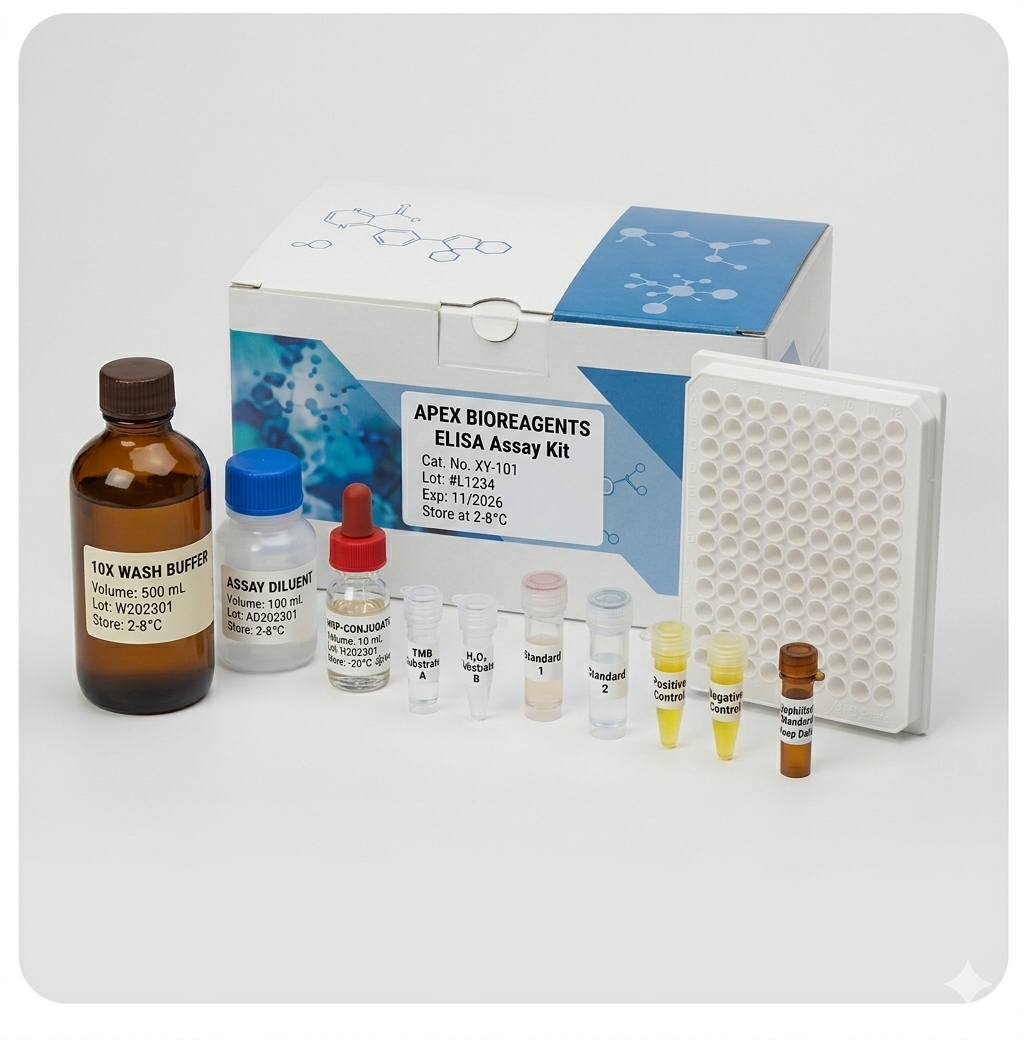
Human Cytokine (M1/M2/MDSC Cytokines) ELISA Kit
Multiplex Human Cytokine ELISA Kit (M1/M2/MDSC Cytokines)
Reactivity
Human
Synonyms
Cytokine (M1/M2/MDSC Cytokines); N/A; Multiplex Human Cytokine ELISA Kit (M1/M2/MDSC Cytokines); Cytokine (M1/M2/MDSC Cytokines) elisa kit
Reactivity
Human
Assay Type
Quantitative Sandwich
Samples
Cell Culture Supernatant and Other Biological Samples
Related Product Information for Cytokine (M1/M2/MDSC Cytokines) elisa kit
Background: Myeloid derived suppressor cells (MOSCs) are myelogenous cells that are capable of negatively regulating T cell immunity. MOSCs are important components of tumor micro-environment. Increased numbers of MOSCs are found in pathological conditions such as malignancy, chronic infection and inflammation. MOSCs cannot be classified by a standard leukocyte linage marker since MOSCs are comprised of various myeloid originated cells at immature status including myeloid progenitor cells, immature monocytes, immature dendritic cells, and immature granulocytes. MOSCs in human are broadly defined as lin(-/low)C033(+) HLA-OR(-)C011 b(+) cells with altered enzyme and cytokine profile and immunosuppressive function. While C033 (+) and C011 b (+) denote myeloid origin in human, Gr-1 (+) and C011 b (+) define myeloid origin in MOSCs in mouse. In chronic inflammation caused by cancer, the interaction between tumor cells and MOSCs causes MOSCs to expand and increase its potential in T cell inhibition (Sevko et al. 2013). MOSCs have been recognized as one of the major mechanisms of tumor evasion from host immunity and are recently evaluated as target for cancer treatment (Sinha et al. 2005). Cytokines are believed to playa critical role in MOSC development and differentiation. GM-CSF and IL-6 have been shown to stimulate MOSC expansion in vivo and in vitro ((Lechner et al. 2010, Morales et al. 2010). T-helper 2 cytokines, IL-4 and IL-13, are the major polarization signals for MOSC to differentiate toward the more T-cell inhibitory M2 phenotype of MOSCs (Bronte et al. 2003, Sinha et al. 2005). Additionally, interleukin 4 receptor alpha (IL-4Ra), the common receptor for IL-4 and IL-13, has been found to be up-regulated in MOSCs (Mandruzzato et al. 2009). One of the main characteristics of M2 MOSCs is the up-regulation of IL-10 and down-regUlation of IL-12 (Bunt et al. 2007). IL-10 inhibits cell immunity by decreasing the secretion of T helper 1 type cytokines and the expression of MHC class II antigens and costimulatory molecules. It has also been postulated that M2 MOSC-mediated T- cell inhibition is the consequence of increased production of arginases and reactive oxygen species by MOSCs (Zea et al. 2005, Rodriguez et al. 2006) and the enzymes secreted by MOSCs block the synthesis of zeta chain in T-cell receptor complex, and sequester cystine to limit the availability of cysteine to T-cells. Signal transduction through calcium binding protein S100A8/A9 and signal transduction and activator of transcription (STAT) is likely implicated in MOSC activities (Zhao et al. 2012). M2 MOSCs inhibit effector T cells but promote Regulatory T cells. The increased expression of cytokines and chemokines such as VEGF, MCP-1 and MIF in the tumor microenvironment is believed to promote the infiltration of MOSCs and stimulate tumor angiogenesis and metastasis (Bellamy et al. 2001, Huang et al. 2007, and Simpson et al. 2013). IFN-y is a potent activator for MOSCs to develop into the more tumoricidal and virucidal M1 phenotype. IFN-y and other M1 polarizing signals up-regulate IL-12 and TNF-a in M1 MOSC. M1-polarized MOSCs express elevated signature markers such as inducible Nitric Oxide Synthase (iNOS), nitric oxide (NO), TNF-a, IFN-y (Yang et al. 2013). Nitric oxide (NO) and TNF-a play important roles in clearing bacterial, and certain fungal, viral, and parasitic invasions as well as in the necrosis of some tumors.
Principle of the Assay: This enzyme linked immunosorbent assay (ELISA) applies a technique called a quantitative sandwich immunoassay. The microwells on the 8-well strips enclosed in the kit have been precoated with monoclonal antibodies specific to GM-CSF, IFN-y, IL-4, IL-6, IL-10, IL-12, MCAF (also known as MCP-1), and TNF-a respectively. Standards or samples are then added to the strips, and the biotin-conjugated detection antibody mixture will be added late on. The above cytokines, if present, will bind and become immobilized by the antibody pre-coated on the wells and then be "sandwiched" by biotin conjugate. The microtiter plate wells are thoroughly washed to remove unbound components of the sample. In order to quantitatively determine the amount of cytokine present in the sample, Avidin conjugated to Horseradish Peroxidase (HRP) is added to each microplate well and incubated. Avidin is a tetramer containing four identical subunits that each has a high affinity-binding site for biotin. The wells are thoroughly washed to remove all unbound HRP-conjugated Avidin. A TMB (3, 3' 5, 5' tetramethyl-benzidine) substrate solution is added to each well. The enzyme (HRP) and substrate are allowed to react over a short incubation period. Only those wells that contain coating antibody and the specific cytokine, biotin-conjugated antibody and enzyme-conjugated Avidin will develop a blue colour. The intensity of colour development is proportional to the concentration of the specific cytokine presented in the each wells. The enzyme-substrate reaction is terminated by the addition of a sulphuric acid solution and the colour will change to yellow. The intensity is measured spectrophotometrically at a wavelength of 450nm ± 2 nm. Samples were tested together with standards diluted with a similar matrix, or one of the Calibrator Diluent provided with the kit. This allows the operator to produce Optical Density (0.0) versus cytokine concentration (pg/mL). The concentration of cytokines in the samples is then determined by comparing the 0.0. of the samples to the standards.
Intended Uses: This multiplex ELISA kit for M1/M2/MOSC cytokines is designed for semi-quantitative and simultaneous determination of cytokines relevant to the proliferation of Myeloid Derived Suppressor Cells (MOSCs) and their differentiation toward M1 or M2 phenotype. The kit simultaneously determines granulocyte macrophage colony stimulating factor (GM-CSF), interferon-y (IFN-y) interleukin-4 (lL-4), interleukin-6 (IL-6), interleukin-10 (IL-10), interleukin-12 (IL-12), monocyte chemotactic and activating factor (MCAF, also known as MCP-1), and tumor necrosis factor-a (TNF-a) in cell culture supernatant and other biological samples. In combination with other Anogen quantitative cytokine ELISA kits, the M1/M2/MOSC cytokine multiplex ELISA kit is expected to be useful for the investigation of the relationship of cytokine expression and MOSC induced inmmunosuppression in various disease models.
References
1. Yagnik G, Rutowski MJ, Shah SS, Aghi MK. Stratifying nonfunctional pituitary adenomas into two groups distinguished by macrophage subtypes. Oncotarget 2019; 22(10):2212-2223.
Customer Reviews
Loading reviews...
Share Your Experience
Similar Products


Product Notes
The Human Cytokine (M1/M2/MDSC Cytokines) (Catalog #AAA77974) is an ELISA Kit and is intended for research purposes only. The product is available for immediate purchase. The AAA77974 ELISA Kit recognizes Human Cytokine (M1/M2/MDSC Cytokines). It is sometimes possible for the material contained within the vial of "Cytokine (M1/M2/MDSC Cytokines), ELISA Kit" to become dispersed throughout the inside of the vial, particularly around the seal of said vial, during shipment and storage. We always suggest centrifuging these vials to consolidate all of the liquid away from the lid and to the bottom of the vial prior to opening. Please be advised that certain products may require dry ice for shipping and that, if this is the case, an additional dry ice fee may also be required.Precautions
All products in the AAA Biotech catalog are strictly for research-use only, and are absolutely not suitable for use in any sort of medical, therapeutic, prophylactic, in-vivo, or diagnostic capacity. By purchasing a product from AAA Biotech, you are explicitly certifying that said products will be properly tested and used in line with industry standard. AAA Biotech and its authorized distribution partners reserve the right to refuse to fulfill any order if we have any indication that a purchaser may be intending to use a product outside of our accepted criteria.Disclaimer
Though we do strive to guarantee the information represented in this datasheet, AAA Biotech cannot be held responsible for any oversights or imprecisions. AAA Biotech reserves the right to adjust any aspect of this datasheet at any time and without notice. It is the responsibility of the customer to inform AAA Biotech of any product performance issues observed or experienced within 30 days of receipt of said product. To see additional details on this or any of our other policies, please see our Terms & Conditions page.Item has been added to Shopping Cart
If you are ready to order, navigate to Shopping Cart and get ready to checkout.