


 Application Data
(Published clone specific image: Flow cytometric analysis of AM from NO2-exposed and control rats. Rats were exposed to NO2 for the indicated times and BAL cells were stained with antibodies to ED7, ED9, RM-4, and OX-6. To overcome autofluorescence signals, primary antibodies were detected using a biotin-PE/streptavidin-anti-streptavidin enhancing system and labeling of AM was analyzed by flow cytometry following gating by help of forward and sideward scatter properties. Shown are representative results of at least six animals per group.From: Garn H, Siese A, Stumpf S, Wensing A, Renz H, Gemsa D. Phenotypical and functional characterization of alveolar macrophage subpopulations in the lungs of NO2-exposed rats. Respir Res. 2006 Jan 6;7:4.)
Application Data
(Published clone specific image: Flow cytometric analysis of AM from NO2-exposed and control rats. Rats were exposed to NO2 for the indicated times and BAL cells were stained with antibodies to ED7, ED9, RM-4, and OX-6. To overcome autofluorescence signals, primary antibodies were detected using a biotin-PE/streptavidin-anti-streptavidin enhancing system and labeling of AM was analyzed by flow cytometry following gating by help of forward and sideward scatter properties. Shown are representative results of at least six animals per group.From: Garn H, Siese A, Stumpf S, Wensing A, Renz H, Gemsa D. Phenotypical and functional characterization of alveolar macrophage subpopulations in the lungs of NO2-exposed rats. Respir Res. 2006 Jan 6;7:4.)
Mouse CD172a Monoclonal Antibody | anti-CD172a antibody
MOUSE ANTI RAT CD172a:RPE
Flow Cytometry: Maximum Dilution: Neat
Perservative Stabilisers
Shelf Life: 12 months from date of reconstitution.
Application Data
(Published clone specific image: Flow cytometric analysis of AM from NO2-exposed and control rats. Rats were exposed to NO2 for the indicated times and BAL cells were stained with antibodies to ED7, ED9, RM-4, and OX-6. To overcome autofluorescence signals, primary antibodies were detected using a biotin-PE/streptavidin-anti-streptavidin enhancing system and labeling of AM was analyzed by flow cytometry following gating by help of forward and sideward scatter properties. Shown are representative results of at least six animals per group.From: Garn H, Siese A, Stumpf S, Wensing A, Renz H, Gemsa D. Phenotypical and functional characterization of alveolar macrophage subpopulations in the lungs of NO2-exposed rats. Respir Res. 2006 Jan 6;7:4.)
Application Data
(Immunoperoxidase staining of rat lymph node cryosection with Mouse anti Rat CD172a antibody, clone ED9 followed by horseradish peroxidase conjugated Goat anti Mouse IgG1 as a detection reagent. Low power)
Application Data
(Immunoperoxidase staining of rat lymph node cryosection with Mouse anti Rat CD172a antibody, clone ED9 followed by horseradish peroxidase conjugated Goat anti Mouse IgG1 as a detection reagent. Low power)
Application Data
(Immunofluorescence staining of rat lymph node cryosection with Mouse anti Rat CD172a antibody, clone ED9 , red in A and Mouse anti Rat CD4 , green in B. C is the merged image with nuclei counterstained blue using DAPI. Low power)
Application Data
(Immunofluorescence staining of rat lymph node cryosection with Mouse anti Rat CD172a antibody, clone ED9 , red in A and Mouse anti Rat CD4 , green in B. C is the merged image with nuclei counterstained blue using DAPI. Low power)
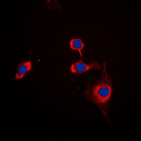
Application Data
(Immunofluorescence staining of rat lymph node cryosection with Mouse anti Rat CD172a antibody, clone ED9 , red in A and Mouse anti Rat CD4 , green in B. C is the merged image with nuclei counterstained blue using DAPI. High power)
Application Data
(Immunofluorescence staining of rat lymph node cryosection with Mouse anti Rat CD172a antibody, clone ED9 , red in A and Mouse anti Rat CD4 , green in B. C is the merged image with nuclei counterstained blue using DAPI. High power)
Application Data
(Immunoperoxidase staining of rat lymph node cryosection with Mouse anti Rat CD172a antibody, clone ED9 followed by horseradish peroxidase conjugated Goat anti Mouse IgG1 as a detection reagent. High power)
Application Data
(Immunoperoxidase staining of rat lymph node cryosection with Mouse anti Rat CD172a antibody, clone ED9 followed by horseradish peroxidase conjugated Goat anti Mouse IgG1 as a detection reagent. High power)
Application Data
(Immunoperoxidase staining of rat lymph node cryosection with Mouse anti Rat CD172a antibody, clone ED9 followed by horseradish peroxidase conjugated Goat anti Mouse IgG1 as a detection reagent. Medium power)
Application Data
(Immunoperoxidase staining of rat lymph node cryosection with Mouse anti Rat CD172a antibody, clone ED9 followed by horseradish peroxidase conjugated Goat anti Mouse IgG1 as a detection reagent. Medium power)
Application Data
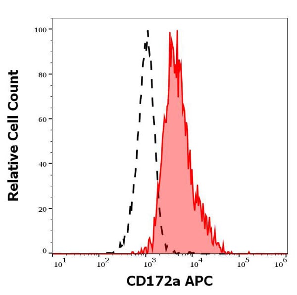
(Published clone specific image: Flow cytometric analysis of ED7 and ED9 expression of AM following magnetic bead separation. AM of 3 days NO2-exposed rats were separated due to their expression of the cell surface molecule ED7 using magnetic bead separation. Susbsequently, ED7 (left) and ED9 (right) expression was analyzed in unseparated AM (A), ED7-positive AM (B), and ED7-negative AM (C). Numbers right of each histogram represent the mean fluorescence of the respective cell population. The figure clearly demonstrates that ED7-positive AM show a lower ED9 expression compared to ED7-negative AM. Shown is a representative data set of more than twenty animals.From: Garn H, Siese A, Stumpf S, Wensing A, Renz H, Gemsa D. Phenotypical and functional characterization of alveolar macrophage subpopulations in the lungs of NO2-exposed rats. Respir Res. 2006 Jan 6;7:4.)
Application Data
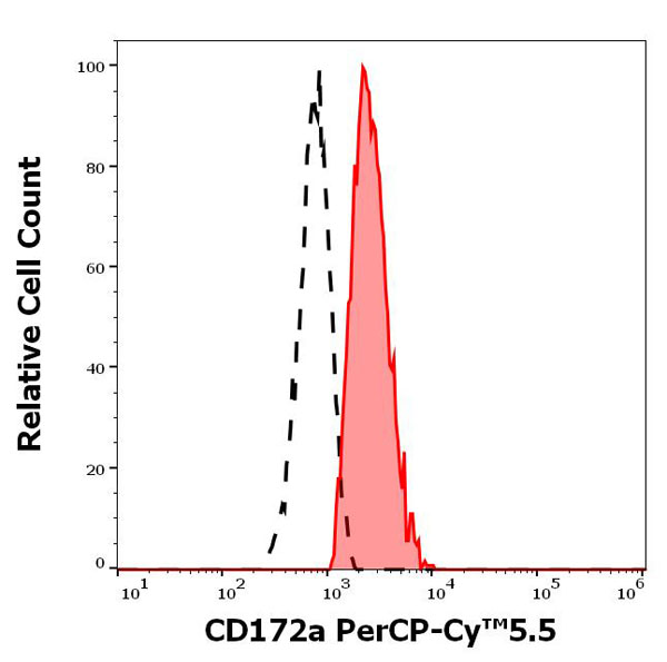
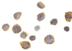
(Staining of rat peritoneal macrophages with Mouse anti Rat CD172a:RPE)
Application Data
(Staining of rat peritoneal macrophages with Mouse anti Rat CD172a:RPE)
NCBI and Uniprot Product Information
Customer Reviews
Loading reviews...
Share Your Experience
Similar Products


Product Notes
The CD172a sirpa (Catalog #AAA12056) is an Antibody produced from Mouse and is intended for research purposes only. The product is available for immediate purchase. AAA Biotech's CD172a can be used in a range of immunoassay formats including, but not limited to, FCM/FACS (Flow Cytometry). Flow Cytometry: Use 10ul of the suggested working dilution to label 106 cells in 100ul. Flow Cytometry: Maximum Dilution: Neat. Researchers should empirically determine the suitability of the CD172a sirpa for an application not listed in the data sheet. Researchers commonly develop new applications and it is an integral, important part of the investigative research process. It is sometimes possible for the material contained within the vial of "CD172a, Monoclonal Antibody" to become dispersed throughout the inside of the vial, particularly around the seal of said vial, during shipment and storage. We always suggest centrifuging these vials to consolidate all of the liquid away from the lid and to the bottom of the vial prior to opening. Please be advised that certain products may require dry ice for shipping and that, if this is the case, an additional dry ice fee may also be required.Precautions
All products in the AAA Biotech catalog are strictly for research-use only, and are absolutely not suitable for use in any sort of medical, therapeutic, prophylactic, in-vivo, or diagnostic capacity. By purchasing a product from AAA Biotech, you are explicitly certifying that said products will be properly tested and used in line with industry standard. AAA Biotech and its authorized distribution partners reserve the right to refuse to fulfill any order if we have any indication that a purchaser may be intending to use a product outside of our accepted criteria.Disclaimer
Though we do strive to guarantee the information represented in this datasheet, AAA Biotech cannot be held responsible for any oversights or imprecisions. AAA Biotech reserves the right to adjust any aspect of this datasheet at any time and without notice. It is the responsibility of the customer to inform AAA Biotech of any product performance issues observed or experienced within 30 days of receipt of said product. To see additional details on this or any of our other policies, please see our Terms & Conditions page.Frequently Asked Questions
What cell types is rat CD172a (SIRPα) a marker for in immunology studies?
CD172a is a marker for myeloid lineage cells, including macrophages, dendritic cells, neutrophils, and bone marrow progenitors. Additionally, it is expressed on neurons in the central nervous system and retina. CD172a serves as a key "don't eat me" signal when complexed with CD47 on cell surfaces.
Can this mouse anti-rat CD172a antibody be used in flow cytometry?
Yes, this monoclonal antibody is optimized for flow cytometry applications. It binds rat CD172a with high specificity, enabling researchers to identify and sort CD172a-positive myeloid populations, study myeloid cell activation states, and analyze immune cell composition in rat models of inflammation and disease.
Is CD172a expression associated with macrophages or dendritic cells?
Yes, CD172a is highly expressed on both macrophages and dendritic cells, as well as other myeloid-derived cells. The CD172a-CD47 interaction regulates macrophage phagocytosis, with high CD172a expression on these cells being essential for immune surveillance and recognition of "altered" vs. "self" cells in inflammatory contexts.
Does this antibody work for immunohistochemistry or immunofluorescence?
Yes, this antibody is validated for immunohistochemistry (IHC) and immunofluorescence (IF) applications. It enables tissue-level visualization of CD172a-expressing immune cells in rat organs, facilitating studies of myeloid cell infiltration, tissue-resident macrophage distribution, and immune cell positioning in inflammatory pathology.
How does CD172a function in cell–cell signaling or immune regulation?
CD172a functions as an inhibitory signal receptor that, upon binding CD47 on target cells, suppresses macrophage phagocytosis and pro-inflammatory responses. This CD172a-CD47 "don't eat me" axis is crucial for immune regulation, and CD172a-blocking antibodies enhance anti-tumor immunity by preventing immune evasion.
Is mouse anti-rat CD172a suitable for studying inflammation or myeloid cell profiling?
Absolutely, CD172a is a standard marker for myeloid cell profiling, and blocking CD172a-CD47 interactions enhances inflammatory responses and anti-tumor immunity. This antibody enables a comprehensive assessment of myeloid cell expansion, activation status, and inflammatory infiltration in rat inflammation, infection, and cancer models.
Item has been added to Shopping Cart
If you are ready to order, navigate to Shopping Cart and get ready to checkout.