 Application Data
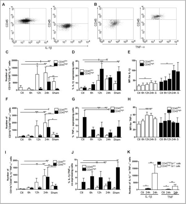
(Published customer image: Cytokine expression in segregated populations of cells following stroke. (A, B) Dot plots showing CD11b+CD45high macrophages/granulocytes (upper right quadrants) and CD11b+CD45dim microglia (bottom right quadrants) expressing IL-1beta (A) or TNF-a (B). (C-J) Bar graphs showing numbers and proportions of IL-1beta (C, D), TNF-a (F, G) and IL-1beta/TNF-a co-expressing (I, J) CD11b+CD45dim microglia and CD11b+CD45high macrophages/granulocytes in unmanipulated control mice (n = 10), in mice 6 (n = 7), 12 (n = 7), or 24 hours after pMCAO (n = 10), and in sham-operated mice 24 hours after pMCAO (n = 7). (E, H) Comparison of the MFI values for IL-1beta (E) and TNF-a (H) in viable CD11b+CD45dim microglia and CD11b+CD45high macrophages/granulocytes in unmanipulated mice, in mice 6, 12, or 24 hours after pMCAO, and in sham-operated mice 24 hours after pMCAO. Macrophages/granulocytes express significantly more IL-1beta than do microglial in unmanipulated mice, in mice 6, 12, or 24 hours after pMCAO, and in sham-operated mice 24 hours after pMCAO (E), whereas microglial cells express significantly higher levels of TNF-a than do macrophages/granulocytes at 12 h and 24 hours, and in sham-operated mice 24 hours after pMCAO (H). (K) CD11b+CD45highGr1- macrophages and not CD11b+CD45highGr1+ granulocytes are the main producers of IL-1beta and TNF-a 24 hours after pMCAO. *P < 0.05, **P < 0.01, and ***P < 0.001.From: http://www.jneuroinflammation.com/content/5/1/46.)
Application Data
(Published customer image: Cytokine expression in segregated populations of cells following stroke. (A, B) Dot plots showing CD11b+CD45high macrophages/granulocytes (upper right quadrants) and CD11b+CD45dim microglia (bottom right quadrants) expressing IL-1beta (A) or TNF-a (B). (C-J) Bar graphs showing numbers and proportions of IL-1beta (C, D), TNF-a (F, G) and IL-1beta/TNF-a co-expressing (I, J) CD11b+CD45dim microglia and CD11b+CD45high macrophages/granulocytes in unmanipulated control mice (n = 10), in mice 6 (n = 7), 12 (n = 7), or 24 hours after pMCAO (n = 10), and in sham-operated mice 24 hours after pMCAO (n = 7). (E, H) Comparison of the MFI values for IL-1beta (E) and TNF-a (H) in viable CD11b+CD45dim microglia and CD11b+CD45high macrophages/granulocytes in unmanipulated mice, in mice 6, 12, or 24 hours after pMCAO, and in sham-operated mice 24 hours after pMCAO. Macrophages/granulocytes express significantly more IL-1beta than do microglial in unmanipulated mice, in mice 6, 12, or 24 hours after pMCAO, and in sham-operated mice 24 hours after pMCAO (E), whereas microglial cells express significantly higher levels of TNF-a than do macrophages/granulocytes at 12 h and 24 hours, and in sham-operated mice 24 hours after pMCAO (H). (K) CD11b+CD45highGr1- macrophages and not CD11b+CD45highGr1+ granulocytes are the main producers of IL-1beta and TNF-a 24 hours after pMCAO. *P < 0.05, **P < 0.01, and ***P < 0.001.From: http://www.jneuroinflammation.com/content/5/1/46.)
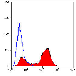
Rat CD45 Monoclonal Antibody | anti-CD45 antibody
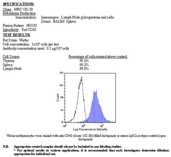
RAT ANTI MOUSE CD45
Purified IgG - liquid
Immunohistology - Frozen: Minimum Dilution: 1/50; Maximum Dilution: 1/100; Application Note: The epitope recognised by this antibody is reported to be sensitive to formaldehyde fixation and tissue processing. We recommends the use of acetone fixation for frozen sections.
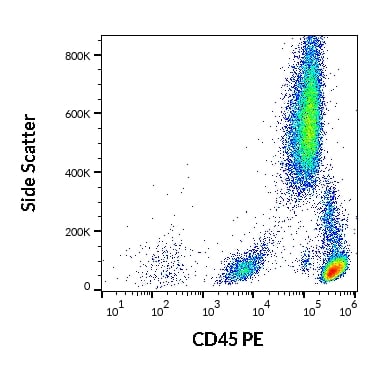
Flow Cytometry: Minimum Dilution: 1/50; Maximum Dilution: 1/100
0.1% Bovine Serum Albumin
Preparation
Short Term: 2-8°C (up to 4 weeks)
store the remaining aliquots at -20°C.
Application Data
(Published customer image: Cytokine expression in segregated populations of cells following stroke. (A, B) Dot plots showing CD11b+CD45high macrophages/granulocytes (upper right quadrants) and CD11b+CD45dim microglia (bottom right quadrants) expressing IL-1beta (A) or TNF-a (B). (C-J) Bar graphs showing numbers and proportions of IL-1beta (C, D), TNF-a (F, G) and IL-1beta/TNF-a co-expressing (I, J) CD11b+CD45dim microglia and CD11b+CD45high macrophages/granulocytes in unmanipulated control mice (n = 10), in mice 6 (n = 7), 12 (n = 7), or 24 hours after pMCAO (n = 10), and in sham-operated mice 24 hours after pMCAO (n = 7). (E, H) Comparison of the MFI values for IL-1beta (E) and TNF-a (H) in viable CD11b+CD45dim microglia and CD11b+CD45high macrophages/granulocytes in unmanipulated mice, in mice 6, 12, or 24 hours after pMCAO, and in sham-operated mice 24 hours after pMCAO. Macrophages/granulocytes express significantly more IL-1beta than do microglial in unmanipulated mice, in mice 6, 12, or 24 hours after pMCAO, and in sham-operated mice 24 hours after pMCAO (E), whereas microglial cells express significantly higher levels of TNF-a than do macrophages/granulocytes at 12 h and 24 hours, and in sham-operated mice 24 hours after pMCAO (H). (K) CD11b+CD45highGr1- macrophages and not CD11b+CD45highGr1+ granulocytes are the main producers of IL-1beta and TNF-a 24 hours after pMCAO. *P < 0.05, **P < 0.01, and ***P < 0.001.From: http://www.jneuroinflammation.com/content/5/1/46.)
Application Data
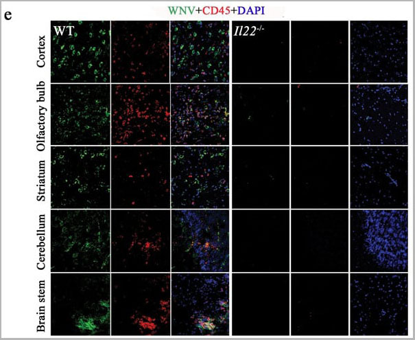
(Published customer image:e) Cryosectioned brain samples from day 6 p.i. were stained for WNV envelope protein (green), CD45 (as a pan-leukocyte marker; red), and DAPI (blue) and utilized for laser scanning confocal microscopy (20X images are shown and are representative of n = 5 mice per group).From: Wang P, Bai F, Zenewicz LA, Dai J, Gate D, et al. (2012) IL-22 Signaling Contributes to West Nile Encephalitis Pathogenesis. PLoS ONE 7(8): e44153.)
Application Data
(Published customer image:e) Cryosectioned brain samples from day 6 p.i. were stained for WNV envelope protein (green), CD45 (as a pan-leukocyte marker; red), and DAPI (blue) and utilized for laser scanning confocal microscopy (20X images are shown and are representative of n = 5 mice per group).From: Wang P, Bai F, Zenewicz LA, Dai J, Gate D, et al. (2012) IL-22 Signaling Contributes to West Nile Encephalitis Pathogenesis. PLoS ONE 7(8): e44153.)
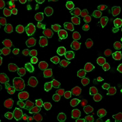
Application Data
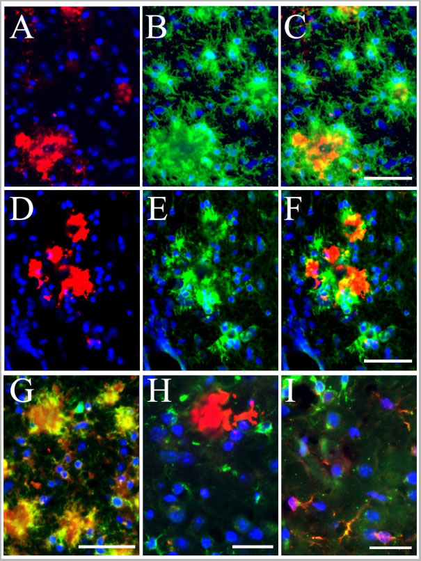
(Published customer image: Colocalization of activated microglial cells with Abeta plaques and IL-1beta immunoreactivity in the TgAPPsw mice with reduced GRK5. Panels A-C, IF staining of Abeta+ plaques (A, red) and surrounding CD45+ microglial cells (B, green), as well as their merged view (C) in the double mice. Scale bar: 50 um for panels A-C. Panels D-F, IF staining of Abeta+ plaques (D, red) and surrounding CD11b+ microglial cells (E, green), as well as their merged view (F) in the double mice. Scale bar: 50 um for panels D-F. Panel G, an example of merged view for CD45 (green) and CD11b/c (clone OX42, red) co-staining of the microglial cells. The image showed that, at least in this particular experimental paradigm, the CD45 antibody stained more specifically for the microglial cell profiles; while the OX42 antibody, in addition to its positive staining of the microglial cells, also non-specifically decorated the plaques. Scale bar: 45 um. Panel H, an example of merged view for Abeta+ plaques (red) and surrounding IL-1beta immunoreactivity (green) in the double mice. Scale bar: 30 um. Panel I, colocalization of CD45+ microglial cells (green) with IL-1beta immunoreactivity (red) in the double mice. Scale bar: 30 um. Blue indicates reference DAPI staining of nuclei.From: Li et al. Journal of Neuroinflammation 2008 5:24.)
Application Data
(Published customer image: Colocalization of activated microglial cells with Abeta plaques and IL-1beta immunoreactivity in the TgAPPsw mice with reduced GRK5. Panels A-C, IF staining of Abeta+ plaques (A, red) and surrounding CD45+ microglial cells (B, green), as well as their merged view (C) in the double mice. Scale bar: 50 um for panels A-C. Panels D-F, IF staining of Abeta+ plaques (D, red) and surrounding CD11b+ microglial cells (E, green), as well as their merged view (F) in the double mice. Scale bar: 50 um for panels D-F. Panel G, an example of merged view for CD45 (green) and CD11b/c (clone OX42, red) co-staining of the microglial cells. The image showed that, at least in this particular experimental paradigm, the CD45 antibody stained more specifically for the microglial cell profiles; while the OX42 antibody, in addition to its positive staining of the microglial cells, also non-specifically decorated the plaques. Scale bar: 45 um. Panel H, an example of merged view for Abeta+ plaques (red) and surrounding IL-1beta immunoreactivity (green) in the double mice. Scale bar: 30 um. Panel I, colocalization of CD45+ microglial cells (green) with IL-1beta immunoreactivity (red) in the double mice. Scale bar: 30 um. Blue indicates reference DAPI staining of nuclei.From: Li et al. Journal of Neuroinflammation 2008 5:24.)
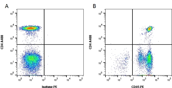
Application Data
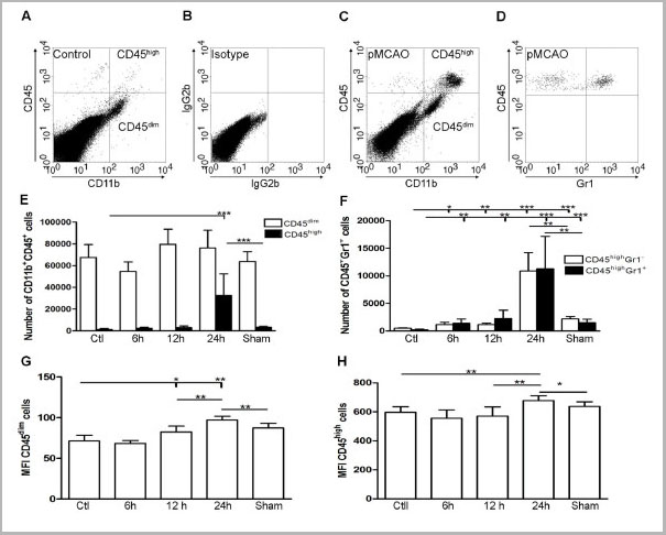
(Published customer image: Inflammatory response following permanent MCA occlusion. (A-C) Dot plots of viable CD11b+CD45high macrophages/granulocytes (top right quadrants) and CD11b+CD45dim microglia (bottom right quadrants) in cortex from unmanipulated control mice (A, B), and mice exposed to pMCAO with 24 hour survival (C). (D) At 24 hours, flow cytometric analysis of the CD11b+CD45high profiles showed that approximately half of the population consisted of CD45highGr1+ granulocytes. (E) Quantification of CD11b+CD45dim and CD11b+CD45high cells in unmanipulated control mice (n = 10), in mice 6 (n = 7), 12 (n = 7), or 24 hours after pMCAO (n = 10), and in sham-operated mice 24 hours after pMCAO (n = 7). (F) Bar graphs showing equal recruitment of CD11b+CD45highGr1- macrophages and CD11b-CD45highGr1+ granulocytes in unmanipulated mice, in mice 6, 12, or 24 hours after pMCAO, and in sham-operated mice 24 hours after pMCAO. (G, H) Bar graphs showing the mean fluorescent intensity (MFI) of CD45 expression by CD45dim microglia (G) and CD45high macrophages/granulocytes (H). *P < 0.05, **P < 0.01, and ***P < 0.001.From: Clausen et al. Journal of Neuroinflammation 2008 5:46.)
Application Data
(Published customer image: Inflammatory response following permanent MCA occlusion. (A-C) Dot plots of viable CD11b+CD45high macrophages/granulocytes (top right quadrants) and CD11b+CD45dim microglia (bottom right quadrants) in cortex from unmanipulated control mice (A, B), and mice exposed to pMCAO with 24 hour survival (C). (D) At 24 hours, flow cytometric analysis of the CD11b+CD45high profiles showed that approximately half of the population consisted of CD45highGr1+ granulocytes. (E) Quantification of CD11b+CD45dim and CD11b+CD45high cells in unmanipulated control mice (n = 10), in mice 6 (n = 7), 12 (n = 7), or 24 hours after pMCAO (n = 10), and in sham-operated mice 24 hours after pMCAO (n = 7). (F) Bar graphs showing equal recruitment of CD11b+CD45highGr1- macrophages and CD11b-CD45highGr1+ granulocytes in unmanipulated mice, in mice 6, 12, or 24 hours after pMCAO, and in sham-operated mice 24 hours after pMCAO. (G, H) Bar graphs showing the mean fluorescent intensity (MFI) of CD45 expression by CD45dim microglia (G) and CD45high macrophages/granulocytes (H). *P < 0.05, **P < 0.01, and ***P < 0.001.From: Clausen et al. Journal of Neuroinflammation 2008 5:46.)
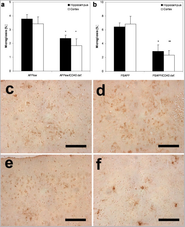
Application Data
(Published customer image Percentage of microgliosis (mean +/- s.e.m) by area in (a) Tg APPsw/CD40 def. mice versus Tg APPsw mice and in (b) Tg PSAPP/CD40 def. mice versus Tg PSAPP mice at 22 to 24 months of age calculated by quantitative image analysis. Post hoc comparison between groups are indicated by the marked bars (* p < 0.05; ** p < 0.01). Representative photographs of brain area in (c) Tg APPsw, (e) Tg APPsw/CD40 def., (d) Tg PSAPP and (f) Tg PSAPP/CD40 def. mice stained with CD45 antibody (each bar represents 0.1 mm).Laporte et al..)
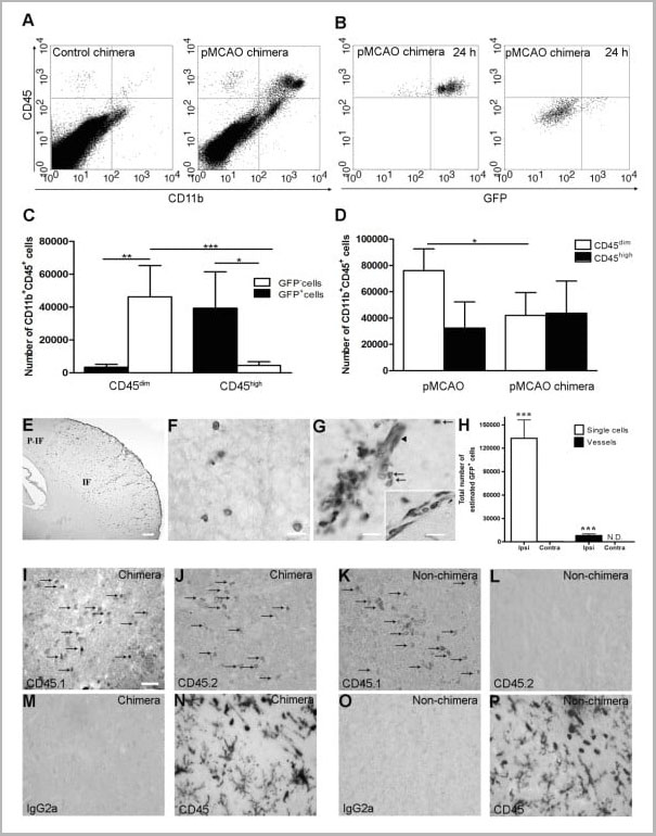
Application Data
(Published customer image: Infiltration of GFP+ BM-cells in infarct and peri-infarct regions. (A-B) Dot plots of viable macrophages/granulocytes (CD11b+CD45high, top right quadrants) and microglia (CD11b+CD45dim, bottom right quadrants) in cortex from BM-chimeric unmanipulated mice and mice exposed to pMCAO. (C) Bar graph showing mean numbers of CD11b+CD45dim microglia and CD11b+CD45high macrophages/granulocytes in BM-chimeric mice 24 hours after pMCAO, subdivided based on expression of GFP (n = 5). Approximately 92% of of the CD45high population were GFP+. (D) Estimation and comparison of mean numbers of CD11b+CD45dim microglia in non-chimeric (n = 10) versus BM-chimeric mice (n = 5) 24 hours after of pMCAO shows significantly fewer CD11b+CD45dim microglial cells in irradiated mice. (E) Overview, showing distribution of infiltrating GFP+ BM-derived cells into infarct (IF) and peri-infarct (P-IF) regions 24 hours after pMCAO. (E-G) By 24 hours, GFP+ single cells (F) and vessel-associated aggregates of GFP+ cells (arrows in G) were observed in infarct and peri-infarct regions. Some of the vessel-associated cells were round, leukocyte-like cells (arrows) while others were elongated cells lining the vasculature (arrow heads in G and in insert). (H) Bar graph showing mean numbers of single GFP+ cells and vessel-associated aggregates of GFP+ cells in ipsi- and contralateral cortex 24 hours after surgery (n = 10). (I-P) Immunohistochemical staining of CD45.1 (I, K), CD45.2 (J, L), IgG2a (M, O) and CD45 (N, P) in ischemic tissue in BM-chimeric (I, J, M, N) and non-chimeric mice (K, L, O, P) 24 hours after pMCAO. N.D, none detected. Scale bars: 200 um (A), 10 um (B, C). 50 um (I-P) *P < 0.05, **P < 0.01, and ***P < 0.001.From: Clausen et al. Journal of Neuroinflammation 2008 5:46.)
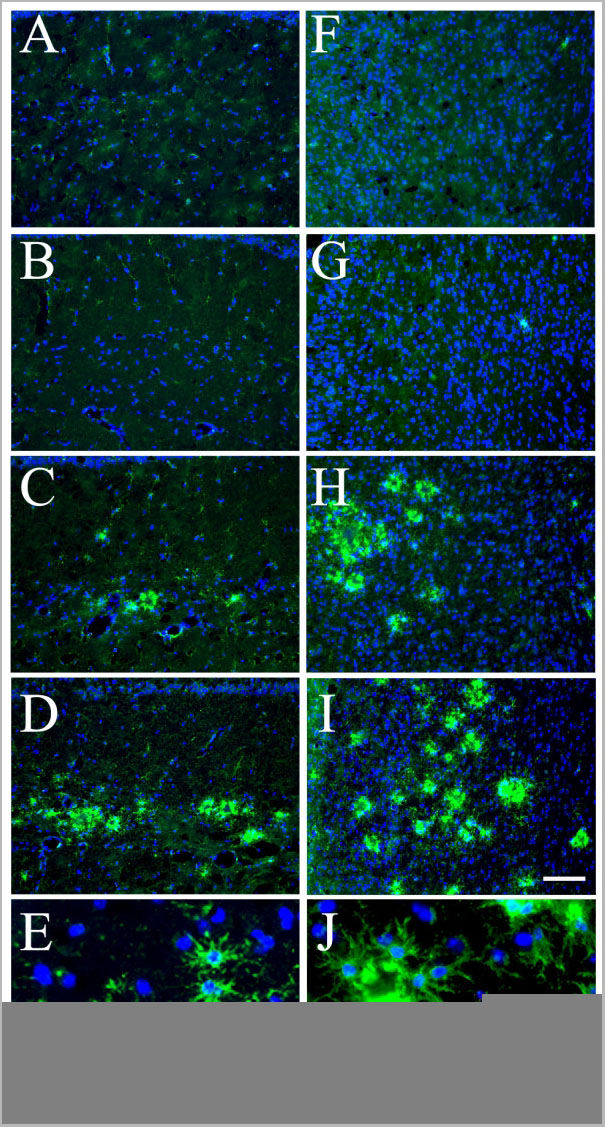
Application Data
(Published customer image: Increased CD45+ microglial cells in hippocampus and cortex of the TgAPPsw mice with reduced GRK5. Representative IF results with anti-CD45 staining (green) in hippocampus (A-D) and cortex (F-I) of WT (A &F), heterozygote GRK5KO (B &G), TgAPPsw (C &H), and the double mice (D &I), respectively. Scale bar in panel I is for panels A-D and F-I: 100 um. Panels E &J, examples of high magnification views of CD45+-microglial cells in hippocampus (E) and cortex (J) of the double mice that show details of activated microglial morphology. Scale bar in panel J is for panels E &J: 20 um. Blue indicates reference DAPI staining of nuclei.From: Li et al. Journal of Neuroinflammation 2008 5:24.)
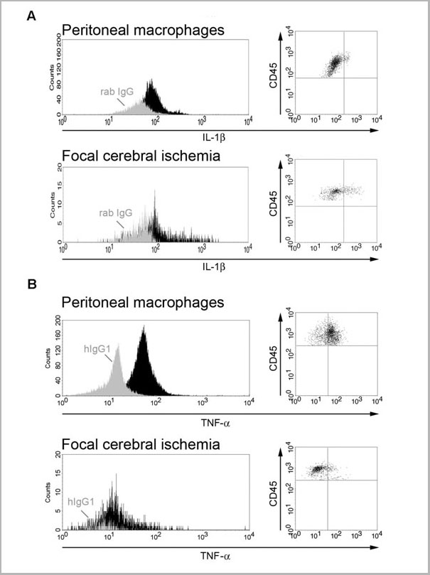
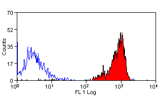
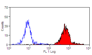
Application Data
(Published customer image: Sensitivity of cytokine detection using flow cytometry. Histograms and dot plots of IL-1beta (A) and TNF-a (B) expression in LPS-activated peritoneal macrophages versus macrophages/granulocytes isolated from cortex 24 hours after pMCAO. Light colored histograms represent cells stained with isotype control antibodies and filled histograms represent cells stained with antibodies for either IL-1beta (A) or TNF-a (B).From: Clausen et al. Journal of Neuroinflammation 2008 5:46)
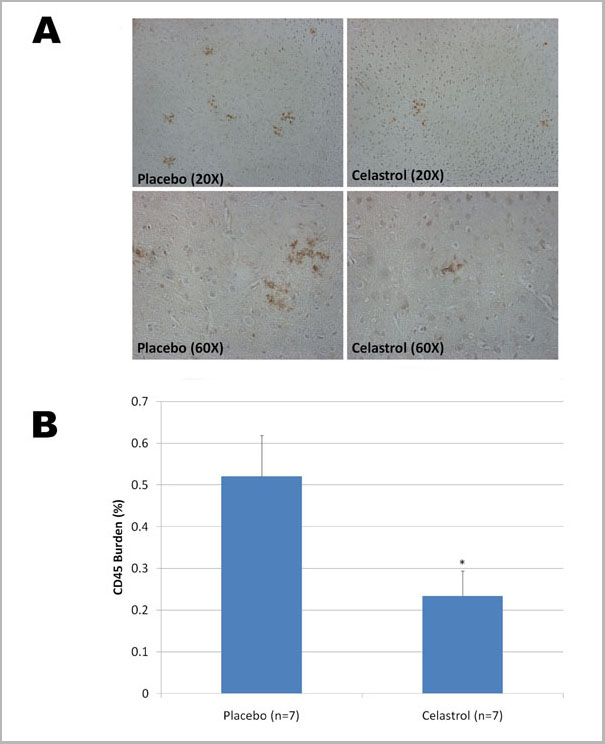
Application Data
(Published customer image: Chronic effects of celastrol on microgliosis in Tg PS1/APPsw mice. A) Representative photomicrographs (taken with a 20x and 60x objective providing a respective magnification of 200x and 600x respectively) depicting the presence of CD45 reactive microglia around Abeta deposits in Tg PS1/APPsw treated with a placebo and celastrol. B) Histogram representing the burden of activated microglia (CD45 positive) in the cortex of Tg PS1/APPsw mice treated with placebo and celastrol pellets. Statistically significant difference in microgliosis burden (P < 0.03) was observed between placebo and celastrol treated mice. (* P < 0.05)..)
NCBI and Uniprot Product Information
Customer Reviews
Loading reviews...
Share Your Experience
Similar Products


Product Notes
The CD45 ptprc (Catalog #AAA11896) is an Antibody produced from Rat and is intended for research purposes only. The product is available for immediate purchase. AAA Biotech's CD45 can be used in a range of immunoassay formats including, but not limited to, IHC (Immunohistochemistry), FCM/FACS (Flow Cytometry), IF (Immunofluorescence), IP (Immunoprecipitation). Flow Cytometry: Use 10ul of the suggested working dilution to label 106 cells in 100ul. Immunohistology - Frozen: Minimum Dilution: 1/50; Maximum Dilution: 1/100; Application Note: The epitope recognised by this antibody is reported to be sensitive to formaldehyde fixation and tissue processing. We recommends the use of acetone fixation for frozen sections. Flow Cytometry: Minimum Dilution: 1/50; Maximum Dilution: 1/100. Researchers should empirically determine the suitability of the CD45 ptprc for an application not listed in the data sheet. Researchers commonly develop new applications and it is an integral, important part of the investigative research process. It is sometimes possible for the material contained within the vial of "CD45, Monoclonal Antibody" to become dispersed throughout the inside of the vial, particularly around the seal of said vial, during shipment and storage. We always suggest centrifuging these vials to consolidate all of the liquid away from the lid and to the bottom of the vial prior to opening. Please be advised that certain products may require dry ice for shipping and that, if this is the case, an additional dry ice fee may also be required.Precautions
All products in the AAA Biotech catalog are strictly for research-use only, and are absolutely not suitable for use in any sort of medical, therapeutic, prophylactic, in-vivo, or diagnostic capacity. By purchasing a product from AAA Biotech, you are explicitly certifying that said products will be properly tested and used in line with industry standard. AAA Biotech and its authorized distribution partners reserve the right to refuse to fulfill any order if we have any indication that a purchaser may be intending to use a product outside of our accepted criteria.Disclaimer
Though we do strive to guarantee the information represented in this datasheet, AAA Biotech cannot be held responsible for any oversights or imprecisions. AAA Biotech reserves the right to adjust any aspect of this datasheet at any time and without notice. It is the responsibility of the customer to inform AAA Biotech of any product performance issues observed or experienced within 30 days of receipt of said product. To see additional details on this or any of our other policies, please see our Terms & Conditions page.Item has been added to Shopping Cart
If you are ready to order, navigate to Shopping Cart and get ready to checkout.