


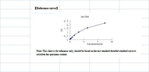
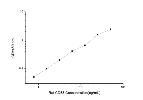
 Application Data
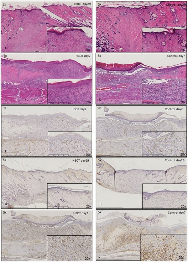
(Published customer image: Histological staining of control and HBOT wounds at post-wounding days 7 and 29. A -D) H&E staining. E -H) CD34 immunohistochemistry. I+J) CD68 immunohistochemistry.From: uk B, Tong M, Fijneman EMG, van Neck JW (2014) Hyperbaric Oxygen Therapy to Treat Diabetes Impaired Wound Healing in Rats. PLoS ONE 9(10): e108533.)
Application Data
(Published customer image: Histological staining of control and HBOT wounds at post-wounding days 7 and 29. A -D) H&E staining. E -H) CD34 immunohistochemistry. I+J) CD68 immunohistochemistry.From: uk B, Tong M, Fijneman EMG, van Neck JW (2014) Hyperbaric Oxygen Therapy to Treat Diabetes Impaired Wound Healing in Rats. PLoS ONE 9(10): e108533.)
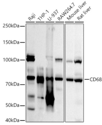
Mouse CD68 Monoclonal Antibody | anti-CD68 antibody
MOUSE ANTI RAT CD68:FITC
Purified IgG conjugated to Fluorescein Isothiocyanate Isomer 1 (FITC) - liquid
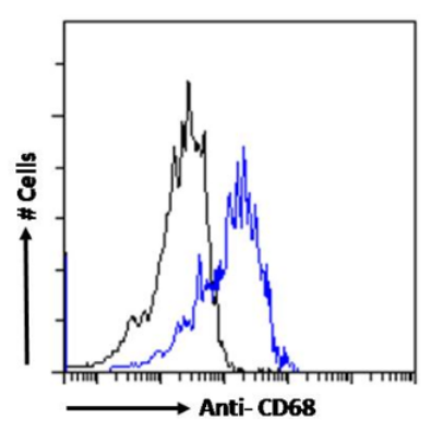
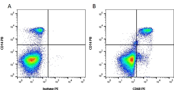
Flow Cytometry: Maximum Dilution: Neat; Application Note: Membrane permeabilisation is required for this application. We recommends the use of Leucoperm for this purpose.
1% Bovine Serum Albumin
Preparation
Shelf Life: 18 months from date of despatch.
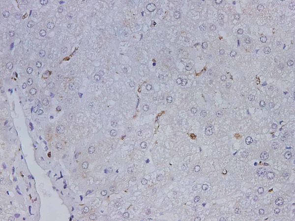
Application Data
(Published customer image: Histological staining of control and HBOT wounds at post-wounding days 7 and 29. A -D) H&E staining. E -H) CD34 immunohistochemistry. I+J) CD68 immunohistochemistry.From: uk B, Tong M, Fijneman EMG, van Neck JW (2014) Hyperbaric Oxygen Therapy to Treat Diabetes Impaired Wound Healing in Rats. PLoS ONE 9(10): e108533.)
Application Data
(Staining of rat peritoneal macrophages cells with Mouse anti Rat CD68:RPE . Permeabilised with Leucoperm (Fix & Perm))
Application Data
(Staining of rat peritoneal macrophages cells with Mouse anti Rat CD68:RPE . Permeabilised with Leucoperm (Fix & Perm))
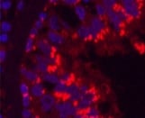
Application Data
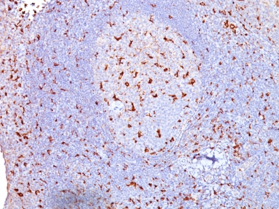
(Immunofluorescence staining of rat lymph node cryosection with Mouse anti Rat CD68 antibody , red in A and Mouse anti Rat CD4 , green in B. C is the merged image with nuclei counter-stained blue using DAPI. Medium power)
Application Data
(Immunofluorescence staining of rat lymph node cryosection with Mouse anti Rat CD68 antibody , red in A and Mouse anti Rat CD4 , green in B. C is the merged image with nuclei counter-stained blue using DAPI. Medium power)
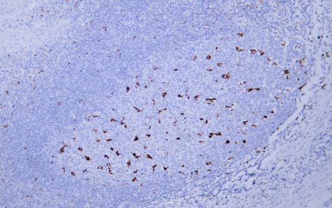
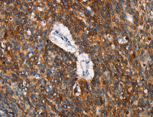
Application Data
(Published customer image: Immunohistochemical stains of ED1 and TRAIL in brain sections treated with AAV2/IL1. Immunohistochemistry for CD68 in the optic nerve. There is a cellular accumulation indicated by DAPI at the crushed sites of the optic nerve. Some of the cells are positively stained with CD68 suggesting that there is a recruitment of microglia/macrophages to the crushed sites. Bar = 100 um.From: Suzuki H, Oku H, Horie T, Morishita S, Tonari M, et al. (2014) Changes in Expression of Aquaporin-4 and Aquaporin-9 in Optic Nerve after Crushing in Rats. PLoS ONE 9(12): e114694.)
Application Data
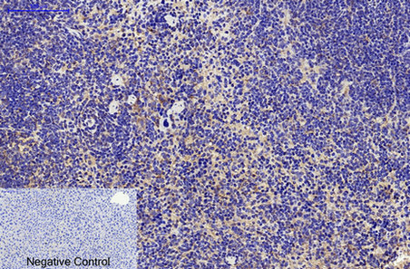
(Immunoperoxidase staining of rat lymph node cryosection with Mouse anti Rat CD68 antibody followed by horseradish peroxidase conjugated Goat anti Mouse IgG1 as a detection reagent. Low power)
Application Data
(Immunoperoxidase staining of rat lymph node cryosection with Mouse anti Rat CD68 antibody followed by horseradish peroxidase conjugated Goat anti Mouse IgG1 as a detection reagent. Low power)
Application Data
(Published customer image: Western blotting of ED1 (Marker of activated microglia) and TRAIL (tumor necrosis factor-related apoptosis-inducing ligand). Brains were treated with PBS or rAAV2/IL12 and then implanted with tumor on the last day of Week 2 post viral vector injection. The brains were used for Western blotting analysis of the expression of ED1, 34 kDa, and TRAIL, 104 kDa, from microglial cells on the last day of week 1, 2, and 3 following tumor implantation. The brain slices for Western blotting analysis are shown at the bottom. All brains were harvested on the last day of week 1, week 2, and week 3 post tumor implantation. The scale bars indicate 2 mm in brain sections.From: The treatment of glioblastoma multiforme through activation of microglia and TRAIL induced by rAAV2-mediated IL-12 in a syngeneic rat model. Chiu TL, Wang MJ, Su CC. J Biomed Sci. 2012 Apr 22;19:45. doi: 10.1186/1423-0127-19-45.)
Application Data
(Staining of rat peritoneal macrophages with Mouse anti Rat CD68: Alexa Fluor 647 following permeabilisation with Leucoperm)
Application Data
(Staining of rat peritoneal macrophages with Mouse anti Rat CD68: Alexa Fluor 647 following permeabilisation with Leucoperm)
Application Data
(Published customer image: Immunohistochemistry of ED1 stain in brain sections treated with rAAV2/IL12 (A, B, C, D, n = 4), AAV2/GFP (E, F, n = 2), or PBS (G, H, n = 2), accompanied with tumor implantation; and that in the brain section treated with nothing (n = 1). Immunohistochemistry of brain sections was performed for ED1 on the last day of week 3 after tumor implantation. The brain sections were stained with hematoxylin for nuclei and ED1 for activated microglial cells. The 1st column shows the brain sections pictured before immunostaining; the 2nd column shows the brain sections pictured after staining; the 3rd to 6th columns show pictures taken at four quadrants (black squares in 2nd column) of the tumor adjacent to the normal tissue in the right hemisphere. ED1-positive cells show dark brown. The scale bars indicate 2 mm in 1st and 2nd columns and 100 um in 3rd-6th columns.From: Chiu TL, Wang MJ, Su CC. The treatment of glioblastoma multiforme through activation of microglia and TRAIL induced by rAAV2-mediated IL-12 in a syngeneic rat model. J Biomed Sci. 2012 Apr 22;19:45.)
Application Data
(Immunofluorescence staining of rat lymph node cryosection with Mouse anti Rat CD68 antibody , red in A and Mouse anti Rat CD4 , green in B. C is the merged image with nuclei counter-stained blue using DAPI. High power)
Application Data
(Immunofluorescence staining of rat lymph node cryosection with Mouse anti Rat CD68 antibody , red in A and Mouse anti Rat CD4 , green in B. C is the merged image with nuclei counter-stained blue using DAPI. High power)
Application Data
(Immunofluorescence staining of rat lymph node cryosection with Mouse anti Rat CD68 antibody , red in A and Mouse anti Rat CD4 , green in B. C is the merged image with nuclei counter-stained blue using DAPI. Low power)
Application Data
(Immunofluorescence staining of rat lymph node cryosection with Mouse anti Rat CD68 antibody , red in A and Mouse anti Rat CD4 , green in B. C is the merged image with nuclei counter-stained blue using DAPI. Low power)
NCBI and Uniprot Product Information
Customer Reviews
Loading reviews...
Share Your Experience
Similar Products


Product Notes
The CD68 (Catalog #AAA12148) is an Antibody produced from Mouse and is intended for research purposes only. The product is available for immediate purchase. AAA Biotech's CD68 can be used in a range of immunoassay formats including, but not limited to, FCM/FACS (Flow Cytometry). Flow Cytometry: Use 10ul of the suggested working dilution to label 106 cells in 100ul. Flow Cytometry: Maximum Dilution: Neat; Application Note: Membrane permeabilisation is required for this application. We recommends the use of Leucoperm for this purpose. Researchers should empirically determine the suitability of the CD68 for an application not listed in the data sheet. Researchers commonly develop new applications and it is an integral, important part of the investigative research process. It is sometimes possible for the material contained within the vial of "CD68, Monoclonal Antibody" to become dispersed throughout the inside of the vial, particularly around the seal of said vial, during shipment and storage. We always suggest centrifuging these vials to consolidate all of the liquid away from the lid and to the bottom of the vial prior to opening. Please be advised that certain products may require dry ice for shipping and that, if this is the case, an additional dry ice fee may also be required.Precautions
All products in the AAA Biotech catalog are strictly for research-use only, and are absolutely not suitable for use in any sort of medical, therapeutic, prophylactic, in-vivo, or diagnostic capacity. By purchasing a product from AAA Biotech, you are explicitly certifying that said products will be properly tested and used in line with industry standard. AAA Biotech and its authorized distribution partners reserve the right to refuse to fulfill any order if we have any indication that a purchaser may be intending to use a product outside of our accepted criteria.Disclaimer
Though we do strive to guarantee the information represented in this datasheet, AAA Biotech cannot be held responsible for any oversights or imprecisions. AAA Biotech reserves the right to adjust any aspect of this datasheet at any time and without notice. It is the responsibility of the customer to inform AAA Biotech of any product performance issues observed or experienced within 30 days of receipt of said product. To see additional details on this or any of our other policies, please see our Terms & Conditions page.Item has been added to Shopping Cart
If you are ready to order, navigate to Shopping Cart and get ready to checkout.