 Application Data
(Rat anti Mouse CD8? antibody, clone KT15 use for the detection of CD8 positive T cells from tumors by flow cytometry and immunofluorescence.Image caption:Vaccination with pFap suppresses TAM, MDSC, Treg, and enhances DC and CTL recruitment. Primary tumors were isolated from mice 25 days after orthotopic challenge and immune cells were identified in primary tumors using antibodies for the following: (A) tumor-associated macrophages (TAMs): F4/80 (red), (B) myeloid derived suppressor cells (MDSCs): CD11b (red, nuclear) and Gr-1 (green, nuclear), (C) T regulatory cells (Tregs, white arrowheads): CD4 (red, cell surface) and FOXP3 (green, nuclear), (D) dendritic cells (DC): 33D1 (red), and (E) cytotoxic T lymphocytes (CTL): CD8 (red) Scale bar for all panels=100 ?m.From: Liao D, Luo Y, Markowitz D, Xiang R, Reisfeld RA (2009)Cancer Associated Fibroblasts Promote Tumor Growth and Metastasis by Modulating the Tumor Immune Microenvironment in a 4T1 Murine Breast Cancer Model.PLoS ONE 4(11): e7965.This image is from an open access article distributed under the terms of the Creative Commons Attribution License.)
Application Data
(Rat anti Mouse CD8? antibody, clone KT15 use for the detection of CD8 positive T cells from tumors by flow cytometry and immunofluorescence.Image caption:Vaccination with pFap suppresses TAM, MDSC, Treg, and enhances DC and CTL recruitment. Primary tumors were isolated from mice 25 days after orthotopic challenge and immune cells were identified in primary tumors using antibodies for the following: (A) tumor-associated macrophages (TAMs): F4/80 (red), (B) myeloid derived suppressor cells (MDSCs): CD11b (red, nuclear) and Gr-1 (green, nuclear), (C) T regulatory cells (Tregs, white arrowheads): CD4 (red, cell surface) and FOXP3 (green, nuclear), (D) dendritic cells (DC): 33D1 (red), and (E) cytotoxic T lymphocytes (CTL): CD8 (red) Scale bar for all panels=100 ?m.From: Liao D, Luo Y, Markowitz D, Xiang R, Reisfeld RA (2009)Cancer Associated Fibroblasts Promote Tumor Growth and Metastasis by Modulating the Tumor Immune Microenvironment in a 4T1 Murine Breast Cancer Model.PLoS ONE 4(11): e7965.This image is from an open access article distributed under the terms of the Creative Commons Attribution License.)
Rat anti-Mouse CD8 Alpha Monoclonal Antibody | anti-CD8Alpha antibody
Rat Anti Mouse CD8 Alpha: Amethyst Orange
Use 10ul of the suggested working dilution to label 106 cells in 100ul.
The Fc region of monoclonal antibodies may bind non-specifically to cells expressing low affinity Fc receptors. This may be reduced by using SeroBlock FcR (BUF041A/B).
1% Bovine Serum Albumin
This product should be stored undiluted.
Storage in frost-free freezers is not recommended.
This product is photosensitive and should be protected from light.
Avoid repeated freezing and thawing as this may denature the antibody.
This product contains a precipitate we recommend microcentrifugation before use.
Application Data
(Rat anti Mouse CD8? antibody, clone KT15 use for the detection of CD8 positive T cells from tumors by flow cytometry and immunofluorescence.Image caption:Vaccination with pFap suppresses TAM, MDSC, Treg, and enhances DC and CTL recruitment. Primary tumors were isolated from mice 25 days after orthotopic challenge and immune cells were identified in primary tumors using antibodies for the following: (A) tumor-associated macrophages (TAMs): F4/80 (red), (B) myeloid derived suppressor cells (MDSCs): CD11b (red, nuclear) and Gr-1 (green, nuclear), (C) T regulatory cells (Tregs, white arrowheads): CD4 (red, cell surface) and FOXP3 (green, nuclear), (D) dendritic cells (DC): 33D1 (red), and (E) cytotoxic T lymphocytes (CTL): CD8 (red) Scale bar for all panels=100 ?m.From: Liao D, Luo Y, Markowitz D, Xiang R, Reisfeld RA (2009)Cancer Associated Fibroblasts Promote Tumor Growth and Metastasis by Modulating the Tumor Immune Microenvironment in a 4T1 Murine Breast Cancer Model.PLoS ONE 4(11): e7965.This image is from an open access article distributed under the terms of the Creative Commons Attribution License.)
Application Data
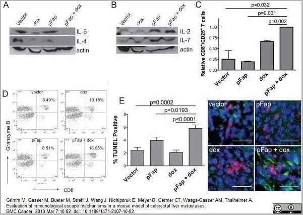
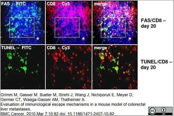
(Rat anti Mouse CD8? antibody, clone KT15 used for the detection of CD8 positive lymphocytes in a mouse model of colorectal liver metastasis by immunofluorescence on acetone treated liver cryosections.Image caption:Immunofluorescence: Representative images of increased CD8+/FAS (a) and CD8+/TUNEL (b) expression on tumor infiltrating lymphocytes at the margin of liver metastases on day 20. FITC green Fluorescein isothiocyanate, Cy3 red and DAPI 4?,6-Diamidino-2-phenylindoldihydrochlorid blue - nuclear countersaining. Case demonstrates area of the TUNEL staining in serial cryostat sections. To adjust a better contrast TUNEL stained sections were not counterstained with DAPI. Asterisk with indication line shows metastatic tumor cells. Magnifications ×250 (top) and ×400 (bottom).From: Grimm M, Gasser M, Bueter M, Strehl J, Wang J, Nichiporuk E, Meyer D, Germer CT, Waaga-Gasser AM, Thalheimer A.Evaluation of immunological escape mechanisms in a mouse model of colorectal liver metastases.BMC Cancer. 2010 Mar 7;10:82.This image is from an open access article distributed under the terms of the Creative Commons Attribution License.)
Application Data
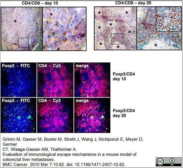
(Rat anti Mouse CD8? antibody, clone KT15 used for the detection of CD8 positive lymphocytes in a mouse model of colorectal liver metastasis by immunohistochemistry and immunofluorescence on acetone treated liver cryosections.Image caption:(a) Immunohistochemistry and (b) immunofluorescence double staining: Representative image of CD4 (red)/CD8 (blue) (top) expression and Foxp3 (FITC)/CD4 (Cy3) (bottom) expression in liver metastases on day 10 (n = 7) compared to increased CD4 (red)/CD8 (blue) expression and Foxp3 (FITC)/CD4 (Cy3) expression on day 20. Immunohistochemistry: Nova red brick red color, Vector Blue blue color. Haemalaun blue color-nuclear counterstaining. Asterisk with indication lines show metastases next to normal tissue (×100 and ×400). Immunofluorescence: FITC green fluorescein isothiocyanate, Cy3 red and DAPI 4?,6-diamidino-2-phenylindole blue - nuclear counterstaining (×250).From: Grimm M, Gasser M, Bueter M, Strehl J, Wang J, Nichiporuk E, Meyer D, Germer CT, Waaga-Gasser AM, Thalheimer A.Evaluation of immunological escape mechanisms in a mouse model of colorectal liver metastases.BMC Cancer. 2010 Mar 7;10:82.This image is from an open access article distributed under the terms of the Creative Commons Attribution License.)
Application Data
(Rat anti Mouse CD8? antibody, clone KT15 used for the detection of CD8 positive lymphocytes in a mouse model of colorectal liver metastasis by immunohistochemistry and immunofluorescence on acetone treated liver cryosections.Image caption:(a) Immunohistochemistry and (b) immunofluorescence double staining: Representative image of CD4 (red)/CD8 (blue) (top) expression and Foxp3 (FITC)/CD4 (Cy3) (bottom) expression in liver metastases on day 10 (n = 7) compared to increased CD4 (red)/CD8 (blue) expression and Foxp3 (FITC)/CD4 (Cy3) expression on day 20. Immunohistochemistry: Nova red brick red color, Vector Blue blue color. Haemalaun blue color-nuclear counterstaining. Asterisk with indication lines show metastases next to normal tissue (×100 and ×400). Immunofluorescence: FITC green fluorescein isothiocyanate, Cy3 red and DAPI 4?,6-diamidino-2-phenylindole blue - nuclear counterstaining (×250).From: Grimm M, Gasser M, Bueter M, Strehl J, Wang J, Nichiporuk E, Meyer D, Germer CT, Waaga-Gasser AM, Thalheimer A.Evaluation of immunological escape mechanisms in a mouse model of colorectal liver metastases.BMC Cancer. 2010 Mar 7;10:82.This image is from an open access article distributed under the terms of the Creative Commons Attribution License.)
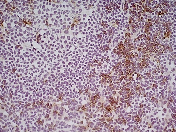
Application Data
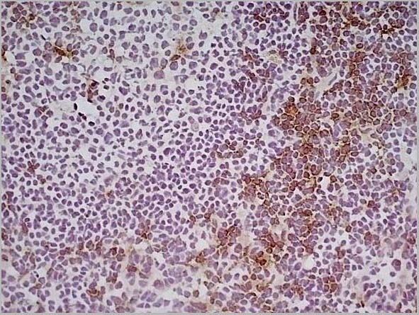
(Immunoperoxidase staining of mouse lymph node cryosection stained with Rat antii Mouse CD8? antibody, clone KT15 followed by horseradish peroxidase conjugated Goat anti Rat IgG s a detection reagent. High power)
Application Data
(Immunoperoxidase staining of mouse lymph node cryosection stained with Rat antii Mouse CD8? antibody, clone KT15 followed by horseradish peroxidase conjugated Goat anti Rat IgG s a detection reagent. High power)
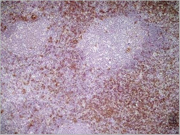
Application Data
(Immunoperoxidase staining of mouse lymph node cryosection stained with Rat antii Mouse CD8? antibody, clone KT15 followed by horseradish peroxidase conjugatedGoat anti Rat IgG s a detection reagent. Medium power)
Application Data
(Immunoperoxidase staining of mouse lymph node cryosection stained with Rat antii Mouse CD8? antibody, clone KT15 followed by horseradish peroxidase conjugatedGoat anti Rat IgG s a detection reagent. Medium power)
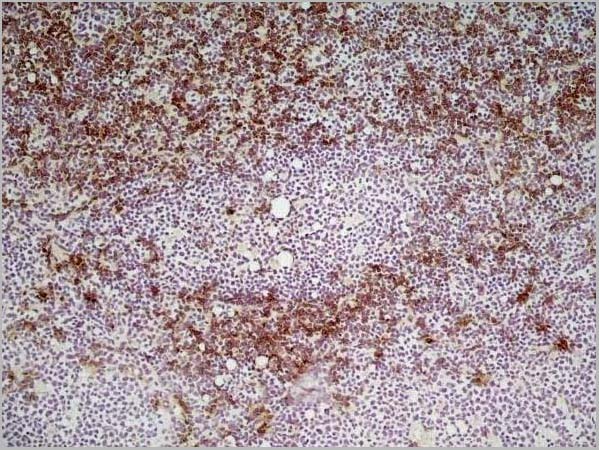
Application Data
(Immunoperoxidase staining of mouse lymph node cryosection stained with Rat antii Mouse CD8? antibody, clone KT15 followed by horseradish peroxidase conjugated Goat anti Rat IgG s a detection reagent. Low power)
Application Data
(Immunoperoxidase staining of mouse lymph node cryosection stained with Rat antii Mouse CD8? antibody, clone KT15 followed by horseradish peroxidase conjugated Goat anti Rat IgG s a detection reagent. Low power)
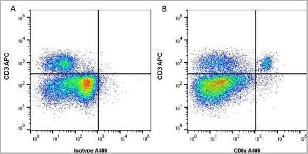
Application Data
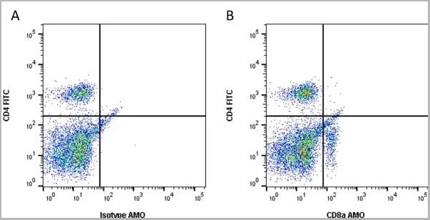
(Figure A. A647 conjugated rat anti mouse CD3 and A488 conjugated rat IgG2a isotype control Figure B. A647 conjugated rat anti mouse CD3 and A488 conjugated rat anti mouse CD8a . All experiments performed on red cell lysed murine splenocytes gated on mononuclear cells. Data acquired on the ZE5 cell analyzer.)
Application Data
(Figure A. A647 conjugated rat anti mouse CD3 and A488 conjugated rat IgG2a isotype control Figure B. A647 conjugated rat anti mouse CD3 and A488 conjugated rat anti mouse CD8a . All experiments performed on red cell lysed murine splenocytes gated on mononuclear cells. Data acquired on the ZE5 cell analyzer.)
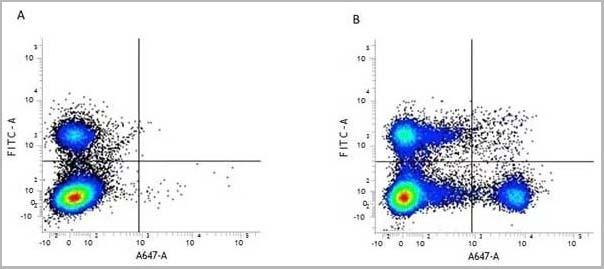
Application Data
(Figure A. FITC conjugated rat anti mouse CD4 and Alexa647 conjugated Rat IgG2b isotype control . Figure B. FITC conjugated rat anti mouse CD4 and Alexa647 conjugated rat anti mouse CD8 alpha . All experiments performed on murine splenocytes in the presence of murine SeroBlock .)
Application Data
(Figure A. FITC conjugated rat anti mouse CD4 and Alexa647 conjugated Rat IgG2b isotype control . Figure B. FITC conjugated rat anti mouse CD4 and Alexa647 conjugated rat anti mouse CD8 alpha . All experiments performed on murine splenocytes in the presence of murine SeroBlock .)
Application Data
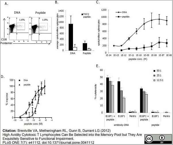
(Rat anti Mouse CD4 antibody, clone KT15 used for the detection of CD8 positive lymphocytes by flow cytometry.Image caption:Peptide boost leads to loss of high avidity responses in vivo. A, schematic of immunization regime. Mice immunized with DNA were boosted at day 63 with DNA or peptide, or with peptide at day 42 followed by DNA at day 63. Responses were analyzed at day 70 for frequency (B) and avidity (C) by measuring responses to increasing peptide concentration in IFN? elispot assay, D) normalization of responses to increasing peptide concentration in IFN? elispot assay. E, Analysis of antigen specific CTL by pentamer and CD8 staining of immunized splenocytes. F, Analysis of memory phenotype of antigen specific CTL by combination staining for CD62L and CD127 markers. Data is representative of at least three independent experiments.From: Brentville VA, Metheringham RL, Gunn B, Durrant LG (2012)High Avidity Cytotoxic T Lymphocytes Can Be Selected into the Memory Pool but They Are Exquisitely Sensitive to Functional Impairment.PLoS ONE 7(7): e41112.This image is from an open access article distributed under the terms of the Creative Commons Attribution License.)
Application Data
(Figure A. FITC conjugated Rat anti Mouse CD4 and Amethyst Orange conjugated Rat IgG2a isotype control . Figure B. FITC conjugated Rat anti Mouse CD4 and Amethyst Orange conjugated Rat anti Mouse CD8a . All experiments performed on mouse blood gated on live single cells, in the presence of 10% mouse serum. Data acquired on the ZE5 Cell Analyzer)
Clone KT15 is reported to block T-cell-mediated cytotoxicity in in vitro assays (Zeis, M. et al., 2002).
NCBI and Uniprot Product Information
Similar Products


Product Notes
The CD8Alpha cd8a (Catalog #AAA12273) is an Antibody produced from Rat and is intended for research purposes only. The product is available for immediate purchase. The Rat Anti Mouse CD8 Alpha: Amethyst Orange reacts with Mouse and may cross-react with other species as described in the data sheet. AAA Biotech's CD8 Alpha can be used in a range of immunoassay formats including, but not limited to, Flow Cytometry (FC/FACS). FC/FACS: Min. Dilution: Neat Use 10ul of the suggested working dilution to label 106 cells in 100ul. The Fc region of monoclonal antibodies may bind non-specifically to cells expressing low affinity Fc receptors. This may be reduced by using SeroBlock FcR (BUF041A/B). Researchers should empirically determine the suitability of the CD8Alpha cd8a for an application not listed in the data sheet. Researchers commonly develop new applications and it is an integral, important part of the investigative research process. It is sometimes possible for the material contained within the vial of "CD8 Alpha, Monoclonal Antibody" to become dispersed throughout the inside of the vial, particularly around the seal of said vial, during shipment and storage. We always suggest centrifuging these vials to consolidate all of the liquid away from the lid and to the bottom of the vial prior to opening. Please be advised that certain products may require dry ice for shipping and that, if this is the case, an additional dry ice fee may also be required.Precautions
All products in the AAA Biotech catalog are strictly for research-use only, and are absolutely not suitable for use in any sort of medical, therapeutic, prophylactic, in-vivo, or diagnostic capacity. By purchasing a product from AAA Biotech, you are explicitly certifying that said products will be properly tested and used in line with industry standard. AAA Biotech and its authorized distribution partners reserve the right to refuse to fulfill any order if we have any indication that a purchaser may be intending to use a product outside of our accepted criteria.Disclaimer
Though we do strive to guarantee the information represented in this datasheet, AAA Biotech cannot be held responsible for any oversights or imprecisions. AAA Biotech reserves the right to adjust any aspect of this datasheet at any time and without notice. It is the responsibility of the customer to inform AAA Biotech of any product performance issues observed or experienced within 30 days of receipt of said product. To see additional details on this or any of our other policies, please see our Terms & Conditions page.Item has been added to Shopping Cart
If you are ready to order, navigate to Shopping Cart and get ready to checkout.