 Application Data
(Immunofluorescence staining of rat lymph node cryosection with Mouse anti Rat CD163 antibody , red an A and Mouse anti Rat CD8 MCA48), green in B. C is the merged image with nuclei counter-stained blue using DAPI. High power)
Application Data
(Immunofluorescence staining of rat lymph node cryosection with Mouse anti Rat CD163 antibody , red an A and Mouse anti Rat CD8 MCA48), green in B. C is the merged image with nuclei counter-stained blue using DAPI. High power)
Mouse CD8 ALPHA Monoclonal Antibody | anti-CD8 ALPHA antibody
MOUSE ANTI RAT CD8 ALPHA
Purified IgG - liquid
Immunohistology: This product does not require protein digestion pre-treatment of paraffin embedded sections.This product does not require antigen retrieval using heat treatment prior to staining of paraffin embedded sections.
Flow Cytometry: Maximum Dilution: 1/100
Preparation
Fusion Partners
Shelf Life: 18 months from date of despatch.
Application Data
(Immunofluorescence staining of rat lymph node cryosection with Mouse anti Rat CD163 antibody , red an A and Mouse anti Rat CD8 MCA48), green in B. C is the merged image with nuclei counter-stained blue using DAPI. High power)
Application Data
(Immunofluorescence staining of rat lymph node cryosection with Mouse anti Rat CD163 antibody , red an A and Mouse anti Rat CD8 MCA48), green in B. C is the merged image with nuclei counter-stained blue using DAPI. High power)
Application Data
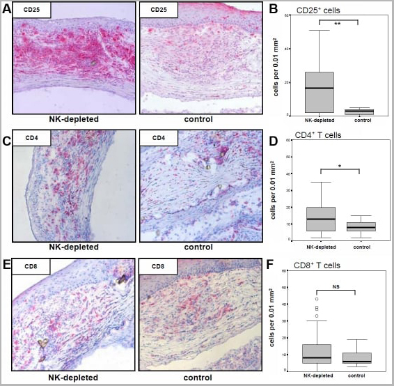
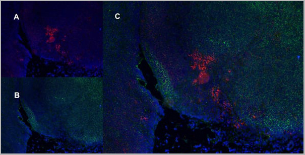
(Published customer image:Analysis of graft-infiltrating T cells. Activated CD25+ T cells and CD4+ and CD8+ T cell subsets were stained at the time points of corneal allograft rejection and calculated within the graft. A, C, and E show representative histological staining for CD25, CD4, and CD8 in grafts of treated and control animals, respectively. CD25+ (B) and CD4+ (D) cells infiltrated to a statistically significantly stronger extent in 3.2.3-treated animals when compared to control treated animals (*p)
Application Data
(Published customer image:Analysis of graft-infiltrating T cells. Activated CD25+ T cells and CD4+ and CD8+ T cell subsets were stained at the time points of corneal allograft rejection and calculated within the graft. A, C, and E show representative histological staining for CD25, CD4, and CD8 in grafts of treated and control animals, respectively. CD25+ (B) and CD4+ (D) cells infiltrated to a statistically significantly stronger extent in 3.2.3-treated animals when compared to control treated animals (*p)
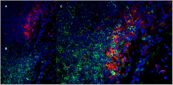
Application Data
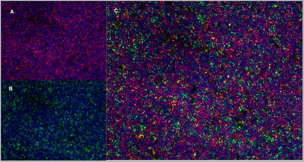
(Immunofluoescence staining of rat lymph node cryosection with Mouse anti Rat CD11b antibody , red in A and Mouse anti Rat CD8 , green in B. C is the merged image with nuclei counter-stained in blue using DAPI. High power)
Application Data
(Immunofluoescence staining of rat lymph node cryosection with Mouse anti Rat CD11b antibody , red in A and Mouse anti Rat CD8 , green in B. C is the merged image with nuclei counter-stained in blue using DAPI. High power)
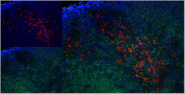
Application Data
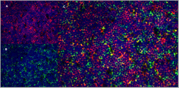
(Immunofluorescence staining of rat lymph node cryosection with Mouse anti Rat CD25 antibody , red in A and Mouse anti Rat CD8 , green in B. C is the merged image with nuclei counterstained blue using DAPI. High power)
Application Data
(Immunofluorescence staining of rat lymph node cryosection with Mouse anti Rat CD25 antibody , red in A and Mouse anti Rat CD8 , green in B. C is the merged image with nuclei counterstained blue using DAPI. High power)
Application Data
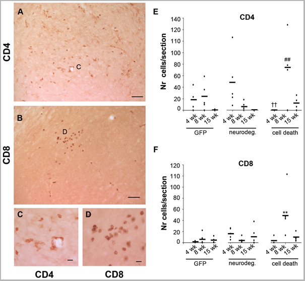
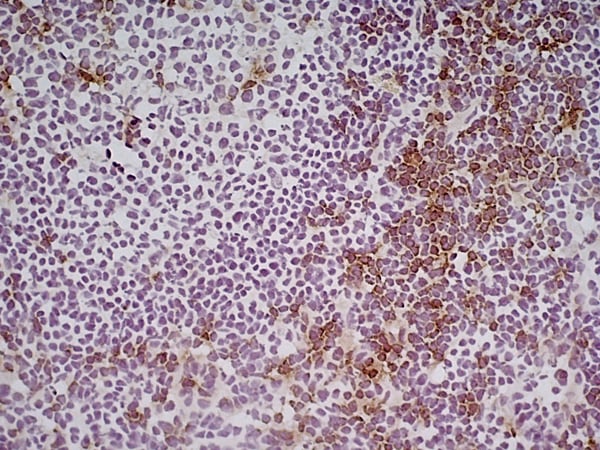
(Published customer image: CD4 and CD8 T cell infiltration. Photos show SN sections of an animal of the cell death group immunostained with antibody against CD4 (A and C) and CD8 (B and D). The small panels show insets in A (C) and in B (D) at higher magnification. Scale: 50 um, applies to A -B, 10 um applies to C -D. (E) Graph shows average (dash) and individual numbers of CD4+ cells found in one SN section per animal of each group plotted per time. Two-way ANOVA [F (8,42) = 4.1, p = 0.001 effect of group and time interaction] followed by Tukey HSD post-hoc analysis. ## or § p)
Application Data
(Published customer image: CD4 and CD8 T cell infiltration. Photos show SN sections of an animal of the cell death group immunostained with antibody against CD4 (A and C) and CD8 (B and D). The small panels show insets in A (C) and in B (D) at higher magnification. Scale: 50 um, applies to A -B, 10 um applies to C -D. (E) Graph shows average (dash) and individual numbers of CD4+ cells found in one SN section per animal of each group plotted per time. Two-way ANOVA [F (8,42) = 4.1, p = 0.001 effect of group and time interaction] followed by Tukey HSD post-hoc analysis. ## or § p)
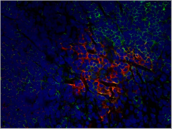
Application Data
(Immunofluorescence staining of rat lymph node cryosection with Mouse anti Rat CD25 antibody , red in A and Mouse anti Rat CD8 , green in B. C is the merged image with nuclei counterstained blue using DAPI. Low powe)
Application Data
(Immunofluorescence staining of rat lymph node cryosection with Mouse anti Rat CD25 antibody , red in A and Mouse anti Rat CD8 , green in B. C is the merged image with nuclei counterstained blue using DAPI. Low powe)
Application Data
(Immunofluorescence staining of rat lymph node cryosection with Mouse anti Rat CD25 antibody , red in A and Mouse anti Rat CD8 , green in B. C is the merged image with nuclei counterstained blue using DAPI. Low power)
Application Data
(Immunofluorescence staining of rat lymph node cryosection with Mouse anti Rat CD25 antibody , red in A and Mouse anti Rat CD8 , green in B. C is the merged image with nuclei counterstained blue using DAPI. Low power)
Application Data
(Immunofluorescence staining of rat lymph node cryosection with Mouse anti Rat CD25 antibody , red in A and Mouse anti Rat CD8 , green in B. C is the merged image with nuclei counterstained blue using DAPI. High power)
Application Data
(Immunofluorescence staining of rat lymph node cryosection with Mouse anti Rat CD25 antibody , red in A and Mouse anti Rat CD8 , green in B. C is the merged image with nuclei counterstained blue using DAPI. High power)
Application Data
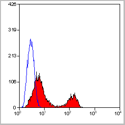
(Staining of rat peripheral blood cells with Mouse anti Rat CD8 Alpha Chain:Alexa Fluor 488)
Application Data
(Staining of rat peripheral blood cells with Mouse anti Rat CD8 Alpha Chain:Alexa Fluor 488)
Application Data
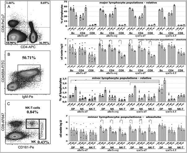
(Published customer image: Qualitative and quantitative flow cytometric analysis of lymphocyte populations in draining lymph nodes. A: Representative FACS plot of CD4+ CD8+ staining used to count T-helper cells, cytotoxic T-cells and CD4+ CD8+ double positive T-lymphocytes. Events acquired: 2x10⁵. B: FACS plot example for B-cell detection. C: Representative FACS plot for NK cell assessment. NK-T cell were confirmed by CD3 expression (not shown). Bar diagrams: Cumulative results for the quantification of major and minor lymphocyte populations in draining LN of cornea transplanted animals. An asterisk (*) indicates statistical significance at p=0.05 determined by Mann -Whitney U-Test. Allo-Tx-d7 - animals allo-grafted and analyzed at day 07 post op, n=6; allo-Tx-rej - animals displaying allo rejection of grafted corneas analyzed after the onset of rejection, n=5; syn-Tx-d7 - syngeneically grafted animals analyzed at day 7 post-op, n=3; syn-Tx-LT - syn-grafted long-term survivors analyzed at the end of the observation period at day 42; n=3.Maenz M, Morcos M, Ritter T. A comprehensive flow-cytometric analysis of graft infiltrating lymphocytes, draining lymph nodes and serum during the rejection phase in a fully allogeneic rat cornea transplant model. Mol Vis. 2011 Feb 8;17:420-9.)
Application Data
(Published customer image: Qualitative and quantitative flow cytometric analysis of lymphocyte populations in draining lymph nodes. A: Representative FACS plot of CD4+ CD8+ staining used to count T-helper cells, cytotoxic T-cells and CD4+ CD8+ double positive T-lymphocytes. Events acquired: 2x10⁵. B: FACS plot example for B-cell detection. C: Representative FACS plot for NK cell assessment. NK-T cell were confirmed by CD3 expression (not shown). Bar diagrams: Cumulative results for the quantification of major and minor lymphocyte populations in draining LN of cornea transplanted animals. An asterisk (*) indicates statistical significance at p=0.05 determined by Mann -Whitney U-Test. Allo-Tx-d7 - animals allo-grafted and analyzed at day 07 post op, n=6; allo-Tx-rej - animals displaying allo rejection of grafted corneas analyzed after the onset of rejection, n=5; syn-Tx-d7 - syngeneically grafted animals analyzed at day 7 post-op, n=3; syn-Tx-LT - syn-grafted long-term survivors analyzed at the end of the observation period at day 42; n=3.Maenz M, Morcos M, Ritter T. A comprehensive flow-cytometric analysis of graft infiltrating lymphocytes, draining lymph nodes and serum during the rejection phase in a fully allogeneic rat cornea transplant model. Mol Vis. 2011 Feb 8;17:420-9.)
NCBI and Uniprot Product Information
Similar Products


Product Notes
The CD8 ALPHA cd8a (Catalog #AAA11982) is an Antibody produced from Mouse and is intended for research purposes only. The product is available for immediate purchase. AAA Biotech's CD8 ALPHA can be used in a range of immunoassay formats including, but not limited to, Immunohistology Frozen, Flow cytometry (FC/FACS), Immunoprecipitation (IP), Immunohistology Paraffin. Flow Cytometry: Use 10ul of the suggested working dilution to label 106 cells in 100ul. Immunohistology: This product does not require protein digestion pre-treatment of paraffin embedded sections.This product does not require antigen retrieval using heat treatment prior to staining of paraffin embedded sections. Flow Cytometry: Maximum Dilution: 1/100. Researchers should empirically determine the suitability of the CD8 ALPHA cd8a for an application not listed in the data sheet. Researchers commonly develop new applications and it is an integral, important part of the investigative research process. It is sometimes possible for the material contained within the vial of "CD8 ALPHA, Monoclonal Antibody" to become dispersed throughout the inside of the vial, particularly around the seal of said vial, during shipment and storage. We always suggest centrifuging these vials to consolidate all of the liquid away from the lid and to the bottom of the vial prior to opening. Please be advised that certain products may require dry ice for shipping and that, if this is the case, an additional dry ice fee may also be required.Precautions
All products in the AAA Biotech catalog are strictly for research-use only, and are absolutely not suitable for use in any sort of medical, therapeutic, prophylactic, in-vivo, or diagnostic capacity. By purchasing a product from AAA Biotech, you are explicitly certifying that said products will be properly tested and used in line with industry standard. AAA Biotech and its authorized distribution partners reserve the right to refuse to fulfill any order if we have any indication that a purchaser may be intending to use a product outside of our accepted criteria.Disclaimer
Though we do strive to guarantee the information represented in this datasheet, AAA Biotech cannot be held responsible for any oversights or imprecisions. AAA Biotech reserves the right to adjust any aspect of this datasheet at any time and without notice. It is the responsibility of the customer to inform AAA Biotech of any product performance issues observed or experienced within 30 days of receipt of said product. To see additional details on this or any of our other policies, please see our Terms & Conditions page.Item has been added to Shopping Cart
If you are ready to order, navigate to Shopping Cart and get ready to checkout.