 Application Data
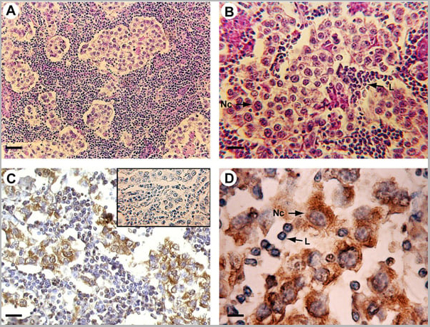
(Published customer image: Mouse anti Human cytochrome p450 aromatase antibody, clone H4 used for the detaction of aromatase in human tissues by Immunohistochemistry on paraffin sectionsImage caption:Morphology and P450 arom immunoreactivity of tumoral region in human testis with seminoma. A-B: Haematoxylin-eosin staining. C-D: Strong P450 arom immunoreactivity in cytoplasm of neoplastic cells (Nc) and unstained lymphocytes (L). Insert: absorption control. Scale bars: A, 20 um; B-C, 12.5 um; D, 5 um.From: Rago V, Romeo F, Aquila S, Montanaro D, And S, Carpino A. Cytochrome P450 aromatase expression in human seminoma. Reprod Biol Endocrinol. 2005 Dec 22;3:72.)
Application Data
(Published customer image: Mouse anti Human cytochrome p450 aromatase antibody, clone H4 used for the detaction of aromatase in human tissues by Immunohistochemistry on paraffin sectionsImage caption:Morphology and P450 arom immunoreactivity of tumoral region in human testis with seminoma. A-B: Haematoxylin-eosin staining. C-D: Strong P450 arom immunoreactivity in cytoplasm of neoplastic cells (Nc) and unstained lymphocytes (L). Insert: absorption control. Scale bars: A, 20 um; B-C, 12.5 um; D, 5 um.From: Rago V, Romeo F, Aquila S, Montanaro D, And S, Carpino A. Cytochrome P450 aromatase expression in human seminoma. Reprod Biol Endocrinol. 2005 Dec 22;3:72.)
Mouse CYTOCHROME P450 AROMATASE Monoclonal Antibody | anti-CYP19A1 antibody
MOUSE ANTI HUMAN CYTOCHROME P450 AROMATASE
Concentrated Tissue Culture Supernatant - liquid
Immunohistology: This product does not require antigen retrieval using heat treatment prior to staining of paraffin sections.
Western Blotting: Maximum Dilution: 1/250
Shelf Life: 18 months from date of despatch.
Application Data
(Published customer image: Mouse anti Human cytochrome p450 aromatase antibody, clone H4 used for the detaction of aromatase in human tissues by Immunohistochemistry on paraffin sectionsImage caption:Morphology and P450 arom immunoreactivity of tumoral region in human testis with seminoma. A-B: Haematoxylin-eosin staining. C-D: Strong P450 arom immunoreactivity in cytoplasm of neoplastic cells (Nc) and unstained lymphocytes (L). Insert: absorption control. Scale bars: A, 20 um; B-C, 12.5 um; D, 5 um.From: Rago V, Romeo F, Aquila S, Montanaro D, And S, Carpino A. Cytochrome P450 aromatase expression in human seminoma. Reprod Biol Endocrinol. 2005 Dec 22;3:72.)
Application Data
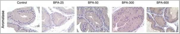
(Published customer image: Mouse anti Human cytochrome p450 aromatase antibody, clone H4 used for the detection of aromatase in rat prostate by immunohistochemistry on paraffin embedded sectionsImage caption:Immunohistochemical staining of aromatase in prostate of control and BPA-treated rats at doses of 25, 50, 300, or 600 ug/Kg/d for 4 days.From: Castro B, S¡nchez P, Torres JM, Preda O, del Moral RG, Ortega E. Bisphenol A exposure during adulthood alters expression of aromatase and 5a-reductase isozymes in rat prostate. PLoS One. 2013;8(2):e55905.)
Application Data
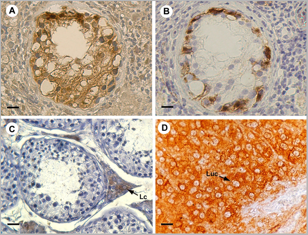
(Published customer image: Mouse anti Human cytochrome p450 aromatase antibody, clone H4 used for the detaction of aromatase in human tissues by Immunohistochemistry on paraffin sectionsImage caption:P450 arom immunoreactivity in testicular region adjacent to seminoma and in controls A: Intense aromatase immunostaining in IGCN cells. B: Placental-like alkaline phosphatase staining of IGCN basal cells C: Strong aromatase immunoreactivity of interstitial Leydig cells in normal testis (Lc) D: Intense immunostaining of luteal cells (Luc) in ovarian tissue Scale bars: A-B, 8 um; C,12.5 um; D, 5 um.From: Rago V, Romeo F, Aquila S, Montanaro D, And S, Carpino A. Cytochrome P450 aromatase expression in human seminoma. Reprod Biol Endocrinol. 2005 Dec 22;3:72.)
Application Data
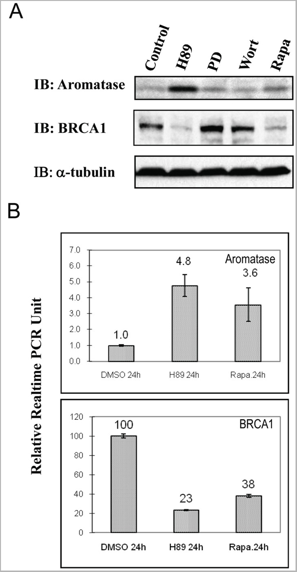
(Published customer image: Mouse anti Human cytochrome p450 aromatase antibody, clone H4 use for the detection of aromatase in KGN cells by western blottingImage caption:Effect of H89 on aromatase and BRCA1 expression in KGN cells. A, The human ovarian granulosa cell line KGN were treated with control (DMSO), 20 mM H89, 50 mM PD98059, 200 nM Wortmannin, 100 nM Rapamycin 100 nM, and cells were harvested 24 hours after treatment. Immunoblotting was performed using specific antibodies against aromatase, BRCA1 and a-tubulin; B, Aromatase and BRCA1 mRNA levels were measured by real-time RT-PCR. Gapdh was used for normalizing the real time PCR results. The data are expressed as a fold (Aromatase mRNA), or a percentage (BRCA1 mRNA) relative to the DMSO-treated control.From: Ghosh S, Lu Y, Hu Y. A Role of CREB in BRCA1 Constitutive Promoter Activity and Aromatase Basal Expression. Int J Biomed Sci. 2008 Dec 15;4(4):260-265.)
Application Data
(Published customer image: Mouse anti Human cytochrome p450 aromatase antibody, clone H4 use for the detection of aromatase in KGN cells by western blottingImage caption:Effect of H89 on aromatase and BRCA1 expression in KGN cells. A, The human ovarian granulosa cell line KGN were treated with control (DMSO), 20 mM H89, 50 mM PD98059, 200 nM Wortmannin, 100 nM Rapamycin 100 nM, and cells were harvested 24 hours after treatment. Immunoblotting was performed using specific antibodies against aromatase, BRCA1 and a-tubulin; B, Aromatase and BRCA1 mRNA levels were measured by real-time RT-PCR. Gapdh was used for normalizing the real time PCR results. The data are expressed as a fold (Aromatase mRNA), or a percentage (BRCA1 mRNA) relative to the DMSO-treated control.From: Ghosh S, Lu Y, Hu Y. A Role of CREB in BRCA1 Constitutive Promoter Activity and Aromatase Basal Expression. Int J Biomed Sci. 2008 Dec 15;4(4):260-265.)
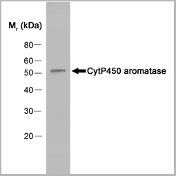
Application Data
(Western blot analysis of human placenta extract probed with Mouse anti Human Cytochrome P450 Aromatase followed by F(ab')2 Rabbit anti Mouse IgG:HRP)
Application Data
(Western blot analysis of human placenta extract probed with Mouse anti Human Cytochrome P450 Aromatase followed by F(ab')2 Rabbit anti Mouse IgG:HRP)
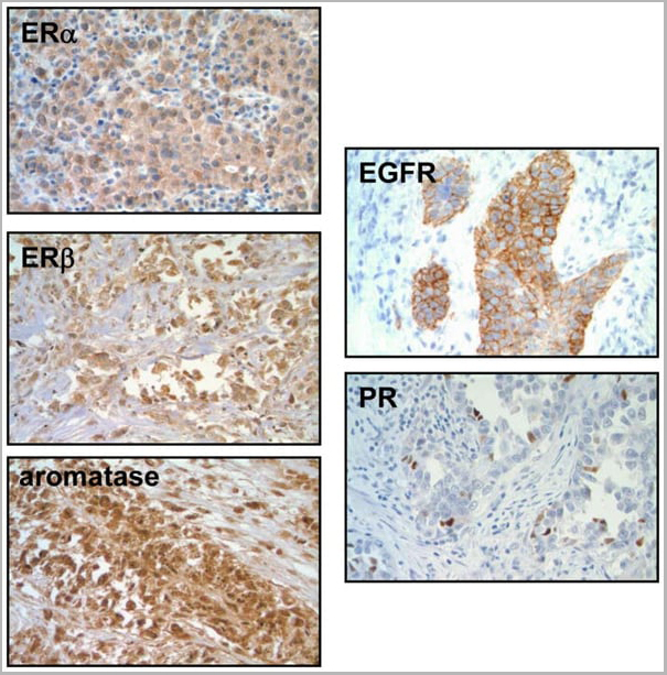
Application Data
(Published customer image: Mouse anti Human cytochrome p450 aromatase antibody, clone H4 used for the detection of aromatase in human lung tumors by immunohistochemistry on formalin fixed paraffin embedded tissue.Image caption:Representative immunohistochemistry of lung tumors for ERa, ERbeta, aromatase, EGFR and PR.From: Stabile LP, Dacic S, Land SR, Lenzner DE, Dhir R, Acquafondata M, Landreneau RJ, Grandis JR, Siegfried JM. Combined analysis of estrogen receptor beta-1 and progesterone receptor expression identifies lung cancer patients with poor outcome. Clin Cancer Res. 2011 Jan 1;17(1):154-64.)
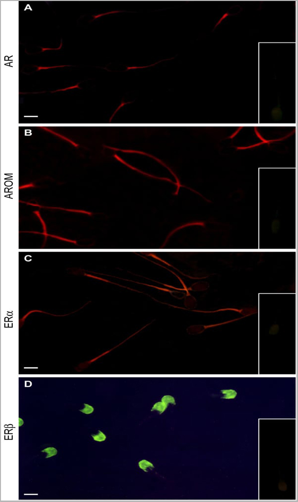
Application Data
(Published customer image: Mouse anti Human cytochrome p450 aromatase antibody, clone H4 used for the detection of aromatase in porcine spermatozoa by immunofluorescenceImage caption:Immunofluorescence labelling of androgen and estrogen receptors in pig spermatozoa: A) AR red fluorescence in sperm proximal mid-piece. B) P450arom red brilliant light in the proximal tail of sperm with a diffuse labelling in the distal tail. C) ERa red fluorescence in the sperm mid-piece, together with a faint labelling in the tail. D) ERbeta green intense light in the sperm acrosomal region. Inserts: immunonegative absorption controls. Scale bars: 5 um.From: Rago V, Aquila S, Panza R, Carpino A. Cytochrome P450arom, androgen and estrogen receptors in pig sperm. Reprod Biol Endocrinol. 2007 Jun 6;5:23.)
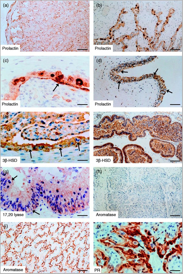
Application Data
(Published customer image: Mouse anti Human cytochrome p450 aromatase antibody, clone H4 used for the detection of aromatase in sheep tissues by immunohistochemistry on paraffin sectionsImage caption:a and b) Low- (a) and higher- (b) power sections of the fetal end of a placentome in G1 stained with the prolactin antiserum and showing the intensely stained binucleate cells scattered along the trophoblast, but with a tendency to be more densely accumulated towards the fetal end (scale bars a=200?um; b=150?um). (c) High-power section of the trophoblast -endometrium interface in a placentome in G2 stained with the prolactin antiserum. Note the patchy staining of the thin, elongated endometrial epithelial cells (arrowed) in close contact with the intensely stained binucleate trophoblast cells (scale bar=40?um). (d) Low-power section of the intercotyledonary allantochorion stained with the prolactin antiserum and showing the relatively dense accumulation of binucleate trophoblast cells (arrowed) which remain unstained in this region of the placenta (scale bar=100?um). (e) High-power section at the edge of a placentome of G1 showing positive staining of the uninucleate trophoblast by the 3beta-HSD antiserum. The binucleate cells (arrowed) remain unstained (scale bar=40?um). (f) Section of the distended endometrial glands at the lateral border of a placentome in G2 stained with 3beta-HSD. The basal portions of the endometrial cells lining the glands stain strongly as does the secretory material adhered to the luminal surface of the cells and the accumulated coagulum, possibly as a result of ˜stickiness' of the material (scale bar=150?um). (g) High-power section of the intercotyledonary region of the allantochorion in G1 showing positive staining of the cytoplasm of the uninucleate, but not binucleate trophoblast by the 17,20 lyase antiserum (scale bar=40?um). (h) Section at the fetal end of a placentome from G2 remaining unstained by the aromatase antiserum (scale bar=150?um). (i) Section of near-term sheep placenta showing strong, positive staining of the uninucleate, but not binucleate, trophoblast by the aromatase antiserum (scale bar=150?um). (j) High-power section at the fetal end of a placentome in G1 showing strong positive staining of the uninucleate trophoblast by the PR antiserum. The binucleate cells (arrowed) remain unstained (scale bar=40?um).From: Wilsher S, Stansfield F, Greenwood RE, Trethowan PD, Anderson RA, Wooding FB, Allen WR. Ovarian and placental morphology and endocrine functions in the pregnant giraffe (Giraffa camelopardalis). Reproduction. 2013 May 21;145(6):541-54.)
Application Data
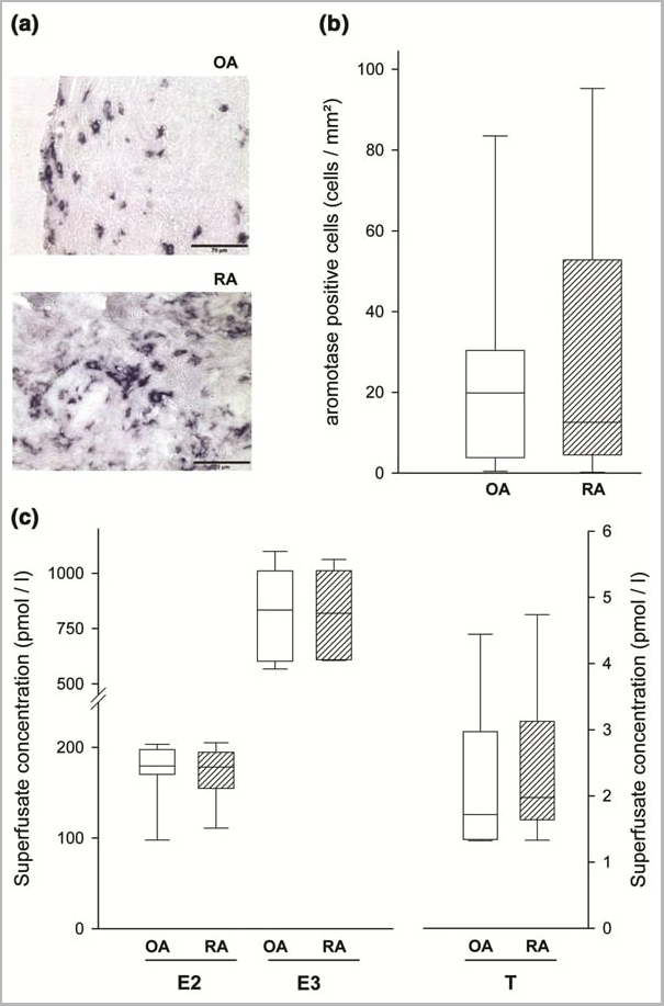
(Published customer image: Mouse anti Human cytochrome p450 aromatase antibody, clone H4 used for the detection of aromatase in human synovial tissue by immunohistochemistry on cryosectionsImage caption:Aromatase expression in synovial tissue and endogenous steroid hormone release from superfused synovium. (a) Immunohistochemistry of aromatase in one OA and one RA patient. Using the respective control antibody revealed no staining of positive cells (not shown). Magnification: 400x. (b) Density of aromatase-positive cells in OA (open bars, n = 20) and RA patients (hatched bars, n = 16). (c) Spontaneously released E2, E3, and free testosterone from standardized superfused pieces of synovial tissue of OA (open bars, n = 20) and RA patients (hatched bars, n = 18). (b,c) Values are given as box blots with the 5th, 25th, 50th (median), 75th, and 95th percentile when applicable. OA, osteoarthritis; RA, rheumatoid arthritis. Other abbreviations are as given in the legend to Fig. 1.From: Schmidt M, Weidler C, Naumann H, Anders S, Sch¶lmerich J, Straub RH. Androgen conversion in osteoarthritis and rheumatoid arthritis synoviocytes--androstenedione and testosterone inhibit estrogen formation and favor production of more potent 5alpha-reduced androgens. Arthritis Res Ther. 2005;7(5):R938-48.)
Application Data
(Published customer image: Mouse anti Human cytochrome p450 aromatase antibody, clone H4 used for the detection of aromatase in human synovial tissue by immunohistochemistry on cryosectionsImage caption:Aromatase expression in synovial tissue and endogenous steroid hormone release from superfused synovium. (a) Immunohistochemistry of aromatase in one OA and one RA patient. Using the respective control antibody revealed no staining of positive cells (not shown). Magnification: 400x. (b) Density of aromatase-positive cells in OA (open bars, n = 20) and RA patients (hatched bars, n = 16). (c) Spontaneously released E2, E3, and free testosterone from standardized superfused pieces of synovial tissue of OA (open bars, n = 20) and RA patients (hatched bars, n = 18). (b,c) Values are given as box blots with the 5th, 25th, 50th (median), 75th, and 95th percentile when applicable. OA, osteoarthritis; RA, rheumatoid arthritis. Other abbreviations are as given in the legend to Fig. 1.From: Schmidt M, Weidler C, Naumann H, Anders S, Sch¶lmerich J, Straub RH. Androgen conversion in osteoarthritis and rheumatoid arthritis synoviocytes--androstenedione and testosterone inhibit estrogen formation and favor production of more potent 5alpha-reduced androgens. Arthritis Res Ther. 2005;7(5):R938-48.)
NCBI and Uniprot Product Information
Similar Products


Product Notes
The CYP19A1 cyp19a1 (Catalog #AAA12113) is an Antibody produced from Mouse and is intended for research purposes only. The product is available for immediate purchase. AAA Biotech's CYTOCHROME P450 AROMATASE can be used in a range of immunoassay formats including, but not limited to, Immunofluorescence (IF), Immunohistology Paraffin, Western Blot (WB). Western Blot: This item detects a band of approximately 55KD in human placental extracts. Immunohistology: This product does not require antigen retrieval using heat treatment prior to staining of paraffin sections. Western Blotting: Maximum Dilution: 1/250. Researchers should empirically determine the suitability of the CYP19A1 cyp19a1 for an application not listed in the data sheet. Researchers commonly develop new applications and it is an integral, important part of the investigative research process. It is sometimes possible for the material contained within the vial of "CYTOCHROME P450 AROMATASE, Monoclonal Antibody" to become dispersed throughout the inside of the vial, particularly around the seal of said vial, during shipment and storage. We always suggest centrifuging these vials to consolidate all of the liquid away from the lid and to the bottom of the vial prior to opening. Please be advised that certain products may require dry ice for shipping and that, if this is the case, an additional dry ice fee may also be required.Precautions
All products in the AAA Biotech catalog are strictly for research-use only, and are absolutely not suitable for use in any sort of medical, therapeutic, prophylactic, in-vivo, or diagnostic capacity. By purchasing a product from AAA Biotech, you are explicitly certifying that said products will be properly tested and used in line with industry standard. AAA Biotech and its authorized distribution partners reserve the right to refuse to fulfill any order if we have any indication that a purchaser may be intending to use a product outside of our accepted criteria.Disclaimer
Though we do strive to guarantee the information represented in this datasheet, AAA Biotech cannot be held responsible for any oversights or imprecisions. AAA Biotech reserves the right to adjust any aspect of this datasheet at any time and without notice. It is the responsibility of the customer to inform AAA Biotech of any product performance issues observed or experienced within 30 days of receipt of said product. To see additional details on this or any of our other policies, please see our Terms & Conditions page.Item has been added to Shopping Cart
If you are ready to order, navigate to Shopping Cart and get ready to checkout.