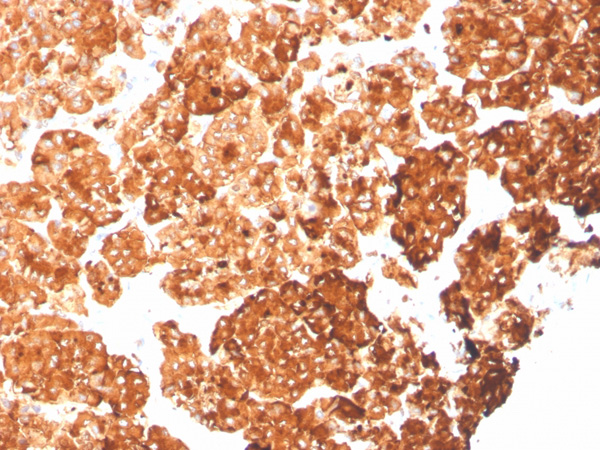
 Application Data
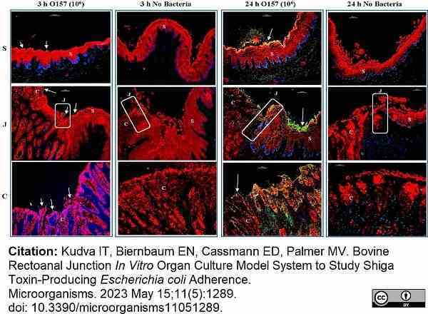
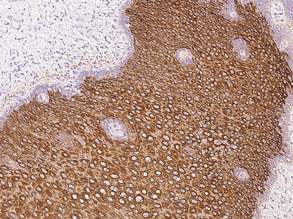
(Published customer image:Mouse anti human cytokeratin (Pan reactive) antibody, clone C-11 used to label cytokeratins in bovine recto-anal junction tissue sections by immunofluorescence.Image caption:Immunofluorescent images of tissue sections from RAJ-IVOC adherence assay trial 1. The RAJ-IVOC were inoculated with either O157 (106 CFU inoculum as shown in parenthesis) or not inoculated (no bacteria) and incubated at 39 °C for 3 or 24 h. Tissue sections of the RAJ-IVOC were then stained with immunofluorescent antibodies targeting the RAJ cells’ cytokeratins and O157, and images were recorded at 100× magnification. The adherent bacteria (shown with arrows), RAJ cells’ cytokeratins, and the nuclei have green, orange–red, and blue fluorescence, respectively. The squamous (S), junction (J, boxed), and columnar (C) regions of the RAJ are indicated along with a 100 um scale bar.)
Application Data
(Published customer image:Mouse anti human cytokeratin (Pan reactive) antibody, clone C-11 used to label cytokeratins in bovine recto-anal junction tissue sections by immunofluorescence.Image caption:Immunofluorescent images of tissue sections from RAJ-IVOC adherence assay trial 1. The RAJ-IVOC were inoculated with either O157 (106 CFU inoculum as shown in parenthesis) or not inoculated (no bacteria) and incubated at 39 °C for 3 or 24 h. Tissue sections of the RAJ-IVOC were then stained with immunofluorescent antibodies targeting the RAJ cells’ cytokeratins and O157, and images were recorded at 100× magnification. The adherent bacteria (shown with arrows), RAJ cells’ cytokeratins, and the nuclei have green, orange–red, and blue fluorescence, respectively. The squamous (S), junction (J, boxed), and columnar (C) regions of the RAJ are indicated along with a 100 um scale bar.)
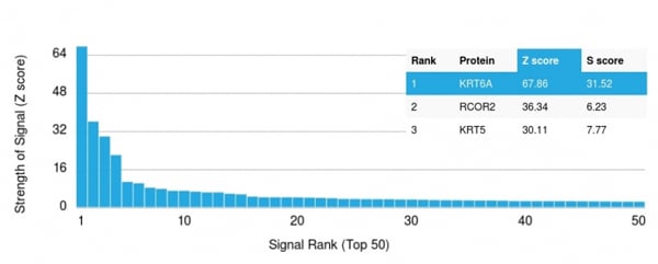
Mouse CYTOKERATIN Monoclonal Antibody | anti-KRT6A antibody
MOUSE ANTI HUMAN CYTOKERATIN (PAN REACTIVE)
Phosphate buffered saline
<0.1% Sodium Azide (NaN3)
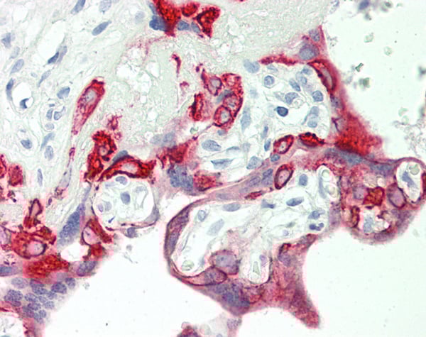
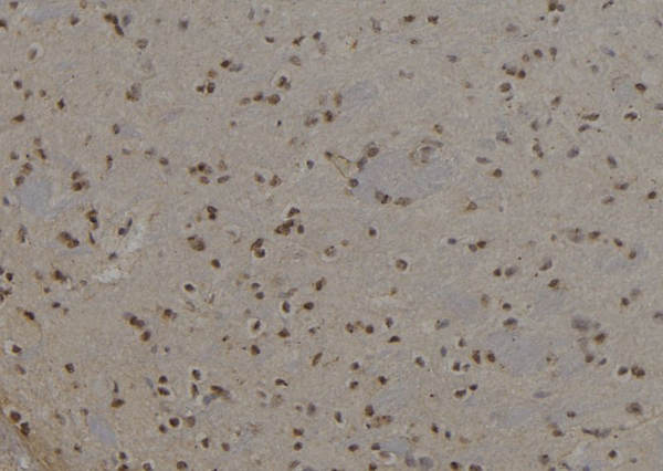
Application Data
(Published customer image:Mouse anti human cytokeratin (Pan reactive) antibody, clone C-11 used to label cytokeratins in bovine recto-anal junction tissue sections by immunofluorescence.Image caption:Immunofluorescent images of tissue sections from RAJ-IVOC adherence assay trial 1. The RAJ-IVOC were inoculated with either O157 (106 CFU inoculum as shown in parenthesis) or not inoculated (no bacteria) and incubated at 39 °C for 3 or 24 h. Tissue sections of the RAJ-IVOC were then stained with immunofluorescent antibodies targeting the RAJ cells’ cytokeratins and O157, and images were recorded at 100× magnification. The adherent bacteria (shown with arrows), RAJ cells’ cytokeratins, and the nuclei have green, orange–red, and blue fluorescence, respectively. The squamous (S), junction (J, boxed), and columnar (C) regions of the RAJ are indicated along with a 100 um scale bar.)
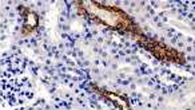
Application Data
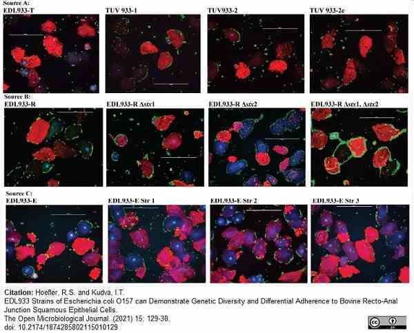
(Published customer image:Mouse anti pan Cytokeratin antibody, clone C-11 (AAA50486) used to label cytokeratins in bovine cells by immunofluorescence.Image caption:Adherence patterns of EDL933 strains and mutants, obtained from sources A, B, and C, on RSE cells. The immunofluorescent stained slides are shown at 40x magnification. O157 have green fluorescence; RSE cells’ cytokeratins have orange-red fluorescence, and nuclei have blue fluorescence.)
Application Data
(Published customer image:Mouse anti pan Cytokeratin antibody, clone C-11 (AAA50486) used to label cytokeratins in bovine cells by immunofluorescence.Image caption:Adherence patterns of EDL933 strains and mutants, obtained from sources A, B, and C, on RSE cells. The immunofluorescent stained slides are shown at 40x magnification. O157 have green fluorescence; RSE cells’ cytokeratins have orange-red fluorescence, and nuclei have blue fluorescence.)
IF (Immunofluorescence)
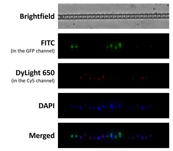
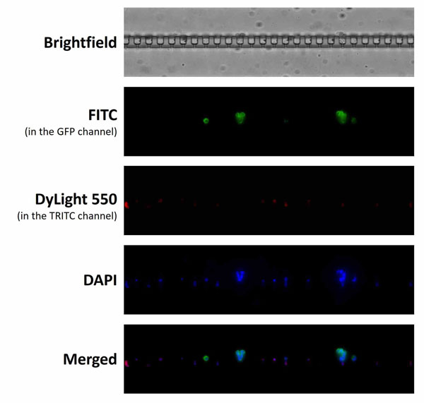
(Immunofluorescence staining of MCF-7 cells spiked in blood samples and captured using the Genesis System and CelSelect SlidesTM Technology. Leukocytes (red) are identified using Mouse anti Human CD45 antibody and detected using a DyLight 550 conjugated Human anti Mouse IgG2a secondary antibody (HCA310D550). Immunolabeling of MCF-7 cells (green) was performed using Mouse anti Human Cytokeratin (Pan reactive) antibody (AAA50486) and detected using a FITC conjugated Goat anti Mouse IgG1 secondary antibody . Imaged using Nikon Ti2 at 20x magnification.)
IF (Immunofluorescence)
(Immunofluorescence staining of MCF-7 cells spiked in blood samples and captured using the Genesis System and CelSelect SlidesTM Technology. Leukocytes (red) are identified using Mouse anti Human CD45 antibody and detected using a DyLight 550 conjugated Human anti Mouse IgG2a secondary antibody (HCA310D550). Immunolabeling of MCF-7 cells (green) was performed using Mouse anti Human Cytokeratin (Pan reactive) antibody (AAA50486) and detected using a FITC conjugated Goat anti Mouse IgG1 secondary antibody . Imaged using Nikon Ti2 at 20x magnification.)
Customer Reviews
Loading reviews...
Share Your Experience
Similar Products


Product Notes
The KRT6A (Catalog #AAA50486) is an Antibody produced from Mouse and is intended for research purposes only. The product is available for immediate purchase. The MOUSE ANTI HUMAN CYTOKERATIN (PAN REACTIVE) reacts with Human, Mammals and may cross-react with other species as described in the data sheet. AAA Biotech's CYTOKERATIN can be used in a range of immunoassay formats including, but not limited to, IP (Immunoprecipitation), IHC (Immunohistochemistry), IHC (Immunohistochemistry), WB (Western Blot), FCM/FACS (Flow Cytometry), IF (Immunofluorescence). Researchers should empirically determine the suitability of the KRT6A for an application not listed in the data sheet. Researchers commonly develop new applications and it is an integral, important part of the investigative research process. It is sometimes possible for the material contained within the vial of "CYTOKERATIN, Monoclonal Antibody" to become dispersed throughout the inside of the vial, particularly around the seal of said vial, during shipment and storage. We always suggest centrifuging these vials to consolidate all of the liquid away from the lid and to the bottom of the vial prior to opening. Please be advised that certain products may require dry ice for shipping and that, if this is the case, an additional dry ice fee may also be required.Precautions
All products in the AAA Biotech catalog are strictly for research-use only, and are absolutely not suitable for use in any sort of medical, therapeutic, prophylactic, in-vivo, or diagnostic capacity. By purchasing a product from AAA Biotech, you are explicitly certifying that said products will be properly tested and used in line with industry standard. AAA Biotech and its authorized distribution partners reserve the right to refuse to fulfill any order if we have any indication that a purchaser may be intending to use a product outside of our accepted criteria.Disclaimer
Though we do strive to guarantee the information represented in this datasheet, AAA Biotech cannot be held responsible for any oversights or imprecisions. AAA Biotech reserves the right to adjust any aspect of this datasheet at any time and without notice. It is the responsibility of the customer to inform AAA Biotech of any product performance issues observed or experienced within 30 days of receipt of said product. To see additional details on this or any of our other policies, please see our Terms & Conditions page.Item has been added to Shopping Cart
If you are ready to order, navigate to Shopping Cart and get ready to checkout.