 Application Data
(Staining of J774 cells with Rat anti Mouse F4/80 antigen Biotin)
Application Data
(Staining of J774 cells with Rat anti Mouse F4/80 antigen Biotin)
Rat F4/80 Monoclonal Antibody | anti-F4/80 antibody
RAT ANTI MOUSE F4/80
Flow Cytometry: Use 10ul of the suggested working dilution to label 106 cells in 100ul
Where this product has not been tested for use in a particular technique this does not necessarily exclude its use in such procedures. Suggested working dilutions are given as a guide only. It is recommended that the user titrates the product for use in their own system using appropriate negative/positive controls
1 Rat anti Mouse F4/80 antibody, clone A3-1 requires pre-treatment of paraffin sections prior to staining. Proteinase K is recommended for tissues fixed for less than 24 hours. Citrate buffer pH 6.0 is recommended for tissues fixed for more than 24 hours. Please view the protocol at Antigen Retrieval Techniques.
Avoid repeated freezing and thawing as this may denature the antibody. Storage in frost-free freezers is not recommended.
Application Data
(Staining of J774 cells with Rat anti Mouse F4/80 antigen Biotin)
Application Data
(Staining of J774 cells with Rat anti Mouse F4/80 antigen Biotin)
Application Data
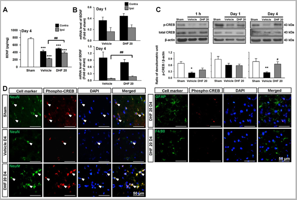
(Published customer image: Post-injury 7,8-dihydroxyflavone treatment increased brain BDNF levels and promoted CREB activation. (A) Bar graphs demonstrating brain derived neurotrophic factor (BDNF) protein concentrations measured by enzyme-linked immunosorbent assay (ELISA) in sham-injured, vehicle-treated, and 20 mg/kg 7,8-dihydroxyflavone (DHF 20)-treated mice at 4 days post-TBI. DHF 20 significantly increased BDNF protein levels in the ipsilateral hemisphere at 4 days post-injury. (B) Bar graphs demonstrating BDNF mRNA expression measured by real -time quantitative reverse transcriptase polymerase chain reaction (RT-PCR) at 1 and 4 days post-injury. DHF 20 significantly increased BDNF mRNA levels in the ipsilateral hemisphere at 4 days post-injury. (C) Western blot analysis of the phospho-CREB level in the ipsilateral hemisphere of sham-injured, vehicle-treated, and DHF 20-treated mice at 1 h, 1 day, and 4 days post-injury. DHF 20 enhanced CREB phosphorylation at 4 days post-injury. (D) Identification of phospho-CREB-positive cells 4 day post-injury in the peri-contusional margin by double immunofluorescence staining. Phospho-CREB is shown in red, and NeuN (neurons), GFAP (astrocytes), or F4/80 (microglia) is shown in green. Co-localization of phospho-CREB with NeuN is shown by yellow labeling. Sections were stained with DAPI (blue) to show all nuclei. Values are mean +/- SEM; *P)
Application Data
(Staining of mouse peritoneal macrophages with Rat anti Mouse F4/80 antigen: Pacific Blue(AAA12225PB))
Application Data
(Staining of mouse peritoneal macrophages with Rat anti Mouse F4/80 antigen: Pacific Blue(AAA12225PB))
Application Data
(Staining of J774 cells with Rat anti Mouse F4/80 antigen:Biotin)
Application Data
(Staining of J774 cells with Rat anti Mouse F4/80 antigen:Biotin)
Application Data
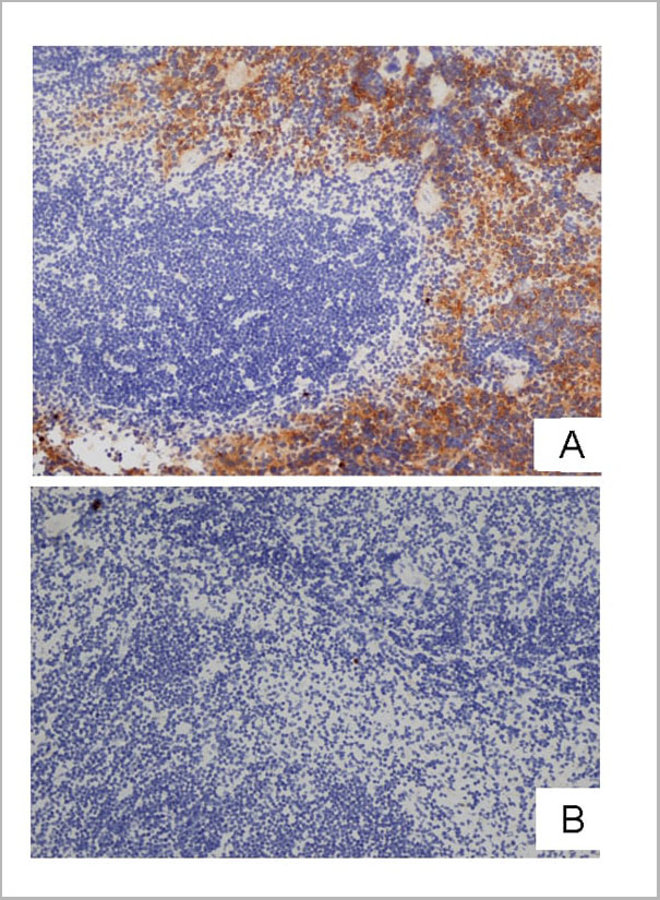
(Frozen mouse spleen stained with A: Rat anti Mouse F4/80 followed by Goat anti Rat IgG:HRP or B: Rat anti Mouse F4/80 preincubated with 2 molar excess of Human anti Idiotypic AAA12225 (HCA154) followed by Goat anti Rat IgG:HRP)
Application Data
(Frozen mouse spleen stained with A: Rat anti Mouse F4/80 followed by Goat anti Rat IgG:HRP or B: Rat anti Mouse F4/80 preincubated with 2 molar excess of Human anti Idiotypic AAA12225 (HCA154) followed by Goat anti Rat IgG:HRP)
Application Data
(Staining of J774 cells with Rat anti Mouse F4/80 antigen:Low Endotoxin)
Application Data
(Staining of J774 cells with Rat anti Mouse F4/80 antigen:Low Endotoxin)
Application Data
(Staining of mouse peritoneal macrophages with Rat anti Mouse F4/80 antigen: RPE - Alexa Fluor 750 (AAA12225P750))
Application Data
(Staining of mouse peritoneal macrophages with Rat anti Mouse F4/80 antigen: RPE - Alexa Fluor 750 (AAA12225P750))
Application Data
(Staining of mouse peritoneal macrophages cells with Rat anti Mouse F4/80 antigen:APC)
Application Data
(Staining of mouse peritoneal macrophages cells with Rat anti Mouse F4/80 antigen:APC)
Application Data
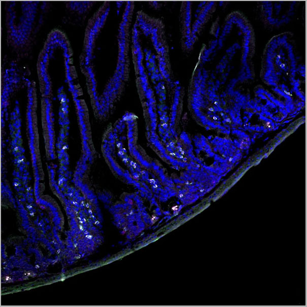
(Immunofluorescence image of mouse small intestine stained with Rat anti Mouse F4/80 , red and Rat anti Mouse CD45 , green; nuclei are stained with DAPI, blue. The image higlights macrophages within the lamina propria)
Application Data
(Immunofluorescence image of mouse small intestine stained with Rat anti Mouse F4/80 , red and Rat anti Mouse CD45 , green; nuclei are stained with DAPI, blue. The image higlights macrophages within the lamina propria)
Application Data
(Staining of mouse peritoneal macrophages with Rat anti Mouse F4/80 antigen:FITC)
Application Data
(Staining of mouse peritoneal macrophages with Rat anti Mouse F4/80 antigen:FITC)
Expression of F4/80 is heterogeneous and is modulated during macrophage maturation and activation. The F4/80 antigen is expressed on a wide range of mature tissue macrophages including Kupffer cells, Langerhans cells, microglia, macrophages located in the gut lamina propria, peritoneal cavity, lung, thymus, bone marrow stroma and macrophages in the red pulp of the spleen (Hume, et al. 1984). F4/80 antigen is also expressed on a subpopulation of dendritic cells but is absent from macrophages located in T cell areas of the spleen and lymph node (Gordon, et al. 1994). The ligands and biological functions of the F4/80 antigen have not been fully determined but a role for F4/80 in the generation of efferent CD8+ve regulatory T cells is proposed (Lin, et al. 2005)
Rat anti mouse F4/80 antibody, clone Cl:A3-1 modulates cytokine levels released in response to Listeria monocytogenes (Warschkau & Kiderlen, 1999).
A Human anti-idiotypic CI:A31 antibody, clone 17867 (HCA154 ) which binds to and blocks activity of Rat anti mouse F4/80 antibody, clone Cl:A3-1 is also available for use as a control in experiments utilizing clone A3-1.
NCBI and Uniprot Product Information
Similar Products


Product Notes
The F4/80 emr1 (Catalog #AAA12225) is an Antibody produced from Rat and is intended for research purposes only. The product is available for immediate purchase. AAA Biotech's F4/80 can be used in a range of immunoassay formats including, but not limited to, Flow Cytometry (FC), Immunohistology Frozen, Immunohistology Paraffin*, Immunohistology Resin (RE), Immunoprecipitation (IP), Immunofluorescence (IF), Radioimmunoassays (RIA), Immunoelectron Microscopy (EM). FC: Neat- 1/10 Flow Cytometry: Use 10ul of the suggested working dilution to label 106 cells in 100ul Where this product has not been tested for use in a particular technique this does not necessarily exclude its use in such procedures. Suggested working dilutions are given as a guide only. It is recommended that the user titrates the product for use in their own system using appropriate negative/positive controls 1 Rat anti Mouse F4/80 antibody, clone A3-1 requires pre-treatment of paraffin sections prior to staining. Proteinase K is recommended for tissues fixed for less than 24 hours. Citrate buffer pH 6.0 is recommended for tissues fixed for more than 24 hours. Please view the protocol at Antigen Retrieval Techniques. . Researchers should empirically determine the suitability of the F4/80 emr1 for an application not listed in the data sheet. Researchers commonly develop new applications and it is an integral, important part of the investigative research process. It is sometimes possible for the material contained within the vial of "F4/80, Monoclonal Antibody" to become dispersed throughout the inside of the vial, particularly around the seal of said vial, during shipment and storage. We always suggest centrifuging these vials to consolidate all of the liquid away from the lid and to the bottom of the vial prior to opening. Please be advised that certain products may require dry ice for shipping and that, if this is the case, an additional dry ice fee may also be required.Precautions
All products in the AAA Biotech catalog are strictly for research-use only, and are absolutely not suitable for use in any sort of medical, therapeutic, prophylactic, in-vivo, or diagnostic capacity. By purchasing a product from AAA Biotech, you are explicitly certifying that said products will be properly tested and used in line with industry standard. AAA Biotech and its authorized distribution partners reserve the right to refuse to fulfill any order if we have any indication that a purchaser may be intending to use a product outside of our accepted criteria.Disclaimer
Though we do strive to guarantee the information represented in this datasheet, AAA Biotech cannot be held responsible for any oversights or imprecisions. AAA Biotech reserves the right to adjust any aspect of this datasheet at any time and without notice. It is the responsibility of the customer to inform AAA Biotech of any product performance issues observed or experienced within 30 days of receipt of said product. To see additional details on this or any of our other policies, please see our Terms & Conditions page.Item has been added to Shopping Cart
If you are ready to order, navigate to Shopping Cart and get ready to checkout.