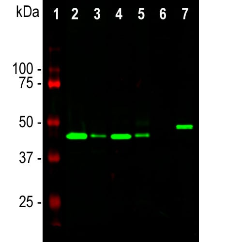
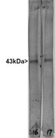
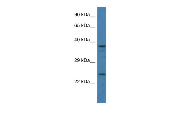
 WB (Western Blot)
(Western blot analysis of different tissue and cell lysates using mouse mAb to GAP43, MO22170, dilution 1:5,000, in green: [1] protein standard (red), [2] rat brain, [3] rat spinal cord, [4] mouse brain, [5] mouse spinal cord, [6] C6 cells, [7] SH-SY5Y cells. The single band at the 43kDa mark corresponds to the GAP43 protein.)
WB (Western Blot)
(Western blot analysis of different tissue and cell lysates using mouse mAb to GAP43, MO22170, dilution 1:5,000, in green: [1] protein standard (red), [2] rat brain, [3] rat spinal cord, [4] mouse brain, [5] mouse spinal cord, [6] C6 cells, [7] SH-SY5Y cells. The single band at the 43kDa mark corresponds to the GAP43 protein.)
Mouse GAP43 Monoclonal Antibody | anti-GAP43 antibody
GAP43
1. Draw of culture medium with aspirator and add 1 mL of 3.7 % formalin in PBS solution to the dish. (make up from 10 mLs Fisher 37% formalin plus 90mls PBS, the Fisher formalin contains 37% formaldehyde plus about 1% methanol which may be relevant sometimes). Let sit at room temp for 1 minute. (can add 0.1% Tween 20 to PBS used here and all subsequent steps to reduce background; probably best not to do this first time round though as it may extract your antigen or help wash your cells off the dish).
2. Take off the formalin/PBS and add 1ml of cold methanol (-20°C, kept in well-sealed bottle in fridge). Let sit for no more than 1 minute.
3. Take off methanol and add 1ml of PBS, not letting the specimen dry out. To block nonspecific antibody binding can add ~10 uL (=1%) of goat serum, and can incubate for 30 minutes. Can then add antibody reagents. Typically, 100 uL of hybridoma tissue culture supernatent or 1ml of mouse ascites fluid or crude serum. Incubate for 1 hour at room temp. (or can go at 37°C for 30 minutes to 1 hour, or can do 4°C overnight, exact time not too critical). Can do very gentle shaking for well adherent cell lines (3T3, Hek293 etc.).
4. Remove primary antibody and replace with 1 mL of PBS. Let sit for 5-10 minutes, replace PBS and repeat twice, to give three washes in PBS.
5. Add 0.5 uLs of secondary antibody. These are fluorescently labeled Goat anti mouse or rabbit antibodies and are conjugated to ALEXA dyes and were originally marketed by Molecular Probes (Eugene Oregon, the ALEXA dyes are sulphonated rhodamine compounds and are much more stable to UV than FITC, TRITC, Texas red etc. Molecular Probes was bought by Invitrogen, which now markets these reagents). Typically make 1:2,000 dilutions of these secondaries in PBS plus 1% goat serum, BSA or non fat milk carrier. Incubate for 1 hour at room temp. (or can go at 37°C for 30 minutes to 1 hour, or can do 4°C overnight). Can do gentle shaking for well adherent cell lines (3T3, HEK293 etc.).
6. Remove secondary antibody and replace with 1 ml of PBS. Let sit for 5-10 minutes, replace PBS and repeat twice, to give three washes in PBS.
7. Drop on one drop of Fisher mounting medium onto dish and apply 22 mm square coverslip. View in the microscope!
WB (Western Blot)
(Western blot analysis of different tissue and cell lysates using mouse mAb to GAP43, MO22170, dilution 1:5,000, in green: [1] protein standard (red), [2] rat brain, [3] rat spinal cord, [4] mouse brain, [5] mouse spinal cord, [6] C6 cells, [7] SH-SY5Y cells. The single band at the 43kDa mark corresponds to the GAP43 protein.)
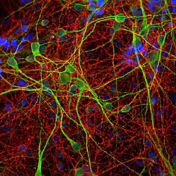
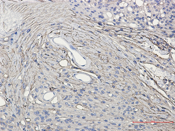
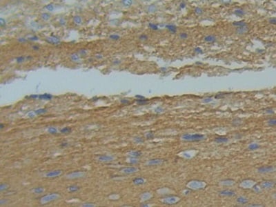
IF (Immunofluorescence)
(Immunofluorescent analysis of cortical neuron-glial cell culture from E20 rat stained with mouse mAb to GAP43, AAA76610, dilution 1:1,000, in red, and costained with chicken pAb to MAP2, dilution 1:10,000, in green. The blue is DAPI staining of nuclear DNA. GAP43 antibody labels protein expressed in the axonal membrane of the neuronal cells, while the MAP2 antibody stains dendrites and perikarya of neurons)
NCBI and Uniprot Product Information
Customer Reviews
Loading reviews...
Share Your Experience
Similar Products


Product Notes
The GAP43 gap43 (Catalog #AAA76610) is an Antibody produced from Mouse and is intended for research purposes only. The product is available for immediate purchase. The GAP43 reacts with Human, Mouse, Rat and may cross-react with other species as described in the data sheet. AAA Biotech's GAP43 can be used in a range of immunoassay formats including, but not limited to, IF (Immunofluorescence), IHC (Immunohistochemistry), WB (Western Blot), ICC (Immunocytochemistry). Researchers should empirically determine the suitability of the GAP43 gap43 for an application not listed in the data sheet. Researchers commonly develop new applications and it is an integral, important part of the investigative research process. It is sometimes possible for the material contained within the vial of "GAP43, Monoclonal Antibody" to become dispersed throughout the inside of the vial, particularly around the seal of said vial, during shipment and storage. We always suggest centrifuging these vials to consolidate all of the liquid away from the lid and to the bottom of the vial prior to opening. Please be advised that certain products may require dry ice for shipping and that, if this is the case, an additional dry ice fee may also be required.Precautions
All products in the AAA Biotech catalog are strictly for research-use only, and are absolutely not suitable for use in any sort of medical, therapeutic, prophylactic, in-vivo, or diagnostic capacity. By purchasing a product from AAA Biotech, you are explicitly certifying that said products will be properly tested and used in line with industry standard. AAA Biotech and its authorized distribution partners reserve the right to refuse to fulfill any order if we have any indication that a purchaser may be intending to use a product outside of our accepted criteria.Disclaimer
Though we do strive to guarantee the information represented in this datasheet, AAA Biotech cannot be held responsible for any oversights or imprecisions. AAA Biotech reserves the right to adjust any aspect of this datasheet at any time and without notice. It is the responsibility of the customer to inform AAA Biotech of any product performance issues observed or experienced within 30 days of receipt of said product. To see additional details on this or any of our other policies, please see our Terms & Conditions page.Item has been added to Shopping Cart
If you are ready to order, navigate to Shopping Cart and get ready to checkout.