
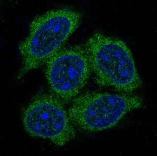
 IF (Immunofluorescence)
(Fluorescence-activated cell sorting analysis using Mouse Anti-HSP70 Monoclonal Antibody, Clone 1H11. Tissue: Jurkat E6.1 cells. Species: Human. Fixation: No fixation. Primary Antibody: Mouse Anti-HSP70 Monoclonal Antibody at 20 ug/ml for 40 min at 4 degree C. Counterstain: Propidium Iodide nuclear stain at 2.5 ug/ml for 5 min at RT. Isotype Control: Anti-mouse FITC at 1:32 for 15 min at RT (blue line). Courtesy of: Dr. Elyse Ireland, Institute of Medicine, University of Chester.)
IF (Immunofluorescence)
(Fluorescence-activated cell sorting analysis using Mouse Anti-HSP70 Monoclonal Antibody, Clone 1H11. Tissue: Jurkat E6.1 cells. Species: Human. Fixation: No fixation. Primary Antibody: Mouse Anti-HSP70 Monoclonal Antibody at 20 ug/ml for 40 min at 4 degree C. Counterstain: Propidium Iodide nuclear stain at 2.5 ug/ml for 5 min at RT. Isotype Control: Anti-mouse FITC at 1:32 for 15 min at RT (blue line). Courtesy of: Dr. Elyse Ireland, Institute of Medicine, University of Chester.)
Mouse HSP70 Monoclonal Antibody | anti-HSP70 antibody
HSP70 Antibody, Clone 1H11: PerCP
IF (Immunofluorescence)
(Fluorescence-activated cell sorting analysis using Mouse Anti-HSP70 Monoclonal Antibody, Clone 1H11. Tissue: Jurkat E6.1 cells. Species: Human. Fixation: No fixation. Primary Antibody: Mouse Anti-HSP70 Monoclonal Antibody at 20 ug/ml for 40 min at 4 degree C. Counterstain: Propidium Iodide nuclear stain at 2.5 ug/ml for 5 min at RT. Isotype Control: Anti-mouse FITC at 1:32 for 15 min at RT (blue line). Courtesy of: Dr. Elyse Ireland, Institute of Medicine, University of Chester.)
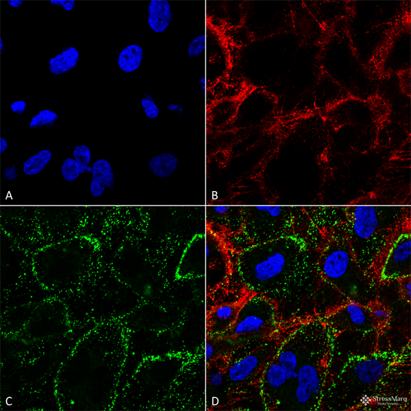
ICC (Immunocytochemistry)
(Immunocytochemistry/Immunofluorescence analysis using Mouse Anti-HSP70 Monoclonal Antibody, Clone 1H11. Tissue: HCT116 cells. Species: Human. Fixation: 4% Formaldehyde. Primary Antibody: Mouse Anti-HSP70 Monoclonal Antibody at 1:100. Counterstain: Wheat germ agglutinin Texas red membrane marker; DAPI (blue) nuclear stain. Localization: Cell surface, cell membrane. (A) DAPI nuclear stain. (B) Wheat germ agglutinin Texas red. (C) HSP70 Antibody. (D) Composite. Courtesy of: Lawrence Hightower, Charles Giardina, and Didem Ozcan from University of Connecticut.)
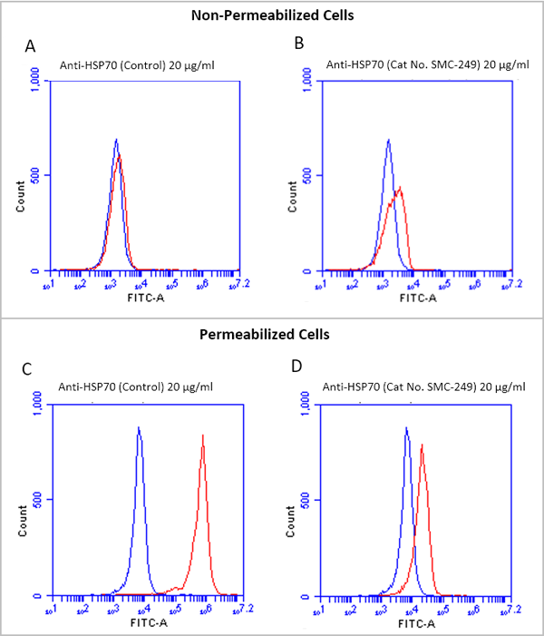
Although HSP70 is ubiquitously expressed, there is not much information about its presence on cell surface. The finding that HSP70 is localized on the cell surface of cancer cells but not normal cells suggests a conformational change of HSP70 in the lower pH environment characteristic of cancer cells (7). The presence of cell membrane embedded HSP70 has been found to increase the stability of cancer cells, thereby protecting tumors from environmental stress (8, 9).
Anti-HSP70 antibody, clone 1H11 is unique from other commercially available antibodies in that it can bind to the extracellular region of the cell membrane embedded HSP70 protein, allowing researchers to differentiate between membrane bound and intracellular HSP70 across cancer cells types.
NCBI and Uniprot Product Information
Customer Reviews
Loading reviews...
Share Your Experience
Similar Products


Product Notes
The HSP70 hspa1a (Catalog #AAA103912) is an Antibody produced from Mouse and is intended for research purposes only. The product is available for immediate purchase. The HSP70 Antibody, Clone 1H11: PerCP reacts with Human; Mouse; Rat; Bovine; Pig and may cross-react with other species as described in the data sheet. AAA Biotech's HSP70 can be used in a range of immunoassay formats including, but not limited to, FCM/FACS (Flow Cytometry), IF (Immunofluorescence), ICC (Immunocytochemistry), WB (Western Blot). Researchers should empirically determine the suitability of the HSP70 hspa1a for an application not listed in the data sheet. Researchers commonly develop new applications and it is an integral, important part of the investigative research process. It is sometimes possible for the material contained within the vial of "HSP70, Monoclonal Antibody" to become dispersed throughout the inside of the vial, particularly around the seal of said vial, during shipment and storage. We always suggest centrifuging these vials to consolidate all of the liquid away from the lid and to the bottom of the vial prior to opening. Please be advised that certain products may require dry ice for shipping and that, if this is the case, an additional dry ice fee may also be required.Precautions
All products in the AAA Biotech catalog are strictly for research-use only, and are absolutely not suitable for use in any sort of medical, therapeutic, prophylactic, in-vivo, or diagnostic capacity. By purchasing a product from AAA Biotech, you are explicitly certifying that said products will be properly tested and used in line with industry standard. AAA Biotech and its authorized distribution partners reserve the right to refuse to fulfill any order if we have any indication that a purchaser may be intending to use a product outside of our accepted criteria.Disclaimer
Though we do strive to guarantee the information represented in this datasheet, AAA Biotech cannot be held responsible for any oversights or imprecisions. AAA Biotech reserves the right to adjust any aspect of this datasheet at any time and without notice. It is the responsibility of the customer to inform AAA Biotech of any product performance issues observed or experienced within 30 days of receipt of said product. To see additional details on this or any of our other policies, please see our Terms & Conditions page.Item has been added to Shopping Cart
If you are ready to order, navigate to Shopping Cart and get ready to checkout.