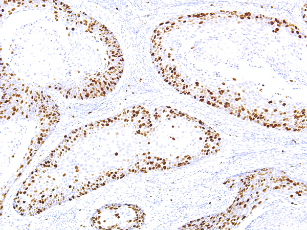
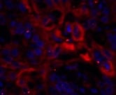
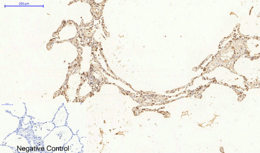
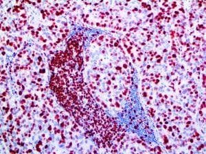
 IHC (Immunohistochemistry)
(Ki-67 [IHC067] on Cervical Cancer - 10X)
IHC (Immunohistochemistry)
(Ki-67 [IHC067] on Cervical Cancer - 10X)
Ki-67 Monoclonal Antibody | anti-Ki-67 antibody
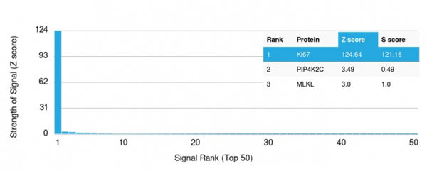
Ki-67
Gene Names
Mki67; Ki67; Ki-67; D630048A14Rik
Synonyms
Ki-67, Antibody; Ki-67; anti-Ki-67 antibody
Clonality
Monoclonal
Form/Format
Predilute Antibody: Diluent Buffer
Concentrate: Tris Buffer, pH 7.3 - 7.7, with 1% BSA and <0.1% Sodium Azide
Concentrate: Tris Buffer, pH 7.3 - 7.7, with 1% BSA and <0.1% Sodium Azide
Sequence Length
2938
Principles and Procedures
Visualization of the antigen present in tissue sections is accomplished in a multi-step immunohistochemical staining process, in conjunction with a horseradish peroxidase (HRP) or alkaline phosphatase (AP) linked detection system. The process involves the addition of the stated antibody (primary antibody) to a tissue slide, followed by a secondary antibody (linked to an enzyme complex) which specifically binds to the primary antibody. A chromogenic substrate is then added which reacts with the enzyme complex, resulting in a colorimetric reaction at the site of the antigen. Results are interpreted using a light microscope.
Note
The recommended working dilution range is 1:50 - 1:200.
Reconstitution
The prediluted antibody does not require any mixing, dilution, reconstitution, or titration; the antibody is ready-to-use and optimized for staining. The concentrated antibody requires dilution in the optimized buffer, to the recommended working dilution range.
Instructions For Use
Recommended Staining Protocols for the Ki-67 [MBS344020] antibody:
Manual Use:
1. Pretreatment: Perform heat-induced epitope retrieval (HIER) at pH 9 for 10 to 30 minutes.
2. Peroxide Block: Block in peroxidase blocking solution for 5 minutes at room temperature. (Not required if using Alkaline Phosphatase System.)
3. Primary Antibody: Apply antibody directly (Predilute) or dilute antibody at 1:50-1:200 (Concentrate) before applying. Incubate antibody for 10 to 30 minutes at room temperature.
4. Secondary Antibody: Incubate for 20 to 30 minutes at room temperature.
5. Substrate Development: Incubate DAB or Fast Red for 5 to 10 minutes at room temperature.
6. Counterstain: Counterstain with hematoxylin for 0.5 to 5 minutes, depending on the hematoxylin used. Rinse with distilled water and blueing solution for 30 seconds.
7. Dehydrate and apply coverslip.
Automated Staining System:
The stated primary antibody has been optimized and validated using the BOND-MAX fully automated IHC & ISH stainer, applying IHC Protocol F. The following edits are recommended for the protocol:
a) Marker Incubation Time: 30 minutes
b) Heat-induced epitope retrieval (HIER) is recommended using Leica Bond ER Solution 2 for 30 minutes.
c) Move Peroxide Block step to after Polymer and before Mixed DAB Refine.
Limitations
1. This antibody is intended for IVD use by qualified laboratories only and is not intended for use in flow cytometry.
2. Due to inevitable variability in immunohistochemical procedures and variables, appropriate positive and negative controls must be used and documented, and the results are to be interpreted by a qualified pathologist. Staining must be conducted in a certified, licensed laboratory, under the supervision and responsibility of the qualified pathologist.
3. Improper handling and processing of tissue samples may compromise the validity and/or analysis of the results.
4. Prediluted antibodies in a ready-to-use, optimally diluted format for use explicitly as instructed. Improper handling and processing of tissue samples and reagents, and any deviation from the recommended procedures outlined herein, may compromise the validity and/or analysis of the results. Due to the potential for variation in tissue processing and fixation, it may be necessary to adjust incubation time for the primary antibody on specific tissue specimens
5. Concentrated antibodies in a format that requires dilution in the optimized buffer, in the context of appropriate validation by the user. Any diluent different than that specified in the package insert must also be validated by the user to ensure proper compatibility with the antibody. Once diluted, any deviation from the recommended procedures outlined herein may compromise the validity and/or analysis of the results.
6. This antibody, when used with the appropriate detection systems and accessories, detects antigen(s) that remain intact through the tissue fixation, processing, and sectioning as described herein. Any deviations from these recommended procedures may compromised the validity and/or analysis of the results.
7. The clinical outcome indicated by staining results must be analyzed accurately by the qualified pathologist, and the patient’s medical history and other histopathological criteria must be taken into account. The user is responsible for interpretation of the results in the context of the patient.
8. Any documented discrepancies or unexplainable results in controls or tissue specimens should be reported to Technical Support Patient results are invalid if analysis of the positive and negative control tissues yields results other than those approved and described herein. The Troubleshooting section of this insert may be referred to for unexplained discrepancies in control tissues.
9. The potential for unexpected results in patient tissue specimens cannot be eliminated due to inherent biological variability in the expression of certain antigens.
10. The potential for false positive results in patient tissue specimens cannot be eliminated due to the possibility of non-immunological binding of substrate reaction products or proteins. False positive results may also occur subject to the type of immunostaining technique used, or due to the activity of pseudoperoxidase, endogenous peroxidase, or endogenous biotin.
11. Due to the effect of autoantibodies or natural antibodies, normal sera from an animal source the same as the secondary antisera may result in false negative or false positive results when used in blocking steps.
12. Non-specific staining with horseradish peroxidase may be observed when using tissues containing hepatitis B surface antigen due to the patient’s infection with the hepatitis B virus.
Warnings and Precautions
1. Ensure proper handling procedures are used with all reagents. Always wear laboratory coats, disposable gloves, and other appropriate laboratory equipment when handling reagents.
2. Do not ingest reagents, and avoid contact with eyes and mucous membranes. Wash eyes with copious amounts of water if contact occurs.
3. All incubation times and temperatures must be validated by the user, as must any storage conditions different than those specified in the package insert.
4. Prediluted antibody is provided in a ready-to-use, optimally diluted format, and any further dilution may result in loss of antigen staining.
5. Concentrated antibody requires dilution in the optimized buffer (refer to Materials and Methods), in the context of appropriate validation by the user.
6. Handle tissue sections, patient specimens, and all materials contacting them as biohazardous materials, using the appropriate precautions.
7. To ensure proper stability of the antibody and validity of results, use proper handling of the reagent and avoid microbial contamination.
Preparation and Storage
Store at 2-8°C. Do not freeze.
When stored correctly, the antibody is stable until the date indicated on the label. To ensure proper stability and delivery of the antibody after each run, replace the cap and immediately place the bottle in a refrigerator in an upright position. Positive and negative controls should be simultaneously run with unknown specimens, as there are no conclusive characteristics to suggest instability of the antibody. If such an indication of instability is suspected, contact Technical Support.
Each tissue section should be fixed with 10% neutral buffered formalin, cut to the applicable thickness (4um), and placed on a glass slide that is positively charged. The prepared slide may then be baked for a minimum of 30 minutes in a 53-65°C oven (do not exceed 24 hours).
Note: Performance evaluation has been shown on human tissues only. Variable results may occur due to extended fixation time or special processes of specific tissue preparations
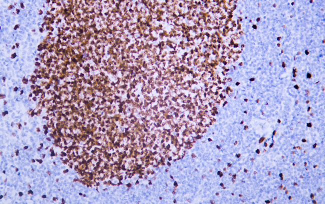
IHC (Immunohistochemistry)
(Ki-67 [IHC067] on Cervical Cancer - 10X)
IHC (Immunohistochemistry)
(Ki-67 [IHC067] on Cervical Cancer - 10X)
Related Product Information for anti-Ki-67 antibody
This antibody is intended for IVD use. The Ki-67 [MBS344020] antibody is intended for qualified laboratories to qualitatively identify by light microscopy the presence of associated antigens in sections of formalin-fixed, paraffin-embedded tissue sections using IHC test methods. Use of this antibody is indicated, subsequent to clinical differential test of diseases, as an aid in the identification of proliferating cells in normal and neoplastic tissues within the context of antibody panels, the patient’s clinical history and other tests evaluated by a qualified pathologist.
Ki-67 is a nuclear, non-histone protein that is expressed only during active phases of the cell cycle (G1, S, G2 and M), but not in the resting phases (G0 and G1 early phase). Although the antigen has also been associated with ribosomal RNA transcription, it is strongly linked to cell proliferation and has thus been indicated as an effective marker in grading the proliferation rate of tumours, including those of the brain, breast, cervix, and prostate.
Ki-67 is a nuclear, non-histone protein that is expressed only during active phases of the cell cycle (G1, S, G2 and M), but not in the resting phases (G0 and G1 early phase). Although the antigen has also been associated with ribosomal RNA transcription, it is strongly linked to cell proliferation and has thus been indicated as an effective marker in grading the proliferation rate of tumours, including those of the brain, breast, cervix, and prostate.
References
1. Mckeever P, et al. J Neuropathol Exp Neurol. 1998; 57:931-6.
2. Coons SW, et al. Neurosurgery. 1997; 41:878-84.
3. Allegra CJ, et al. J Clin Oncol. 2003; 21:241-50.
4. Pathmanathan N, et al. J Clin Pathol. 2013; 66:512-6.
5. Jansen R, et al. Br J Cancer. 1998; 78:460-65.
6. Goodson WH, et al. Breast Cancer Res Treat. 1998; 49:155-164.
7. Rossi S, et al. Am J Clin Pathol. 2005; 124:295-302.
8. Pena LL, et al. J Vet Diag Invest. 1998; 10:237-46.
9. Gibbons D, et al. Comparison Mod Pathol. 1997; 10:409-13.
NCBI and Uniprot Product Information
NCBI GI #
NCBI GeneID
NCBI Official Full Name
Ki-67
NCBI Official Synonym Full Names
antigen identified by monoclonal antibody Ki 67
NCBI Official Symbol
Mki67
NCBI Official Synonym Symbols
Ki67; Ki-67; D630048A14Rik
NCBI Protein Information
proliferation marker protein Ki-67; antigen KI-67
UniProt Protein Name
Proliferation marker protein Ki-67
UniProt Gene Name
Mki67
UniProt Synonym Gene Names
Antigen KI-67 homologCurated; Antigen Ki67 homologCurated
Customer Reviews
Loading reviews...
Share Your Experience
Similar Products


Product Notes
The Ki-67 mki67 (Catalog #AAA58777) is an Antibody and is intended for research purposes only. The product is available for immediate purchase. It is sometimes possible for the material contained within the vial of "Ki-67, Monoclonal Antibody" to become dispersed throughout the inside of the vial, particularly around the seal of said vial, during shipment and storage. We always suggest centrifuging these vials to consolidate all of the liquid away from the lid and to the bottom of the vial prior to opening. Please be advised that certain products may require dry ice for shipping and that, if this is the case, an additional dry ice fee may also be required.Precautions
All products in the AAA Biotech catalog are strictly for research-use only, and are absolutely not suitable for use in any sort of medical, therapeutic, prophylactic, in-vivo, or diagnostic capacity. By purchasing a product from AAA Biotech, you are explicitly certifying that said products will be properly tested and used in line with industry standard. AAA Biotech and its authorized distribution partners reserve the right to refuse to fulfill any order if we have any indication that a purchaser may be intending to use a product outside of our accepted criteria.Disclaimer
Though we do strive to guarantee the information represented in this datasheet, AAA Biotech cannot be held responsible for any oversights or imprecisions. AAA Biotech reserves the right to adjust any aspect of this datasheet at any time and without notice. It is the responsibility of the customer to inform AAA Biotech of any product performance issues observed or experienced within 30 days of receipt of said product. To see additional details on this or any of our other policies, please see our Terms & Conditions page.Item has been added to Shopping Cart
If you are ready to order, navigate to Shopping Cart and get ready to checkout.