 Application Data
(Staining of New Zealand Black mouse peripheral blood granulocytes with Rat anti Mouse Ly-6B.2 conjugated to FITC Data)
Application Data
(Staining of New Zealand Black mouse peripheral blood granulocytes with Rat anti Mouse Ly-6B.2 conjugated to FITC Data)
Rat Ly-6B.2 ALLOANTIGEN Monoclonal Antibody | anti-Ly-6B.2 antibody
RAT ANTI MOUSE Ly-6B.2 ALLOANTIGEN:RPE
Flow Cytometry: Minimum Dilution: Neat; Maximum Dilution: 1/10
Perservative Stabilisers
Shelf Life: 12 months from date of reconstitution.
Application Data
(Staining of New Zealand Black mouse peripheral blood granulocytes with Rat anti Mouse Ly-6B.2 conjugated to FITC Data)
Application Data
(Staining of New Zealand Black mouse peripheral blood granulocytes with Rat anti Mouse Ly-6B.2 conjugated to FITC Data)
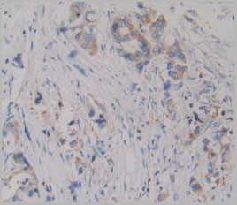
Application Data
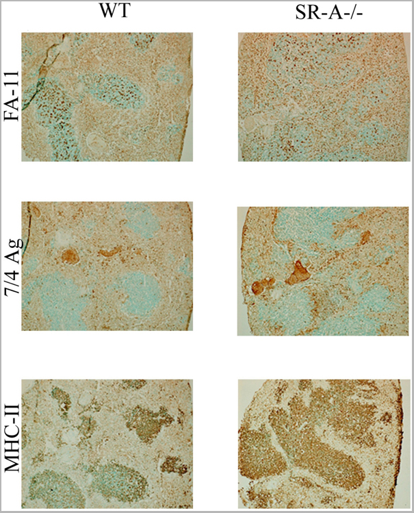
(Published originator image Intact spleen segments from infected animals were fixed in paraformaldehyde, embedded in OCT, sectioned, and stained with various antibodies for the infiltration of macrophages (FA-11), polymorphonuclear neutrophils, and activated monocytes (7/4Ag) and activated macrophages (MHC-II), as described in the text. All data are representative of at least three individual experiments.From: Plüddemann A, Hoe JC, Makepeace K, Moxon ER, Gordon S (2009) The Macrophage Scavenger Receptor A Is Host-Protective in Experimental Meningococcal Septicaemia. PLoS Pathog 5(2): e1000297.)
Application Data
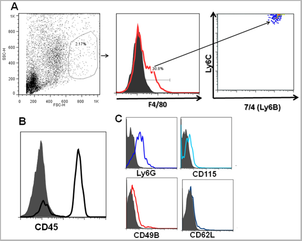
(Published customer image: F4/80 and CD11b+ cells infiltrating the iris after the intracameral injection of antigen also express Ly6C, 7/4 (Ly 6B) and CD45. Preparations of na¯ve iris cells and cells recovered after an intracameral injection of antigen were stained with anti- F4/80 and (A) Ly6C and 7/4 (B) show a characteristic CD45 peak C) Ly6Glo or negative, CD115, CD49b+, CD62Llo. The figures are representative of 2 experiments. All the isotype controls in the histograms are shown as shaded.From: Pais R, Bhowmick S, Chattopadhyay S, Lemire Y, Sharafieh R, et al. (2012) An Intracameral Injection of Antigen Induces In Situ Chemokines and Cytokines Required for the Generation of Circulating Immunoregulatory Monocytes. PLoS ONE 7(8): e43182.)
Application Data
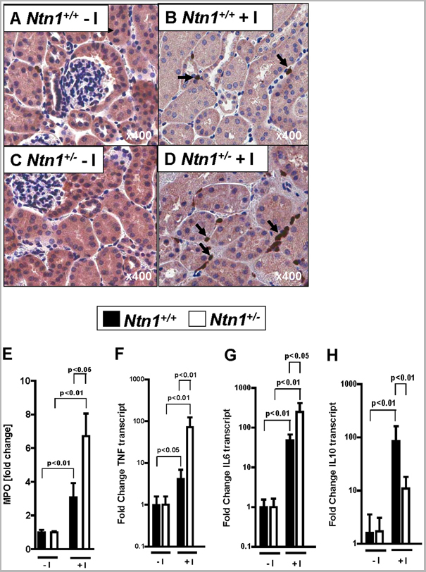
(Published customer image: Renal inflammatory changes in Ntn1+/- mice following ischemia. Ntn1+/- mice and their respective age-, weight-, and gender-matched littermate controls (Ntn1+/+) were subjected to 30 minutes of left renal artery ischemia. (A -D) Neutrophil staining. Arrows indicate neutrophils (magnification 400x). (E) Quantification of neutrophil tissue accumulation by measurement of myeloperoxidase (MPO). (F) TNF-a and (G) interleukin-6 (IL-6) and (H) interleukin-10 (IL-10) were assessed by real-time RT-PCR from renal tissues. Data were calculated relative to beta-actin and are expressed as fold change compared to sham-operated animals without ischemia (-I). Data are representative of four to six independent experiments for each experimental condition (mean +/- SD).From: Grenz A, Dalton JH, Bauerle JD, Badulak A, Ridyard D, et al. (2011) Partial Netrin-1 Deficiency Aggravates Acute Kidney Injury. PLoS ONE 6(5): e14812.)
Application Data
(Published customer image: Renal inflammatory changes in Ntn1+/- mice following ischemia. Ntn1+/- mice and their respective age-, weight-, and gender-matched littermate controls (Ntn1+/+) were subjected to 30 minutes of left renal artery ischemia. (A -D) Neutrophil staining. Arrows indicate neutrophils (magnification 400x). (E) Quantification of neutrophil tissue accumulation by measurement of myeloperoxidase (MPO). (F) TNF-a and (G) interleukin-6 (IL-6) and (H) interleukin-10 (IL-10) were assessed by real-time RT-PCR from renal tissues. Data were calculated relative to beta-actin and are expressed as fold change compared to sham-operated animals without ischemia (-I). Data are representative of four to six independent experiments for each experimental condition (mean +/- SD).From: Grenz A, Dalton JH, Bauerle JD, Badulak A, Ridyard D, et al. (2011) Partial Netrin-1 Deficiency Aggravates Acute Kidney Injury. PLoS ONE 6(5): e14812.)
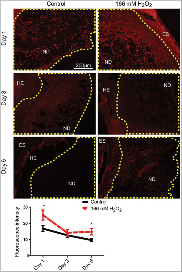
Application Data
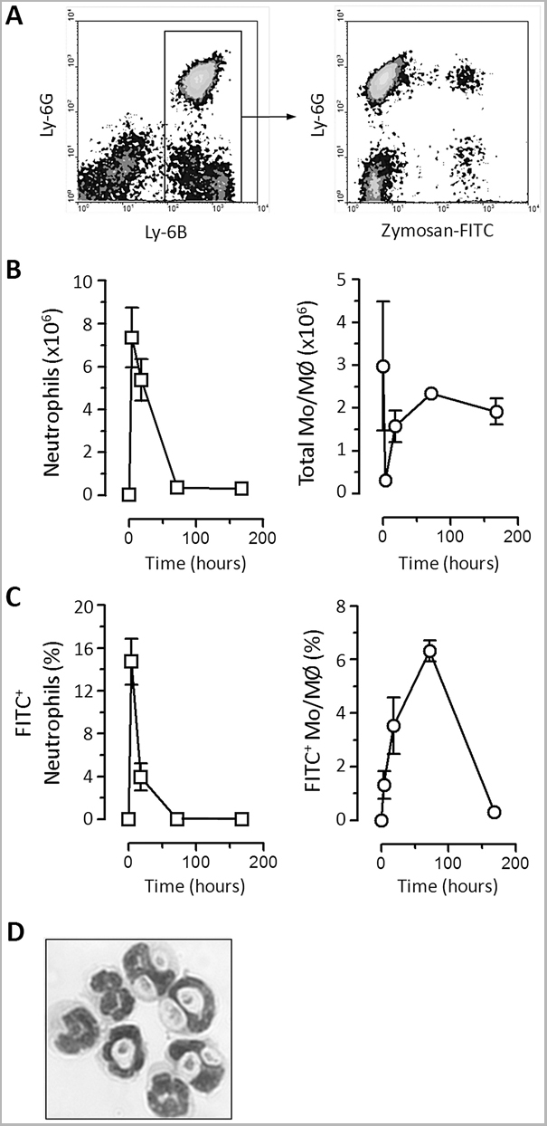
(Published customer image: 166 mM H2O2 increased neutrophil infiltration in day 1 and 6 wounds. Fluorescence intensity of the neodermis was quantified using ImageJ. The area quantified is outlined with the dashed line. Results shown are mean +/- S.E.M, n = 6-7. A representative section from each treatment is shown. ES - Eschar; HE - Hyper-proliferating epidermis; ND - neodermis.*p)
Application Data
(Published customer image: 166 mM H2O2 increased neutrophil infiltration in day 1 and 6 wounds. Fluorescence intensity of the neodermis was quantified using ImageJ. The area quantified is outlined with the dashed line. Results shown are mean +/- S.E.M, n = 6-7. A representative section from each treatment is shown. ES - Eschar; HE - Hyper-proliferating epidermis; ND - neodermis.*p)
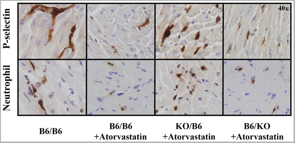
Application Data
(Published customer image: Effect of atorvastatin on platelet and neutrophil infiltration after reperfusion. The infiltration of platelets (upper panel) and neutrophils (lower panel) were evaluated by immunohistochemistry in the 4 chimeric groups that received 40 min of LAD occlusion and 60 min of reperfusion. Platelets and neutrophils were found predominantly in the ischemic area in B6/B6 chimeras. Atorvastatin reduced both platelet and neutrophil infiltration in B6/B6 and B6/KO chimeras, but not in KO/B6 chimeras.From: Tian Y, Linden J, French BA, Yang Z (2014) Atorvastatin at Reperfusion Reduces Myocardial Infarct Size in Mice by Activating eNOS in Bone Marrow-Derived Cells. PLoS ONE 9(12): e114375.)
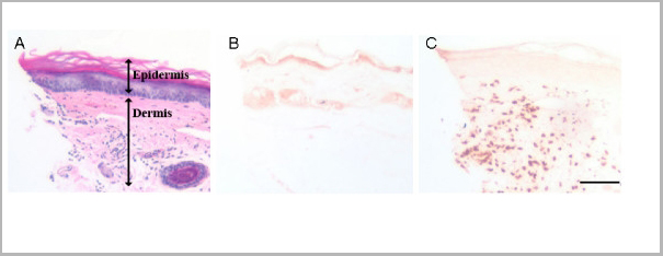
Application Data
(Published customer image: Peri-incisional infiltration of neutrophils, immunohistochemical appearance. Hind paw skin from incised mice was processed for the identification of neutrophils. Panel A displays an H&E stained section of plantar hind paw skin demonstrating a predominantly dermal cellular infiltrate 2 hours after incision. The dermal and epidermal layers are labeled. Panel B displays the appearance of non-incised hind paw skin stained with a neutrophil-specific antibody. Panel C displays a micrograph taken of incised skin stained for neutrophils 2 hours after incision. Note the abundance of darkly staining infiltrating neutrophils in the dermis of this section compared to that displayed in panel B. The scale bar in panel C is 150 um in length. All micrographs were taken using 200x magnification.From: Clark JD, Shi X, Li X, Qiao Y, Liang D, Angst MS, Yeomans DC. Morphine reduces local cytokine expression and neutrophil infiltration after incision. Mol Pain. 2007 Oct 2;3:28.)
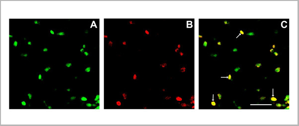
Application Data
(Published customer image: Confocal neutrophil-cytokine double labeling experiments using incised skin. For these studies skin from the wound edges was harvested 2 hours after incision. After sectioning, the tissue was exposed to anti-neutrophil and anti-IL-1beta antibodies followed by the application of CY3 (green fluorescence, neutrophils, panel A) and FITC (red, IL-1beta, panel B) conjugated secondary antibodies. Panel C presents the merged image with arrows pointing to several strongly double labeling cells. The scale bar in panel C is 50 um in length. These micrographs were taken of dermal tissue under 630x magnification.From: Clark JD, Shi X, Li X, Qiao Y, Liang D, Angst MS, Yeomans DC. Morphine reduces local cytokine expression and neutrophil infiltration after incision. Mol Pain. 2007 Oct 2;3:28.)
Similar Products


Product Notes
The Ly-6B.2 (Catalog #AAA12198) is an Antibody produced from Rat and is intended for research purposes only. The product is available for immediate purchase. AAA Biotech's Ly-6B.2 ALLOANTIGEN can be used in a range of immunoassay formats including, but not limited to, Flow cytometry (FC/FACS). Flow Cytometry: Use 10ul of the suggested working dilution to label 106 cells in 100ul. Flow Cytometry: Minimum Dilution: Neat; Maximum Dilution: 1/10. Researchers should empirically determine the suitability of the Ly-6B.2 for an application not listed in the data sheet. Researchers commonly develop new applications and it is an integral, important part of the investigative research process. It is sometimes possible for the material contained within the vial of "Ly-6B.2 ALLOANTIGEN, Monoclonal Antibody" to become dispersed throughout the inside of the vial, particularly around the seal of said vial, during shipment and storage. We always suggest centrifuging these vials to consolidate all of the liquid away from the lid and to the bottom of the vial prior to opening. Please be advised that certain products may require dry ice for shipping and that, if this is the case, an additional dry ice fee may also be required.Precautions
All products in the AAA Biotech catalog are strictly for research-use only, and are absolutely not suitable for use in any sort of medical, therapeutic, prophylactic, in-vivo, or diagnostic capacity. By purchasing a product from AAA Biotech, you are explicitly certifying that said products will be properly tested and used in line with industry standard. AAA Biotech and its authorized distribution partners reserve the right to refuse to fulfill any order if we have any indication that a purchaser may be intending to use a product outside of our accepted criteria.Disclaimer
Though we do strive to guarantee the information represented in this datasheet, AAA Biotech cannot be held responsible for any oversights or imprecisions. AAA Biotech reserves the right to adjust any aspect of this datasheet at any time and without notice. It is the responsibility of the customer to inform AAA Biotech of any product performance issues observed or experienced within 30 days of receipt of said product. To see additional details on this or any of our other policies, please see our Terms & Conditions page.Item has been added to Shopping Cart
If you are ready to order, navigate to Shopping Cart and get ready to checkout.