


 FCM (Flow Cytometry)
(Flow cytometric analysis of Hela cells with PCNA antibody at 1/100 dilution (blue) compared with an unlabelled control (cells without incubation with primary antibody; red). Goat anti mouse IgG (FITC) was used as the secondary antibody.)
FCM (Flow Cytometry)
(Flow cytometric analysis of Hela cells with PCNA antibody at 1/100 dilution (blue) compared with an unlabelled control (cells without incubation with primary antibody; red). Goat anti mouse IgG (FITC) was used as the secondary antibody.)
Mouse PCNA Monoclonal Antibody | anti-PCNA antibody
PCNA Antibody
IHC: 1:21:100
FCM (Flow Cytometry)
(Flow cytometric analysis of Hela cells with PCNA antibody at 1/100 dilution (blue) compared with an unlabelled control (cells without incubation with primary antibody; red). Goat anti mouse IgG (FITC) was used as the secondary antibody.)
FCM (Flow Cytometry)
(Flow cytometric analysis of Hela cells with PCNA antibody at 1/100 dilution (blue) compared with an unlabelled control (cells without incubation with primary antibody; red). Goat anti mouse IgG (FITC) was used as the secondary antibody.)
IHC (Immunohistochemistry)
(Immunohistochemical analysis of paraffin-embedded mouse large intestine tissue using anti-PCNA antibody. Counter stained with hematoxylin.)
IHC (Immunohistochemistry)
(Immunohistochemical analysis of paraffin-embedded mouse large intestine tissue using anti-PCNA antibody. Counter stained with hematoxylin.)
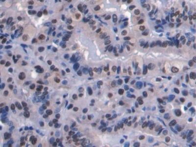
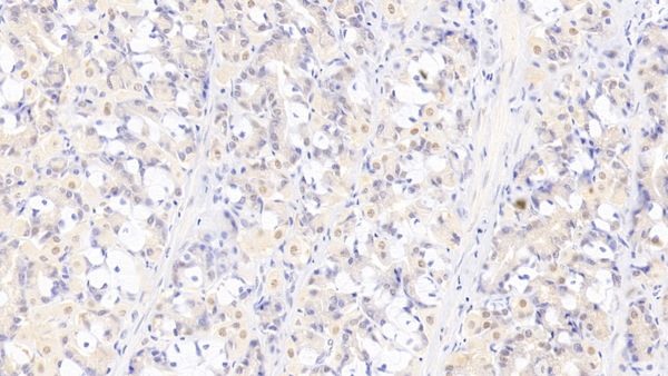
IHC (Immunohistochemistry)
(Immunohistochemical analysis of paraffin-embedded human stomach carcinoma tissue using anti-PCNA antibody. Counter stained with hematoxylin.)
IHC (Immunohistochemistry)
(Immunohistochemical analysis of paraffin-embedded human stomach carcinoma tissue using anti-PCNA antibody. Counter stained with hematoxylin.)
IHC (Immunohistochemistry)
(Immunohistochemical analysis of paraffin-embedded human tonsil tissue using anti-PCNA antibody. Counter stained with hematoxylin.)
IHC (Immunohistochemistry)
(Immunohistochemical analysis of paraffin-embedded human tonsil tissue using anti-PCNA antibody. Counter stained with hematoxylin.)
IHC (Immunohistochemistry)
(Immunohistochemical analysis of paraffin-embedded mouse spleen tissue using anti-PCNA antibody. Counter stained with hematoxylin.)
IHC (Immunohistochemistry)
(Immunohistochemical analysis of paraffin-embedded mouse spleen tissue using anti-PCNA antibody. Counter stained with hematoxylin.)
WB (Western Blot)
(Western blot analysis of PCNA on different cell lysates using anti-PCNA antibody at 1/1000 dilution. Positive control: Line 1: L929 Line 2 :MCF-7 Line 3:PC12 Line 4:Raji Line 5:F9 Line 6:A549)
WB (Western Blot)
(Western blot analysis of PCNA on different cell lysates using anti-PCNA antibody at 1/1000 dilution. Positive control: Line 1: L929 Line 2 :MCF-7 Line 3:PC12 Line 4:Raji Line 5:F9 Line 6:A549)
NCBI and Uniprot Product Information
Customer Reviews
Loading reviews...
Share Your Experience
Similar Products


Product Notes
The PCNA pcna (Catalog #AAA29892) is an Antibody produced from Mouse and is intended for research purposes only. The product is available for immediate purchase. The PCNA Antibody reacts with Human, Mouse, Rat and may cross-react with other species as described in the data sheet. AAA Biotech's PCNA can be used in a range of immunoassay formats including, but not limited to, WB (Western Blot), IHC (Immunohistochemistry), FCM/FACS (Flow Cytometry). WB: 1:1000-1:2000 IHC: 1:21:100. Researchers should empirically determine the suitability of the PCNA pcna for an application not listed in the data sheet. Researchers commonly develop new applications and it is an integral, important part of the investigative research process. It is sometimes possible for the material contained within the vial of "PCNA, Monoclonal Antibody" to become dispersed throughout the inside of the vial, particularly around the seal of said vial, during shipment and storage. We always suggest centrifuging these vials to consolidate all of the liquid away from the lid and to the bottom of the vial prior to opening. Please be advised that certain products may require dry ice for shipping and that, if this is the case, an additional dry ice fee may also be required.Precautions
All products in the AAA Biotech catalog are strictly for research-use only, and are absolutely not suitable for use in any sort of medical, therapeutic, prophylactic, in-vivo, or diagnostic capacity. By purchasing a product from AAA Biotech, you are explicitly certifying that said products will be properly tested and used in line with industry standard. AAA Biotech and its authorized distribution partners reserve the right to refuse to fulfill any order if we have any indication that a purchaser may be intending to use a product outside of our accepted criteria.Disclaimer
Though we do strive to guarantee the information represented in this datasheet, AAA Biotech cannot be held responsible for any oversights or imprecisions. AAA Biotech reserves the right to adjust any aspect of this datasheet at any time and without notice. It is the responsibility of the customer to inform AAA Biotech of any product performance issues observed or experienced within 30 days of receipt of said product. To see additional details on this or any of our other policies, please see our Terms & Conditions page.Frequently Asked Questions
What is PCNA used as a marker for in cell proliferation studies?
PCNA marks actively dividing cells. It binds to the PCNA protein, which appears when cells are making new DNA. This helps researchers find and study cells that are actively dividing.
Is PCNA monoclonal antibody suitable for detecting proliferating cells?
Yes, the PCNA monoclonal antibody is suitable for detecting proliferating cells. It binds to the PCNA protein, which appears when cells are making new DNA. This helps researchers find and study cells that are actively dividing.
Can PCNA antibody be used for IHC or immunofluorescence staining?
Yes, the PCNA antibody can be used for immunohistochemistry (IHC) and immunofluorescence staining. These methods let scientists visualize PCNA in cells or tissues, revealing active cell growth and division.
How does PCNA differ from other proliferation markers like Ki-67?
PCNA shows up mainly during DNA copying, while Ki-67 appears in nearly all active growth phases. Ki-67 tracks overall cell activity, but PCNA zeroes in on DNA replication. Both are valuable but spotlight different stages.
Does PCNA expression change during different cell cycle phases?
Yes, PCNA levels change during the cell cycle. It is highest during the DNA synthesis phase. When cells are not dividing, PCNA levels are low. This change helps scientists understand when cells are actively growing.
Can PCNA antibody be used to assess tumor growth or cancer progression?
Yes, PCNA antibodies help study tumor growth. High PCNA levels usually mean cancer cells are dividing quickly. This helps researchers understand how fast a tumor is growing and how aggressive the cancer might be.
Item has been added to Shopping Cart
If you are ready to order, navigate to Shopping Cart and get ready to checkout.