Filters
▼Clonality
▼Type
▼Reactivity
▼Gene Name
▼Isotype
▼Host
▼Application
▼Clone
▼Polyclonal Antibodies
At AAA Biotech also known as AAA Bio or AAABio, we provide a broad range of purified polyclonal antibodies (pAbs) that are able to all be browsed online through our website. Due to their high specificity and strong binding affinity, these antibodies are ideal for wide swathes of research and experimental applications.
Our polyclonal antibodies can easily support your work, whether you use them for Western Blotting, Immunocytochemistry (with or without Immunofluorescence used in conjunction), Immunohistochemistry, Immunoprecipitation, and ELISA tests. We highly encourage you to browse our range of pAbs and choose the one that best suits your experimental model.
Viewing 7800-7850 of 96812 product results
 FCM/FACS (Flow Cytometry)
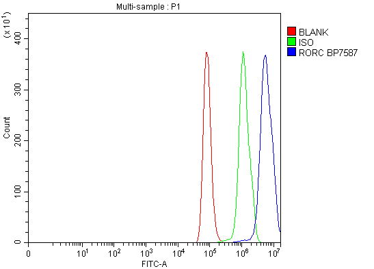
(Figure 2. Flow Cytometry analysis of HeLa cells using anti-ROR gamma/RORC antibody (AAA126026).Overlay histogram showing HeLa cells stained with AAA126026 (Blue line). The cells were blocked with 10% normal goat serum. And then incubated with rabbit anti-ROR gamma/RORC Antibody (AAA126026, 1 ug/1x10^6 cells) for 30 min at 20 degree C. DyLight488 conjugated goat anti-rabbit IgG was used as secondary antibody for 30 minutes at 20 degree C. Isotype control antibody (Green line) was rabbit IgG (1 ug/1x10^6) used under the same conditions. Unlabelled sample (Red line) was also used as a control.)
FCM/FACS (Flow Cytometry)
(Figure 2. Flow Cytometry analysis of HeLa cells using anti-ROR gamma/RORC antibody (AAA126026).Overlay histogram showing HeLa cells stained with AAA126026 (Blue line). The cells were blocked with 10% normal goat serum. And then incubated with rabbit anti-ROR gamma/RORC Antibody (AAA126026, 1 ug/1x10^6 cells) for 30 min at 20 degree C. DyLight488 conjugated goat anti-rabbit IgG was used as secondary antibody for 30 minutes at 20 degree C. Isotype control antibody (Green line) was rabbit IgG (1 ug/1x10^6) used under the same conditions. Unlabelled sample (Red line) was also used as a control.)
ROR gamma/RORC, Polyclonal Antibody (Cat# AAA126026)
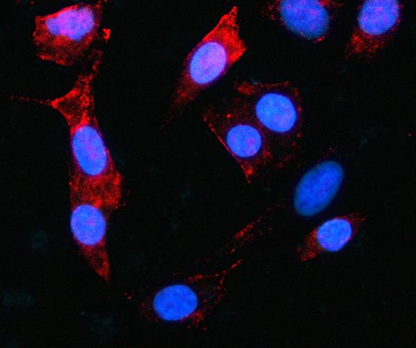
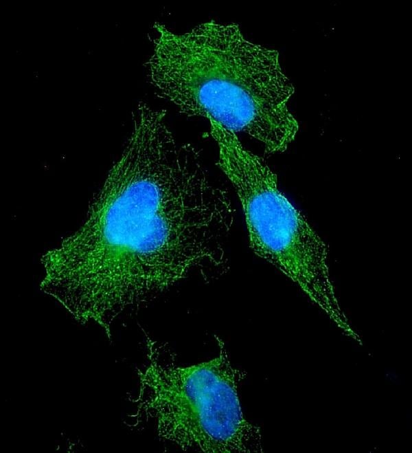
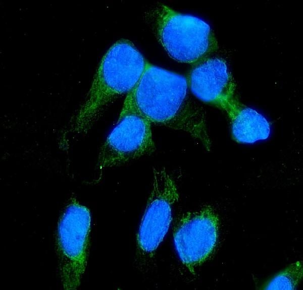
 IF (Immunofluorescence)
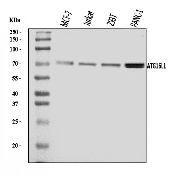
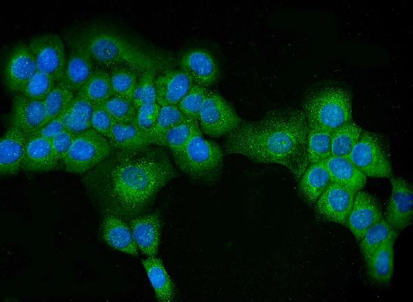
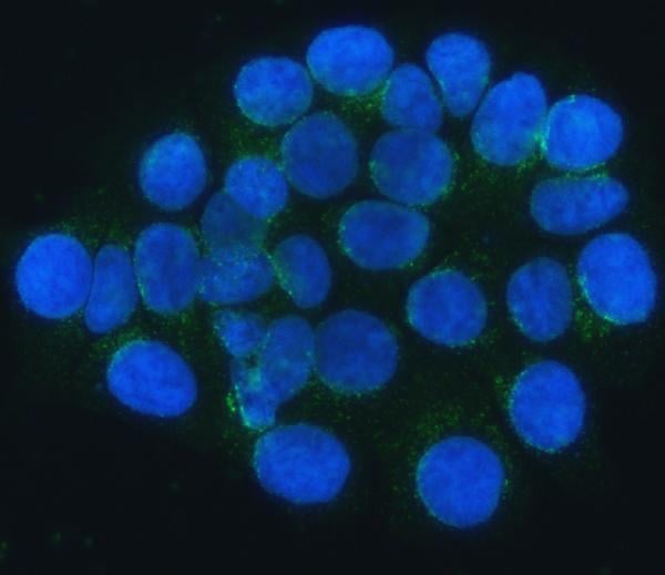
(Figure 5. IF analysis of ATG16L1 using anti-ATG16L1 antibody (AAA126035).ATG16L1 was detected in an immunocytochemical section of A431 cells. Enzyme antigen retrieval was performed using IHC enzyme antigen retrieval reagent (AR0022) for 15 mins. The cells were blocked with 10% goat serum. And then incubated with 5 ug/mL rabbit anti-ATG16L1 Antibody (AAA126035) overnight at 4 degree C. DyLight488 Conjugated Goat Anti-Rabbit IgG was used as secondary antibody at 1:100 dilution and incubated for 30 minutes at 37 degree C. The section was counterstained with DAPI. Visualize using a fluorescence microscope and filter sets appropriate for the label used.)
IF (Immunofluorescence)
(Figure 5. IF analysis of ATG16L1 using anti-ATG16L1 antibody (AAA126035).ATG16L1 was detected in an immunocytochemical section of A431 cells. Enzyme antigen retrieval was performed using IHC enzyme antigen retrieval reagent (AR0022) for 15 mins. The cells were blocked with 10% goat serum. And then incubated with 5 ug/mL rabbit anti-ATG16L1 Antibody (AAA126035) overnight at 4 degree C. DyLight488 Conjugated Goat Anti-Rabbit IgG was used as secondary antibody at 1:100 dilution and incubated for 30 minutes at 37 degree C. The section was counterstained with DAPI. Visualize using a fluorescence microscope and filter sets appropriate for the label used.)
ATG16L1, Polyclonal Antibody (Cat# AAA126035)
 IF (Immunofluorescence)
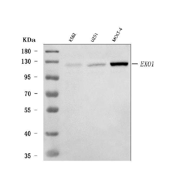
(Figure 2. IF analysis of Exonuclease 1/EXO1 using anti-Exonuclease 1/EXO1 antibody (AAA126036).Exonuclease 1/EXO1 was detected in an immunocytochemical section of MCF-7 cells. Enzyme antigen retrieval was performed using IHC enzyme antigen retrieval reagent (AR0022) for 15 mins. The cells were blocked with 10% goat serum. And then incubated with 5 ug/mL rabbit anti-Exonuclease 1/EXO1 Antibody (AAA126036) overnight at 4 degree C. DyLight488 Conjugated Goat Anti-Rabbit IgG was used as secondary antibody at 1:100 dilution and incubated for 30 minutes at 37 degree C. The section was counterstained with DAPI. Visualize using a fluorescence microscope and filter sets appropriate for the label used.)
IF (Immunofluorescence)
(Figure 2. IF analysis of Exonuclease 1/EXO1 using anti-Exonuclease 1/EXO1 antibody (AAA126036).Exonuclease 1/EXO1 was detected in an immunocytochemical section of MCF-7 cells. Enzyme antigen retrieval was performed using IHC enzyme antigen retrieval reagent (AR0022) for 15 mins. The cells were blocked with 10% goat serum. And then incubated with 5 ug/mL rabbit anti-Exonuclease 1/EXO1 Antibody (AAA126036) overnight at 4 degree C. DyLight488 Conjugated Goat Anti-Rabbit IgG was used as secondary antibody at 1:100 dilution and incubated for 30 minutes at 37 degree C. The section was counterstained with DAPI. Visualize using a fluorescence microscope and filter sets appropriate for the label used.)
Exonuclease 1/EXO1, Polyclonal Antibody (Cat# AAA126036)
 IF (Immunofluorescence)
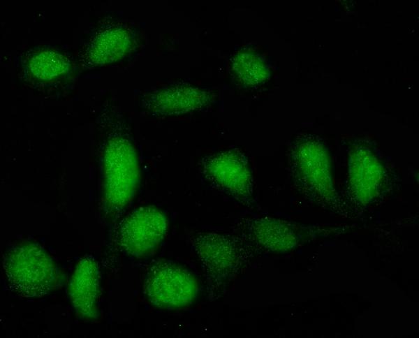
(Figure 2. IF analysis of PTPN22 using anti-PTPN22 antibody (AAA126044).PTPN22 was detected in an immunocytochemical section of U2OS cells. Enzyme antigen retrieval was performed using IHC enzyme antigen retrieval reagent (AR0022) for 15 mins. The cells were blocked with 10% goat serum. And then incubated with 5 ug/mL rabbit anti-PTPN22 Antibody (AAA126044) overnight at 4 degree C. DyLight488 Conjugated Goat Anti-Rabbit IgG was used as secondary antibody at 1:100 dilution and incubated for 30 minutes at 37 degree C. The section was counterstained with DAPI. Visualize using a fluorescence microscope and filter sets appropriate for the label used.)
IF (Immunofluorescence)
(Figure 2. IF analysis of PTPN22 using anti-PTPN22 antibody (AAA126044).PTPN22 was detected in an immunocytochemical section of U2OS cells. Enzyme antigen retrieval was performed using IHC enzyme antigen retrieval reagent (AR0022) for 15 mins. The cells were blocked with 10% goat serum. And then incubated with 5 ug/mL rabbit anti-PTPN22 Antibody (AAA126044) overnight at 4 degree C. DyLight488 Conjugated Goat Anti-Rabbit IgG was used as secondary antibody at 1:100 dilution and incubated for 30 minutes at 37 degree C. The section was counterstained with DAPI. Visualize using a fluorescence microscope and filter sets appropriate for the label used.)
PTPN22, Polyclonal Antibody (Cat# AAA126044)
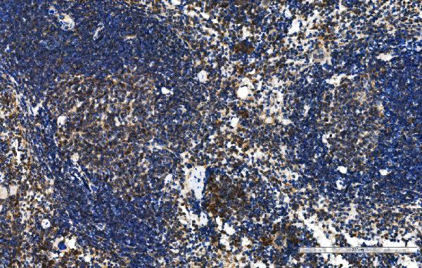
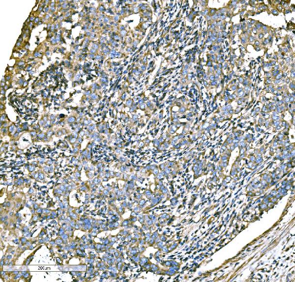
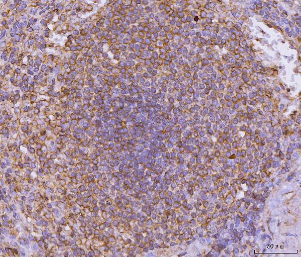
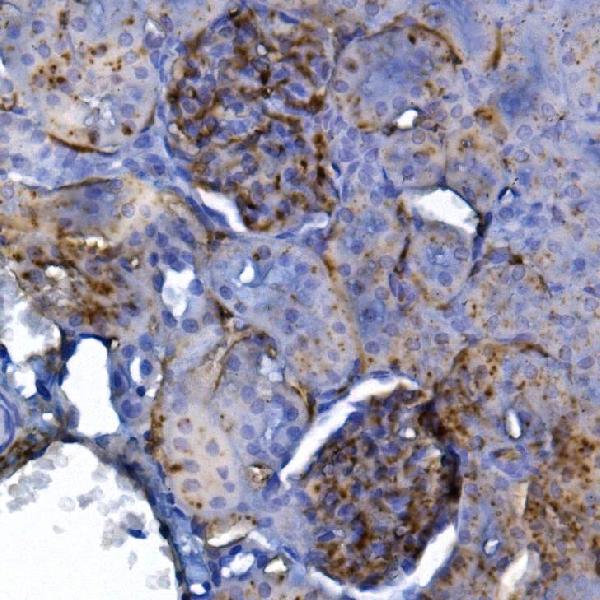
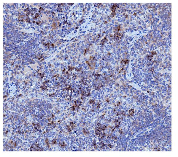
 IHC (Immunohiostchemistry)
(Figure 2. IHC analysis of TFEB using anti-TFEB antibody (AAA126053).TFEB was detected in a paraffin-embedded section of rat lymph nodes tissue. Heat mediated antigen retrieval was performed in EDTA buffer (pH 8.0, epitope retrieval solution). The tissue section was blocked with 10% goat serum. The tissue section was then incubated with 2 ug/ml rabbit anti-TFEB Antibody (AAA126053) overnight at 4 degree C. Peroxidase Conjugated Goat Anti-rabbit IgG was used as secondary antibody and incubated for 30 minutes at 37 degree C. The tissue section was developed using HRP Conjugated Rabbit IgG Super Vision Assay Kit with DAB as the chromogen.)
IHC (Immunohiostchemistry)
(Figure 2. IHC analysis of TFEB using anti-TFEB antibody (AAA126053).TFEB was detected in a paraffin-embedded section of rat lymph nodes tissue. Heat mediated antigen retrieval was performed in EDTA buffer (pH 8.0, epitope retrieval solution). The tissue section was blocked with 10% goat serum. The tissue section was then incubated with 2 ug/ml rabbit anti-TFEB Antibody (AAA126053) overnight at 4 degree C. Peroxidase Conjugated Goat Anti-rabbit IgG was used as secondary antibody and incubated for 30 minutes at 37 degree C. The tissue section was developed using HRP Conjugated Rabbit IgG Super Vision Assay Kit with DAB as the chromogen.)
Cxcr5, Polyclonal Antibody (Cat# AAA126053)
 FCM/FACS (Flow Cytometry)
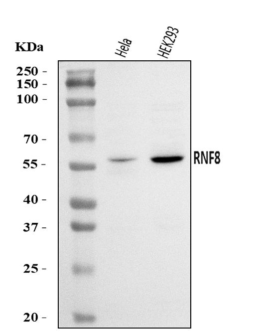
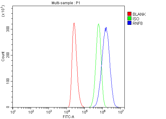
(Figure 2. Flow Cytometry analysis of THP-1 cells using anti-RNF8 antibody (AAA126060).Overlay histogram showing THP-1 cells stained with AAA126060 (Blue line). The cells were blocked with 10% normal goat serum. And then incubated with rabbit anti-RNF8 Antibody (AAA126060, 1 ug/1x10^6 cells) for 30 min at 20 degree C. DyLight488 conjugated goat anti-rabbit IgG was used as secondary antibody for 30 minutes at 20 degree C. Isotype control antibody (Green line) was rabbit IgG (1 ug/1x10^6) used under the same conditions. Unlabelled sample (Red line) was also used as a control.)
FCM/FACS (Flow Cytometry)
(Figure 2. Flow Cytometry analysis of THP-1 cells using anti-RNF8 antibody (AAA126060).Overlay histogram showing THP-1 cells stained with AAA126060 (Blue line). The cells were blocked with 10% normal goat serum. And then incubated with rabbit anti-RNF8 Antibody (AAA126060, 1 ug/1x10^6 cells) for 30 min at 20 degree C. DyLight488 conjugated goat anti-rabbit IgG was used as secondary antibody for 30 minutes at 20 degree C. Isotype control antibody (Green line) was rabbit IgG (1 ug/1x10^6) used under the same conditions. Unlabelled sample (Red line) was also used as a control.)
RNF8, Polyclonal Antibody (Cat# AAA126060)
 FCM/FACS (Flow Cytometry)
(Figure 3. Flow Cytometry analysis of 293T cells using anti-SNAIL/SNAI1 antibody (AAA126063).Overlay histogram showing 293T cells stained with AAA126063 (Blue line).The cells were blocked with 10% normal goat serum. And then incubated with rabbit anti-SNAIL/SNAI1 Antibody (AAA126063,1ug/1x10^6 cells) for 30 min at 20 degree C. DyLight488 conjugated goat anti-rabbit IgG was used as secondary antibody for 30 minutes at 20 degree C. Isotype control antibody (Green line) was rabbit IgG (1ug/1x10^6) used under the same conditions. Unlabelled sample (Red line) was also used as a control.)
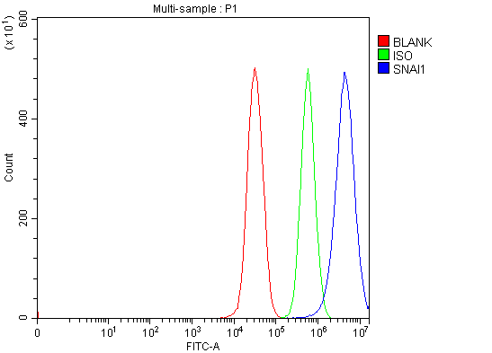
FCM/FACS (Flow Cytometry)
(Figure 3. Flow Cytometry analysis of 293T cells using anti-SNAIL/SNAI1 antibody (AAA126063).Overlay histogram showing 293T cells stained with AAA126063 (Blue line).The cells were blocked with 10% normal goat serum. And then incubated with rabbit anti-SNAIL/SNAI1 Antibody (AAA126063,1ug/1x10^6 cells) for 30 min at 20 degree C. DyLight488 conjugated goat anti-rabbit IgG was used as secondary antibody for 30 minutes at 20 degree C. Isotype control antibody (Green line) was rabbit IgG (1ug/1x10^6) used under the same conditions. Unlabelled sample (Red line) was also used as a control.)
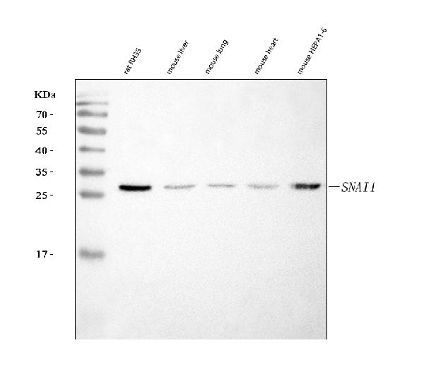
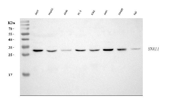
SNAIL/SNAI1, Polyclonal Antibody (Cat# AAA126063)
 FCM/FACS (Flow Cytometry)
(Figure 2. Flow Cytometry analysis of 293T cells using anti-PCH2/TRIP13 antibody (AAA126476).Overlay histogram showing 293T cells stained with AAA126476 (Blue line). The cells were blocked with 10% normal goat serum. And then incubated with rabbit anti-PCH2/TRIP13 Antibody (AAA126476, 1 ug/1x10^6 cells) for 30 min at 20 degree C. DyLight488 conjugated goat anti-rabbit IgG was used as secondary antibody for 30 minutes at 20 degree C. Isotype control antibody (Green line) was rabbit IgG (1 ug/1x10^6) used under the same conditions. Unlabelled sample (Red line) was also used as a control.)
FCM/FACS (Flow Cytometry)
(Figure 2. Flow Cytometry analysis of 293T cells using anti-PCH2/TRIP13 antibody (AAA126476).Overlay histogram showing 293T cells stained with AAA126476 (Blue line). The cells were blocked with 10% normal goat serum. And then incubated with rabbit anti-PCH2/TRIP13 Antibody (AAA126476, 1 ug/1x10^6 cells) for 30 min at 20 degree C. DyLight488 conjugated goat anti-rabbit IgG was used as secondary antibody for 30 minutes at 20 degree C. Isotype control antibody (Green line) was rabbit IgG (1 ug/1x10^6) used under the same conditions. Unlabelled sample (Red line) was also used as a control.)
PCH2/TRIP13, Polyclonal Antibody (Cat# AAA126476)
 FCM/FACS (Flow Cytometry)
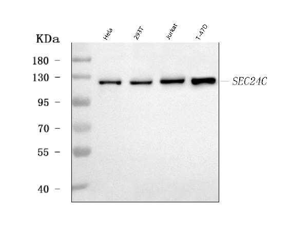
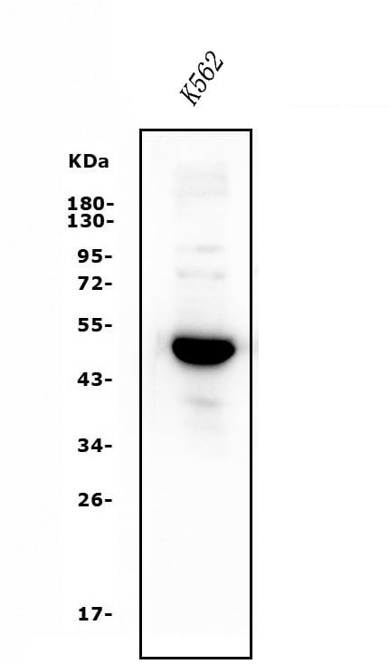
(Figure 3. Flow Cytometry analysis of K562 cells using anti-SEC24C antibody (AAA126493).Overlay histogram showing K562 cells stained with AAA126493 (Blue line). The cells were blocked with 10% normal goat serum. And then incubated with rabbit anti-SEC24C Antibody (AAA126493, 1 ug/1x10^6 cells) for 30 min at 20 degree C. DyLight488 conjugated goat anti-rabbit IgG was used as secondary antibody for 30 minutes at 20 degree C. Isotype control antibody (Green line) was rabbit IgG (1 ug/1x10^6) used under the same conditions. Unlabelled sample (Red line) was also used as a control.)
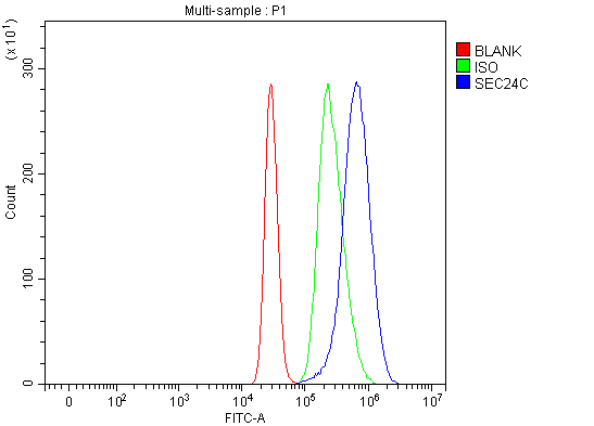
FCM/FACS (Flow Cytometry)
(Figure 3. Flow Cytometry analysis of K562 cells using anti-SEC24C antibody (AAA126493).Overlay histogram showing K562 cells stained with AAA126493 (Blue line). The cells were blocked with 10% normal goat serum. And then incubated with rabbit anti-SEC24C Antibody (AAA126493, 1 ug/1x10^6 cells) for 30 min at 20 degree C. DyLight488 conjugated goat anti-rabbit IgG was used as secondary antibody for 30 minutes at 20 degree C. Isotype control antibody (Green line) was rabbit IgG (1 ug/1x10^6) used under the same conditions. Unlabelled sample (Red line) was also used as a control.)
SEC24C, Polyclonal Antibody (Cat# AAA126493)
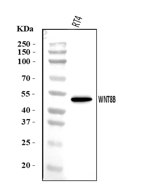
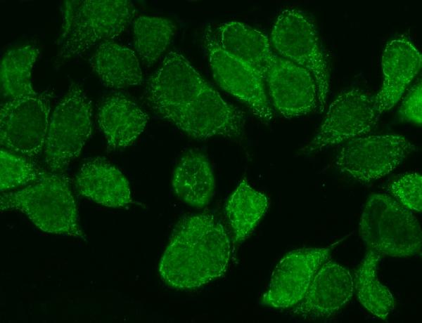 IF (Immunofluorescence)
(Figure 2. IF analysis of WNT8B using anti-WNT8B antibody (AAA126506).WNT8B was detected in an immunocytochemical section of MCF-7 cells. Enzyme antigen retrieval was performed using IHC enzyme antigen retrieval reagent (AR0022) for 15 mins. The cells were blocked with 10% goat serum. And then incubated with 5 ug/mL rabbit anti-WNT8B Antibody (AAA126506) overnight at 4 degree C. DyLight488 Conjugated Goat Anti-Rabbit IgG was used as secondary antibody at 1:100 dilution and incubated for 30 minutes at 37 degree C. The section was counterstained with DAPI. Visualize using a fluorescence microscope and filter sets appropriate for the label used.)
IF (Immunofluorescence)
(Figure 2. IF analysis of WNT8B using anti-WNT8B antibody (AAA126506).WNT8B was detected in an immunocytochemical section of MCF-7 cells. Enzyme antigen retrieval was performed using IHC enzyme antigen retrieval reagent (AR0022) for 15 mins. The cells were blocked with 10% goat serum. And then incubated with 5 ug/mL rabbit anti-WNT8B Antibody (AAA126506) overnight at 4 degree C. DyLight488 Conjugated Goat Anti-Rabbit IgG was used as secondary antibody at 1:100 dilution and incubated for 30 minutes at 37 degree C. The section was counterstained with DAPI. Visualize using a fluorescence microscope and filter sets appropriate for the label used.)
WNT8B, Polyclonal Antibody (Cat# AAA126506)
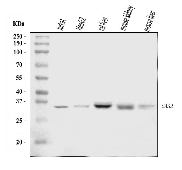
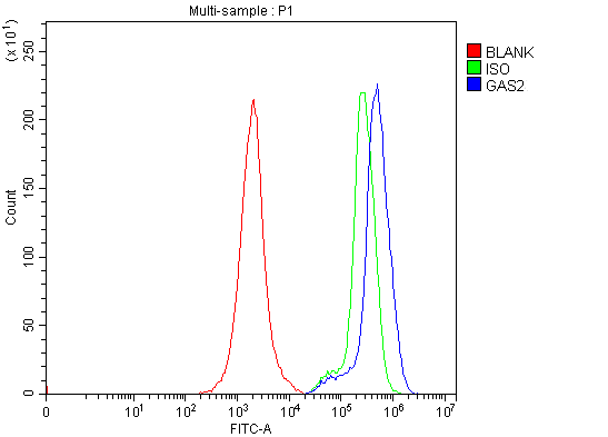 FCM/FACS (Flow Cytometry)
(Figure 2. Flow Cytometry analysis of Jurkat cells using anti-GAS2 antibody (AAA126528).Overlay histogram showing Jurkat cells stained with AAA126528 (Blue line). The cells were blocked with 10% normal goat serum. And then incubated with rabbit anti-GAS2 Antibody (AAA126528, 1 ug/1x10^6 cells) for 30 min at 20 degree C. DyLight488 conjugated goat anti-rabbit IgG was used as secondary antibody for 30 minutes at 20 degree C. Isotype control antibody (Green line) was rabbit IgG (1 ug/1x10^6) used under the same conditions. Unlabelled sample (Red line) was also used as a control.)
FCM/FACS (Flow Cytometry)
(Figure 2. Flow Cytometry analysis of Jurkat cells using anti-GAS2 antibody (AAA126528).Overlay histogram showing Jurkat cells stained with AAA126528 (Blue line). The cells were blocked with 10% normal goat serum. And then incubated with rabbit anti-GAS2 Antibody (AAA126528, 1 ug/1x10^6 cells) for 30 min at 20 degree C. DyLight488 conjugated goat anti-rabbit IgG was used as secondary antibody for 30 minutes at 20 degree C. Isotype control antibody (Green line) was rabbit IgG (1 ug/1x10^6) used under the same conditions. Unlabelled sample (Red line) was also used as a control.)
GAS2, Polyclonal Antibody (Cat# AAA126528)
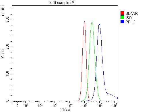 FCM/FACS (Flow Cytometry)
(Figure 3. Flow Cytometry analysis of PC-3 cells using anti-PPIL3 antibody (AAA126548).Overlay histogram showing PC-3 cells stained with AAA126548 (Blue line). The cells were blocked with 10% normal goat serum. And then incubated with rabbit anti-PPIL3 Antibody (AAA126548, 1 ug/1x10^6 cells) for 30 min at 20 degree C. DyLight488 conjugated goat anti-rabbit IgG was used as secondary antibody for 30 minutes at 20 degree C. Isotype control antibody (Green line) was rabbit IgG (1 ug/1x10^6) used under the same conditions. Unlabelled sample (Red line) was also used as a control.)
FCM/FACS (Flow Cytometry)
(Figure 3. Flow Cytometry analysis of PC-3 cells using anti-PPIL3 antibody (AAA126548).Overlay histogram showing PC-3 cells stained with AAA126548 (Blue line). The cells were blocked with 10% normal goat serum. And then incubated with rabbit anti-PPIL3 Antibody (AAA126548, 1 ug/1x10^6 cells) for 30 min at 20 degree C. DyLight488 conjugated goat anti-rabbit IgG was used as secondary antibody for 30 minutes at 20 degree C. Isotype control antibody (Green line) was rabbit IgG (1 ug/1x10^6) used under the same conditions. Unlabelled sample (Red line) was also used as a control.)
PPIL3, Polyclonal Antibody (Cat# AAA126548)
 FCM/FACS (Flow Cytometry)
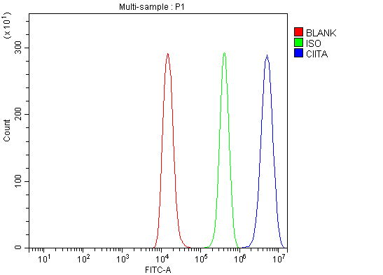
(Figure 2. Flow Cytometry analysis of Raji cells using anti-CIITA antibody (AAA126151).Overlay histogram showing Raji cells stained with AAA126151 (Blue line). The cells were blocked with 10% normal goat serum. And then incubated with rabbit anti-CIITA Antibody (AAA126151, 1 ug/1x10^6 cells) for 30 min at 20 degree C. DyLight488 conjugated goat anti-rabbit IgG was used as secondary antibody for 30 minutes at 20 degree C. Isotype control antibody (Green line) was rabbit IgG (1 ug/1x10^6) used under the same conditions. Unlabelled sample (Red line) was also used as a control.)
FCM/FACS (Flow Cytometry)
(Figure 2. Flow Cytometry analysis of Raji cells using anti-CIITA antibody (AAA126151).Overlay histogram showing Raji cells stained with AAA126151 (Blue line). The cells were blocked with 10% normal goat serum. And then incubated with rabbit anti-CIITA Antibody (AAA126151, 1 ug/1x10^6 cells) for 30 min at 20 degree C. DyLight488 conjugated goat anti-rabbit IgG was used as secondary antibody for 30 minutes at 20 degree C. Isotype control antibody (Green line) was rabbit IgG (1 ug/1x10^6) used under the same conditions. Unlabelled sample (Red line) was also used as a control.)
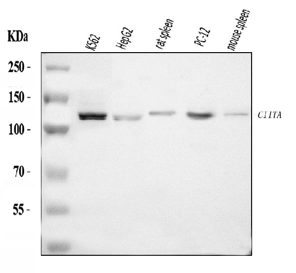
CIITA, Polyclonal Antibody (Cat# AAA126151)
 FCM/FACS (Flow Cytometry)
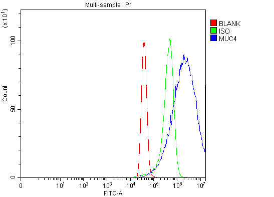
(Figure 2. Flow Cytometry analysis of MCF-7 cells using anti-MUC4 antibody (AAA126162).Overlay histogram showing MCF-7 cells stained with AAA126162 (Blue line). The cells were blocked with 10% normal goat serum. And then incubated with rabbit anti-MUC4 Antibody (AAA126162, 1 ug/1x10^6 cells) for 30 min at 20 degree C. DyLight488 conjugated goat anti-rabbit IgG was used as secondary antibody for 30 minutes at 20 degree C. Isotype control antibody (Green line) was rabbit IgG (1 ug/1x10^6) used under the same conditions. Unlabelled sample (Red line) was also used as a control.)
FCM/FACS (Flow Cytometry)
(Figure 2. Flow Cytometry analysis of MCF-7 cells using anti-MUC4 antibody (AAA126162).Overlay histogram showing MCF-7 cells stained with AAA126162 (Blue line). The cells were blocked with 10% normal goat serum. And then incubated with rabbit anti-MUC4 Antibody (AAA126162, 1 ug/1x10^6 cells) for 30 min at 20 degree C. DyLight488 conjugated goat anti-rabbit IgG was used as secondary antibody for 30 minutes at 20 degree C. Isotype control antibody (Green line) was rabbit IgG (1 ug/1x10^6) used under the same conditions. Unlabelled sample (Red line) was also used as a control.)
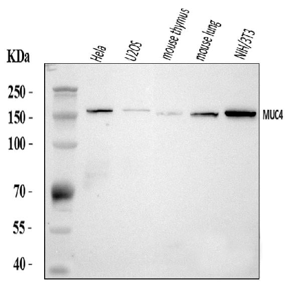
MUC4, Polyclonal Antibody (Cat# AAA126162)
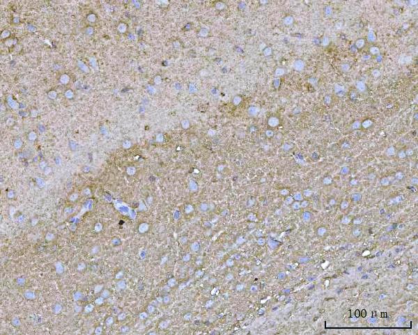
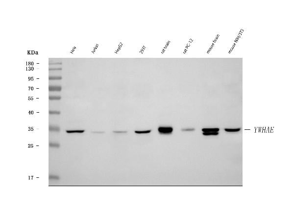
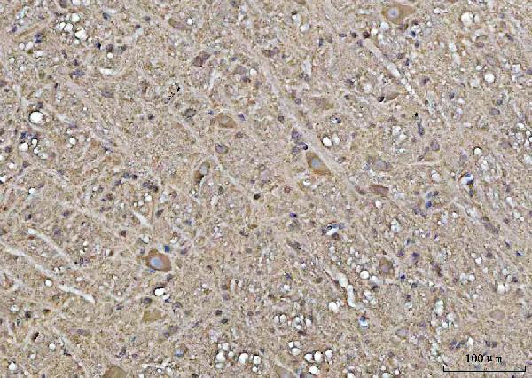 IHC (Immunohistochemistry)
(Figure 5. IHC analysis of YWHAE using anti-YWHAE antibody (AAA126166).YWHAE was detected in a paraffin-embedded section of rat brain tissue tissue. Heat mediated antigen retrieval was performed in EDTA buffer (pH 8.0, epitope retrieval solution). The tissue section was blocked with 10% goat serum. The tissue section was then incubated with 2 ug/ml rabbit anti-YWHAE Antibody (AAA126166) overnight at 4 degree C. Peroxidase Conjugated Goat Anti-rabbit IgG was used as secondary antibody and incubated for 30 minutes at 37 degree C. The tissue section was developed using HRP Conjugated Rabbit IgG Super Vision Assay Kit with DAB as the chromogen.)
IHC (Immunohistochemistry)
(Figure 5. IHC analysis of YWHAE using anti-YWHAE antibody (AAA126166).YWHAE was detected in a paraffin-embedded section of rat brain tissue tissue. Heat mediated antigen retrieval was performed in EDTA buffer (pH 8.0, epitope retrieval solution). The tissue section was blocked with 10% goat serum. The tissue section was then incubated with 2 ug/ml rabbit anti-YWHAE Antibody (AAA126166) overnight at 4 degree C. Peroxidase Conjugated Goat Anti-rabbit IgG was used as secondary antibody and incubated for 30 minutes at 37 degree C. The tissue section was developed using HRP Conjugated Rabbit IgG Super Vision Assay Kit with DAB as the chromogen.)
YWHAE, Polyclonal Antibody (Cat# AAA126166)
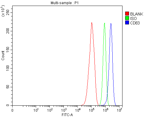 FCM/FACS (Flow Cytometry)
(Figure 2. Flow Cytometry analysis of RT4 cells using anti-ADAM22 antibody (AAA126172).Overlay histogram showing RT4 cells stained with AAA126172 (Blue line). The cells were blocked with 10% normal goat serum. And then incubated with rabbit anti-ADAM22 Antibody (AAA126172, 1 ug/1x10^6 cells) for 30 min at 20 degree C. DyLight488 conjugated goat anti-rabbit IgG was used as secondary antibody for 30 minutes at 20 degree C. Isotype control antibody (Green line) was rabbit IgG (1 ug/1x10^6) used under the same conditions. Unlabelled sample (Red line) was also used as a control.)
FCM/FACS (Flow Cytometry)
(Figure 2. Flow Cytometry analysis of RT4 cells using anti-ADAM22 antibody (AAA126172).Overlay histogram showing RT4 cells stained with AAA126172 (Blue line). The cells were blocked with 10% normal goat serum. And then incubated with rabbit anti-ADAM22 Antibody (AAA126172, 1 ug/1x10^6 cells) for 30 min at 20 degree C. DyLight488 conjugated goat anti-rabbit IgG was used as secondary antibody for 30 minutes at 20 degree C. Isotype control antibody (Green line) was rabbit IgG (1 ug/1x10^6) used under the same conditions. Unlabelled sample (Red line) was also used as a control.)
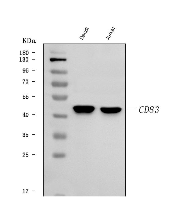
CD83, Polyclonal Antibody (Cat# AAA126172)
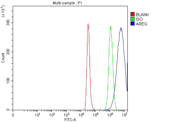 FCM/FACS (Flow Cytometry)
(Figure 2. Flow Cytometry analysis of CACO-2 cells using anti-Amphiregulin/AREG antibody (AAA126174).Overlay histogram showing CACO-2 cells stained with AAA126174 (Blue line). The cells were blocked with 10% normal goat serum. And then incubated with rabbit anti-Amphiregulin/AREG Antibody (AAA126174, 1 ug/1x10^6 cells) for 30 min at 20 degree C. DyLight488 conjugated goat anti-rabbit IgG was used as secondary antibody for 30 minutes at 20 degree C. Isotype control antibody (Green line) was rabbit IgG (1 ug/1x10^6) used under the same conditions. Unlabelled sample (Red line) was also used as a control.)
FCM/FACS (Flow Cytometry)
(Figure 2. Flow Cytometry analysis of CACO-2 cells using anti-Amphiregulin/AREG antibody (AAA126174).Overlay histogram showing CACO-2 cells stained with AAA126174 (Blue line). The cells were blocked with 10% normal goat serum. And then incubated with rabbit anti-Amphiregulin/AREG Antibody (AAA126174, 1 ug/1x10^6 cells) for 30 min at 20 degree C. DyLight488 conjugated goat anti-rabbit IgG was used as secondary antibody for 30 minutes at 20 degree C. Isotype control antibody (Green line) was rabbit IgG (1 ug/1x10^6) used under the same conditions. Unlabelled sample (Red line) was also used as a control.)
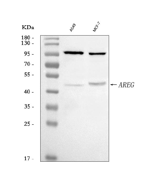
Amphiregulin/AREG, Polyclonal Antibody (Cat# AAA126174)
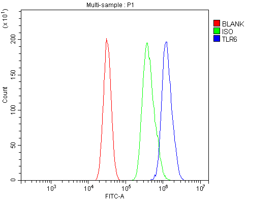 FCM/FACS (Flow Cytometry)
(Figure 2. Flow Cytometry analysis of HEPA1-6 cells using anti-Tlr6 antibody (AAA126188).Overlay histogram showing HEPA1-6 cells stained with AAA126188 (Blue line). The cells were blocked with 10% normal goat serum. And then incubated with rabbit anti-Tlr6 Antibody (AAA126188, 1 ug/1x10^6 cells) for 30 min at 20 degree C. DyLight488 conjugated goat anti-rabbit IgG was used as secondary antibody for 30 minutes at 20 degree C. Isotype control antibody (Green line) was rabbit IgG (1 ug/1x10^6) used under the same conditions. Unlabelled sample (Red line) was also used as a control.)
FCM/FACS (Flow Cytometry)
(Figure 2. Flow Cytometry analysis of HEPA1-6 cells using anti-Tlr6 antibody (AAA126188).Overlay histogram showing HEPA1-6 cells stained with AAA126188 (Blue line). The cells were blocked with 10% normal goat serum. And then incubated with rabbit anti-Tlr6 Antibody (AAA126188, 1 ug/1x10^6 cells) for 30 min at 20 degree C. DyLight488 conjugated goat anti-rabbit IgG was used as secondary antibody for 30 minutes at 20 degree C. Isotype control antibody (Green line) was rabbit IgG (1 ug/1x10^6) used under the same conditions. Unlabelled sample (Red line) was also used as a control.)
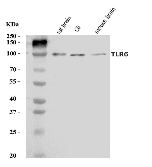
Tlr6, Polyclonal Antibody (Cat# AAA126188)
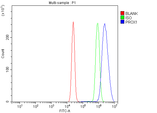 FCM/FACS (Flow Cytometry)
(Figure 2. Flow Cytometry analysis of K562 cells using anti-PROX1 antibody (AAA126192).Overlay histogram showing K562 cells stained with AAA126192 (Blue line). The cells were blocked with 10% normal goat serum. And then incubated with rabbit anti-PROX1 Antibody (AAA126192, 1 ug/1x10^6 cells) for 30 min at 20 degree C. DyLight488 conjugated goat anti-rabbit IgG was used as secondary antibody for 30 minutes at 20 degree C. Isotype control antibody (Green line) was rabbit IgG (1 ug/1x10^6) used under the same conditions. Unlabelled sample (Red line) was also used as a control.)
FCM/FACS (Flow Cytometry)
(Figure 2. Flow Cytometry analysis of K562 cells using anti-PROX1 antibody (AAA126192).Overlay histogram showing K562 cells stained with AAA126192 (Blue line). The cells were blocked with 10% normal goat serum. And then incubated with rabbit anti-PROX1 Antibody (AAA126192, 1 ug/1x10^6 cells) for 30 min at 20 degree C. DyLight488 conjugated goat anti-rabbit IgG was used as secondary antibody for 30 minutes at 20 degree C. Isotype control antibody (Green line) was rabbit IgG (1 ug/1x10^6) used under the same conditions. Unlabelled sample (Red line) was also used as a control.)
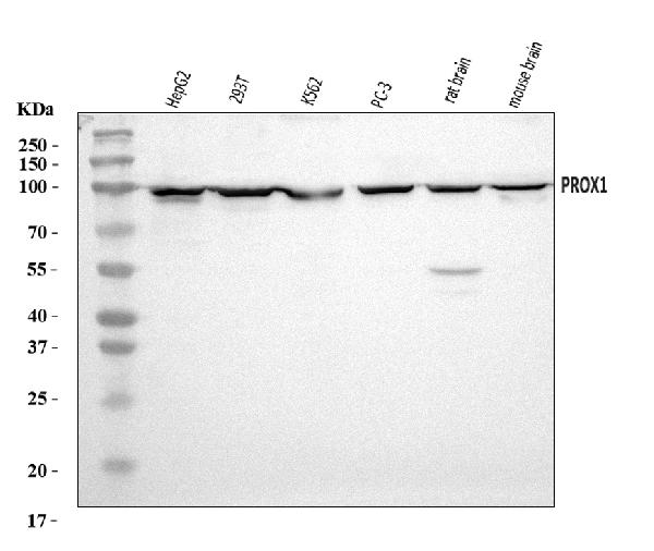
PROX1, Polyclonal Antibody (Cat# AAA126192)
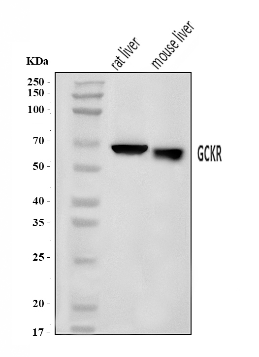
 FCM/FACS (Flow Cytometry)
(Figure 3. Flow Cytometry analysis of U2OS cells using anti-GCKR antibody (AAA126200).Overlay histogram showing U2OS cells stained with AAA126200 (Blue line). The cells were blocked with 10% normal goat serum. And then incubated with rabbit anti-GCKR Antibody (AAA126200, 1 ug/1x10^6 cells) for 30 min at 20 degree C. DyLight488 conjugated goat anti-rabbit IgG was used as secondary antibody for 30 minutes at 20 degree C. Isotype control antibody (Green line) was rabbit IgG (1 ug/1x10^6) used under the same conditions. Unlabelled sample (Red line) was also used as a control.)
FCM/FACS (Flow Cytometry)
(Figure 3. Flow Cytometry analysis of U2OS cells using anti-GCKR antibody (AAA126200).Overlay histogram showing U2OS cells stained with AAA126200 (Blue line). The cells were blocked with 10% normal goat serum. And then incubated with rabbit anti-GCKR Antibody (AAA126200, 1 ug/1x10^6 cells) for 30 min at 20 degree C. DyLight488 conjugated goat anti-rabbit IgG was used as secondary antibody for 30 minutes at 20 degree C. Isotype control antibody (Green line) was rabbit IgG (1 ug/1x10^6) used under the same conditions. Unlabelled sample (Red line) was also used as a control.)
GCKR, Polyclonal Antibody (Cat# AAA126200)
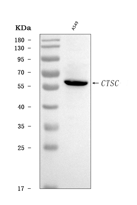
 FCM/FACS (Flow Cytometry)
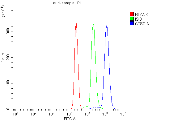
(Figure 2. Flow Cytometry analysis of U937 cells using anti-CTSC antibody (AAA126202).Overlay histogram showing U937 cells stained with AAA126202 (Blue line). The cells were blocked with 10% normal goat serum. And then incubated with rabbit anti-CTSC Antibody (AAA126202, 1 ug/1x10^6 cells) for 30 min at 20 degree C. DyLight488 conjugated goat anti-rabbit IgG was used as secondary antibody for 30 minutes at 20 degree C. Isotype control antibody (Green line) was rabbit IgG (1 ug/1x10^6) used under the same conditions. Unlabelled sample (Red line) was also used as a control.)
FCM/FACS (Flow Cytometry)
(Figure 2. Flow Cytometry analysis of U937 cells using anti-CTSC antibody (AAA126202).Overlay histogram showing U937 cells stained with AAA126202 (Blue line). The cells were blocked with 10% normal goat serum. And then incubated with rabbit anti-CTSC Antibody (AAA126202, 1 ug/1x10^6 cells) for 30 min at 20 degree C. DyLight488 conjugated goat anti-rabbit IgG was used as secondary antibody for 30 minutes at 20 degree C. Isotype control antibody (Green line) was rabbit IgG (1 ug/1x10^6) used under the same conditions. Unlabelled sample (Red line) was also used as a control.)
CTSC, Polyclonal Antibody (Cat# AAA126202)
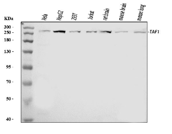
 FCM/FACS (Flow Cytometry)
(Figure 2. Flow Cytometry analysis of THP-1 cells using anti-TAF1 antibody (AAA126204).Overlay histogram showing THP-1 cells stained with AAA126204 (Blue line). The cells were blocked with 10% normal goat serum. And then incubated with rabbit anti-TAF1 Antibody (AAA126204, 1 ug/1x10^6 cells) for 30 min at 20 degree C. DyLight488 conjugated goat anti-rabbit IgG was used as secondary antibody for 30 minutes at 20 degree C. Isotype control antibody (Green line) was rabbit IgG (1 ug/1x10^6) used under the same conditions. Unlabelled sample (Red line) was also used as a control.)
FCM/FACS (Flow Cytometry)
(Figure 2. Flow Cytometry analysis of THP-1 cells using anti-TAF1 antibody (AAA126204).Overlay histogram showing THP-1 cells stained with AAA126204 (Blue line). The cells were blocked with 10% normal goat serum. And then incubated with rabbit anti-TAF1 Antibody (AAA126204, 1 ug/1x10^6 cells) for 30 min at 20 degree C. DyLight488 conjugated goat anti-rabbit IgG was used as secondary antibody for 30 minutes at 20 degree C. Isotype control antibody (Green line) was rabbit IgG (1 ug/1x10^6) used under the same conditions. Unlabelled sample (Red line) was also used as a control.)
TAF1, Polyclonal Antibody (Cat# AAA126204)
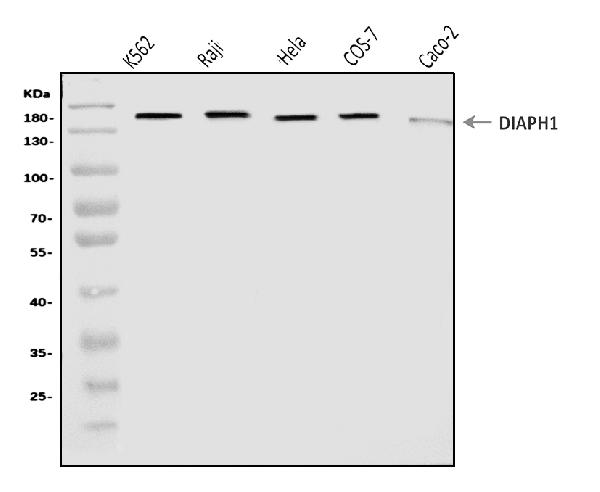
 FCM/FACS (Flow Cytometry)
(Figure 3. Flow Cytometry analysis of K562 cells using anti-DIAPH1 antibody (AAA126212).Overlay histogram showing K562 cells stained with AAA126212 (Blue line). The cells were blocked with 10% normal goat serum. And then incubated with rabbit anti-DIAPH1 Antibody (AAA126212, 1 ug/1x10^6 cells) for 30 min at 20 degree C. DyLight488 conjugated goat anti-rabbit IgG was used as secondary antibody for 30 minutes at 20 degree C. Isotype control antibody (Green line) was rabbit IgG (1 ug/1x10^6) used under the same conditions. Unlabelled sample (Red line) was also used as a control.)
FCM/FACS (Flow Cytometry)
(Figure 3. Flow Cytometry analysis of K562 cells using anti-DIAPH1 antibody (AAA126212).Overlay histogram showing K562 cells stained with AAA126212 (Blue line). The cells were blocked with 10% normal goat serum. And then incubated with rabbit anti-DIAPH1 Antibody (AAA126212, 1 ug/1x10^6 cells) for 30 min at 20 degree C. DyLight488 conjugated goat anti-rabbit IgG was used as secondary antibody for 30 minutes at 20 degree C. Isotype control antibody (Green line) was rabbit IgG (1 ug/1x10^6) used under the same conditions. Unlabelled sample (Red line) was also used as a control.)
DIAPH1, Polyclonal Antibody (Cat# AAA126212)
 FCM/FACS (Flow Cytometry)
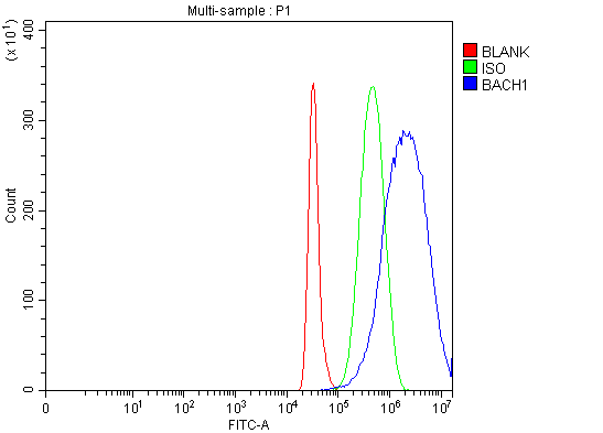
(Figure 2. Flow Cytometry analysis of HepG2 cells using anti-BACH1.3/BACH1 antibody (AAA126217).Overlay histogram showing HepG2 cells stained with AAA126217 (Blue line). The cells were blocked with 10% normal goat serum. And then incubated with rabbit anti-BACH1.3/BACH1 Antibody (AAA126217, 1 ug/1x10^6 cells) for 30 min at 20 degree C. DyLight488 conjugated goat anti-rabbit IgG was used as secondary antibody for 30 minutes at 20 degree C. Isotype control antibody (Green line) was rabbit IgG (1 ug/1x10^6) used under the same conditions. Unlabelled sample (Red line) was also used as a control.)
FCM/FACS (Flow Cytometry)
(Figure 2. Flow Cytometry analysis of HepG2 cells using anti-BACH1.3/BACH1 antibody (AAA126217).Overlay histogram showing HepG2 cells stained with AAA126217 (Blue line). The cells were blocked with 10% normal goat serum. And then incubated with rabbit anti-BACH1.3/BACH1 Antibody (AAA126217, 1 ug/1x10^6 cells) for 30 min at 20 degree C. DyLight488 conjugated goat anti-rabbit IgG was used as secondary antibody for 30 minutes at 20 degree C. Isotype control antibody (Green line) was rabbit IgG (1 ug/1x10^6) used under the same conditions. Unlabelled sample (Red line) was also used as a control.)
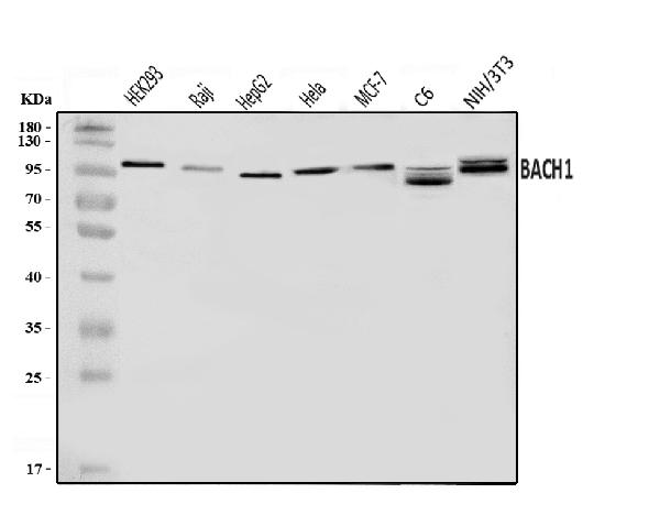
BACH1.3/BACH1, Polyclonal Antibody (Cat# AAA126217)
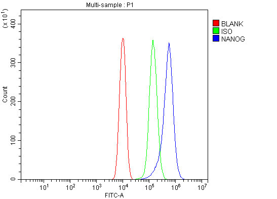 FCM/FACS (Flow Cytometry)
(Figure 3. Flow Cytometry analysis of RAW264.7 cells using anti-Nanog antibody (AAA125966).Overlay histogram showing RAW264.7 cells stained with AAA125966 (Blue line). The cells were blocked with 10% normal goat serum. And then incubated with rabbit anti-Nanog Antibody (AAA125966, 1 ug/1x10^6 cells) for 30 min at 20 degree C. DyLight488 conjugated goat anti-rabbit IgG was used as secondary antibody for 30 minutes at 20 degree C. Isotype control antibody (Green line) was rabbit IgG (1 ug/1x10^6) used under the same conditions. Unlabelled sample (Red line) was also used as a control.)
FCM/FACS (Flow Cytometry)
(Figure 3. Flow Cytometry analysis of RAW264.7 cells using anti-Nanog antibody (AAA125966).Overlay histogram showing RAW264.7 cells stained with AAA125966 (Blue line). The cells were blocked with 10% normal goat serum. And then incubated with rabbit anti-Nanog Antibody (AAA125966, 1 ug/1x10^6 cells) for 30 min at 20 degree C. DyLight488 conjugated goat anti-rabbit IgG was used as secondary antibody for 30 minutes at 20 degree C. Isotype control antibody (Green line) was rabbit IgG (1 ug/1x10^6) used under the same conditions. Unlabelled sample (Red line) was also used as a control.)
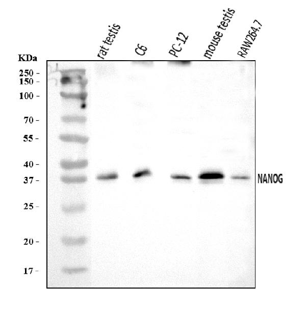
Nanog, Polyclonal Antibody (Cat# AAA125966)
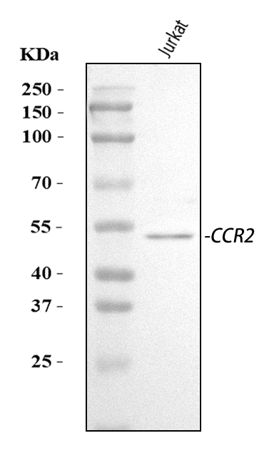
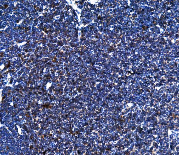
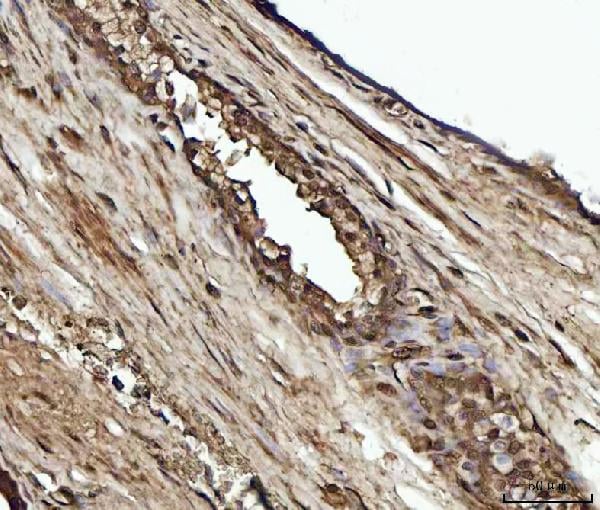
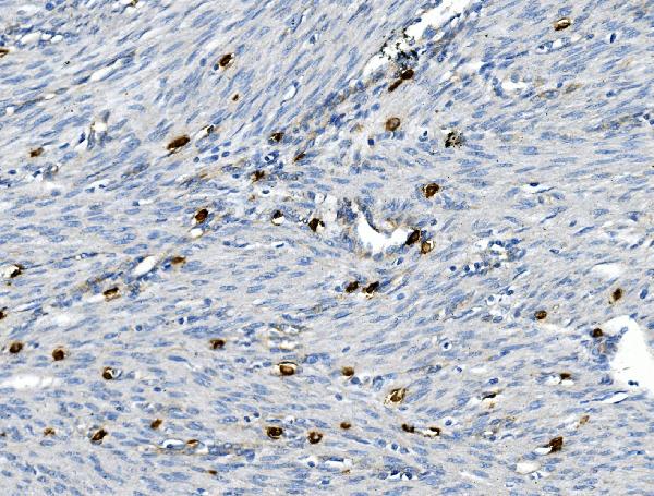
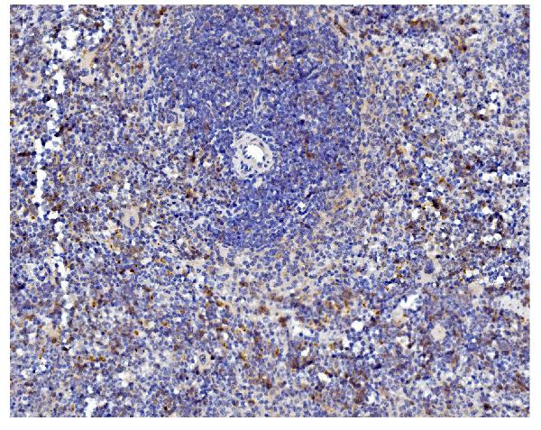
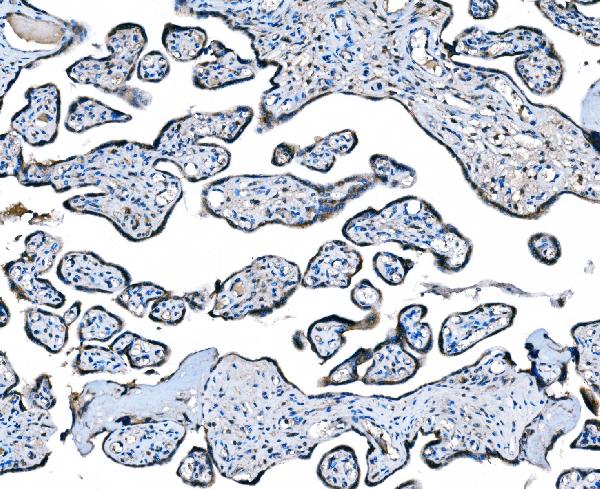 IHC (Immunohistochemistry)
(Figure 4. IHC analysis of CCR2 using anti-CCR2 antibody (AAA125969).CCR2 was detected in a paraffin-embedded section of human placenta tissue. Heat mediated antigen retrieval was performed in EDTA buffer (pH 8.0, epitope retrieval solution). The tissue section was blocked with 10% goat serum. The tissue section was then incubated with 2 ug/ml rabbit anti-CCR2 Antibody (AAA125969) overnight at 4 degree C. Peroxidase Conjugated Goat Anti-rabbit IgG was used as secondary antibody and incubated for 30 minutes at 37 degree C. The tissue section was developed using HRP Conjugated Rabbit IgG Super Vision Assay Kit with DAB as the chromogen.)
IHC (Immunohistochemistry)
(Figure 4. IHC analysis of CCR2 using anti-CCR2 antibody (AAA125969).CCR2 was detected in a paraffin-embedded section of human placenta tissue. Heat mediated antigen retrieval was performed in EDTA buffer (pH 8.0, epitope retrieval solution). The tissue section was blocked with 10% goat serum. The tissue section was then incubated with 2 ug/ml rabbit anti-CCR2 Antibody (AAA125969) overnight at 4 degree C. Peroxidase Conjugated Goat Anti-rabbit IgG was used as secondary antibody and incubated for 30 minutes at 37 degree C. The tissue section was developed using HRP Conjugated Rabbit IgG Super Vision Assay Kit with DAB as the chromogen.)
CCR2, Polyclonal Antibody (Cat# AAA125969)
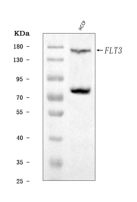
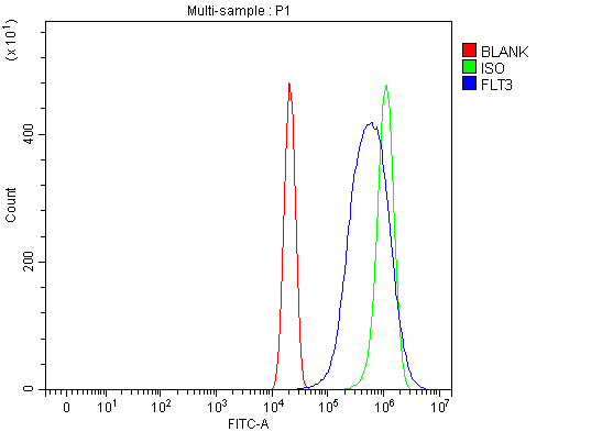 FCM/FACS (Flow Cytometry)
(Figure 3. Flow Cytometry analysis of U2OS cells using anti-CD135/FLT3 antibody (AAA125976).Overlay histogram showing U2OS cells stained with AAA125976 (Blue line). The cells were blocked with 10% normal goat serum. And then incubated with rabbit anti-CD135/FLT3 Antibody (AAA125976, 1 ug/1x10^6 cells) for 30 min at 20 degree C. DyLight488 conjugated goat anti-rabbit IgG was used as secondary antibody for 30 minutes at 20 degree C. Isotype control antibody (Green line) was rabbit IgG (1 ug/1x10^6) used under the same conditions. Unlabelled sample (Red line) was also used as a control.)
FCM/FACS (Flow Cytometry)
(Figure 3. Flow Cytometry analysis of U2OS cells using anti-CD135/FLT3 antibody (AAA125976).Overlay histogram showing U2OS cells stained with AAA125976 (Blue line). The cells were blocked with 10% normal goat serum. And then incubated with rabbit anti-CD135/FLT3 Antibody (AAA125976, 1 ug/1x10^6 cells) for 30 min at 20 degree C. DyLight488 conjugated goat anti-rabbit IgG was used as secondary antibody for 30 minutes at 20 degree C. Isotype control antibody (Green line) was rabbit IgG (1 ug/1x10^6) used under the same conditions. Unlabelled sample (Red line) was also used as a control.)
CD135/FLT3, Polyclonal Antibody (Cat# AAA125976)
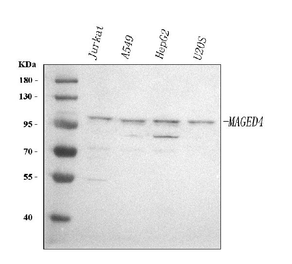
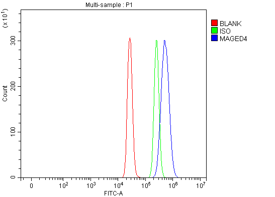 FCM/FACS (Flow Cytometry)
(Figure 5. Flow Cytometry analysis of HepG2 cells using anti-MAGED4 antibody (AAA127895).Overlay histogram showing HepG2 cells stained with AAA127895 (Blue line). To facilitate intracellular staining, cells were fixed with 4% paraformaldehyde and permeabilized with permeabilization buffer. The cells were blocked with 10% normal goat serum. And then incubated with rabbit anti-MAGED4 Antibody (AAA127895, 1ug/1x106 cells) for 30 min at 20 degree C. DyLight488 conjugated goat anti-rabbit IgG was used as secondary antibody for 30 minutes at 20 degree C. Isotype control antibody (Green line) was rabbit IgG (1ug/1x106) used under the same conditions. Unlabelled sample (Red line) was also used as a control.)
FCM/FACS (Flow Cytometry)
(Figure 5. Flow Cytometry analysis of HepG2 cells using anti-MAGED4 antibody (AAA127895).Overlay histogram showing HepG2 cells stained with AAA127895 (Blue line). To facilitate intracellular staining, cells were fixed with 4% paraformaldehyde and permeabilized with permeabilization buffer. The cells were blocked with 10% normal goat serum. And then incubated with rabbit anti-MAGED4 Antibody (AAA127895, 1ug/1x106 cells) for 30 min at 20 degree C. DyLight488 conjugated goat anti-rabbit IgG was used as secondary antibody for 30 minutes at 20 degree C. Isotype control antibody (Green line) was rabbit IgG (1ug/1x106) used under the same conditions. Unlabelled sample (Red line) was also used as a control.)
MAGED4, Polyclonal Antibody (Cat# AAA127895)
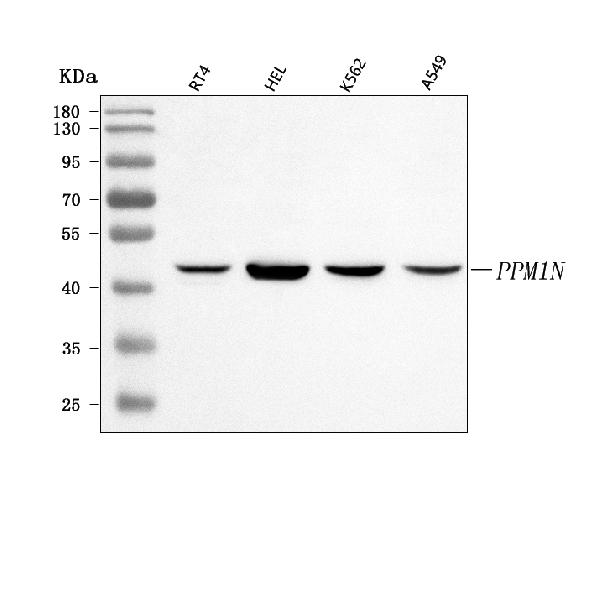
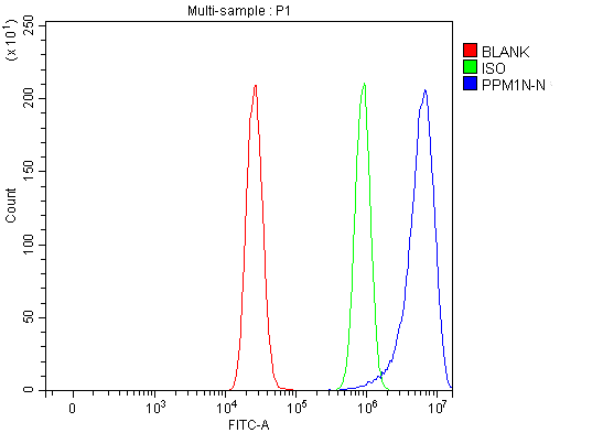 FCM/FACS (Flow Cytometry)
(Figure 3. Flow Cytometry analysis of MCF-7 cells using anti-PPM1N antibody (AAA127902).Overlay histogram showing MCF-7 cells stained with AAA127902 (Blue line). To facilitate intracellular staining, cells were fixed with 4% paraformaldehyde and permeabilized with permeabilization buffer. The cells were blocked with 10% normal goat serum. And then incubated with rabbit anti-PPM1N Antibody (AAA127902, 1ug/1x106 cells) for 30 min at 20 degree C. DyLight488 conjugated goat anti-rabbit IgG was used as secondary antibody for 30 minutes at 20 degree C. Isotype control antibody (Green line) was rabbit IgG (1ug/1x106) used under the same conditions. Unlabelled sample (Red line) was also used as a control.)
FCM/FACS (Flow Cytometry)
(Figure 3. Flow Cytometry analysis of MCF-7 cells using anti-PPM1N antibody (AAA127902).Overlay histogram showing MCF-7 cells stained with AAA127902 (Blue line). To facilitate intracellular staining, cells were fixed with 4% paraformaldehyde and permeabilized with permeabilization buffer. The cells were blocked with 10% normal goat serum. And then incubated with rabbit anti-PPM1N Antibody (AAA127902, 1ug/1x106 cells) for 30 min at 20 degree C. DyLight488 conjugated goat anti-rabbit IgG was used as secondary antibody for 30 minutes at 20 degree C. Isotype control antibody (Green line) was rabbit IgG (1ug/1x106) used under the same conditions. Unlabelled sample (Red line) was also used as a control.)
PPM1N, Polyclonal Antibody (Cat# AAA127902)
 FCM/FACS (Flow Cytometry)
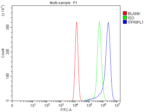
(Figure 3. Flow Cytometry analysis of HEL cells using anti-ITPRIPL1 antibody (AAA127911).Overlay histogram showing HEL cells stained with AAA127911 (Blue line). To facilitate intracellular staining, cells were fixed with 4% paraformaldehyde and permeabilized with permeabilization buffer. The cells were blocked with 10% normal goat serum. And then incubated with rabbit anti-ITPRIPL1 Antibody (AAA127911, 1ug/1x106 cells) for 30 min at 20 degree C. DyLight488 conjugated goat anti-rabbit IgG was used as secondary antibody for 30 minutes at 20 degree C. Isotype control antibody (Green line) was rabbit IgG (1ug/1x106) used under the same conditions. Unlabelled sample (Red line) was also used as a control.)
FCM/FACS (Flow Cytometry)
(Figure 3. Flow Cytometry analysis of HEL cells using anti-ITPRIPL1 antibody (AAA127911).Overlay histogram showing HEL cells stained with AAA127911 (Blue line). To facilitate intracellular staining, cells were fixed with 4% paraformaldehyde and permeabilized with permeabilization buffer. The cells were blocked with 10% normal goat serum. And then incubated with rabbit anti-ITPRIPL1 Antibody (AAA127911, 1ug/1x106 cells) for 30 min at 20 degree C. DyLight488 conjugated goat anti-rabbit IgG was used as secondary antibody for 30 minutes at 20 degree C. Isotype control antibody (Green line) was rabbit IgG (1ug/1x106) used under the same conditions. Unlabelled sample (Red line) was also used as a control.)
ITPRIPL1, Polyclonal Antibody (Cat# AAA127911)
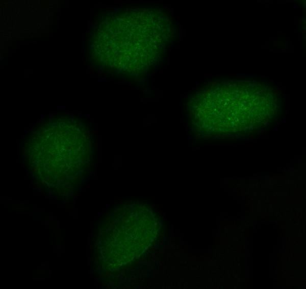
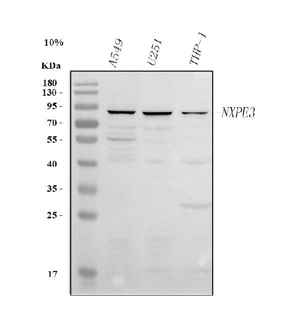
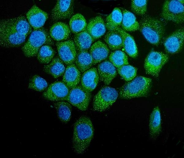
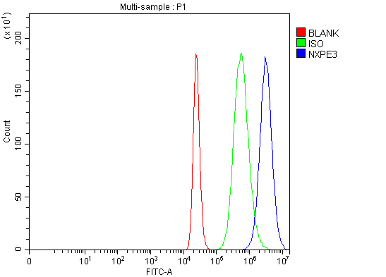 FCM/FACS (Flow Cytometry)
(Figure 3. Flow Cytometry analysis of THP-1 cells using anti-NXPE3 antibody (AAA127918).Overlay histogram showing THP-1 cells stained with AAA127918 (Blue line). The cells were fixed with 4% paraformaldehyde and blocked with 10% normal goat serum. And then incubated with rabbit anti-NXPE3 Antibody (AAA127918, 1ug/1x106 cells) for 30 min at 20 degree C. DyLight488 conjugated goat anti-rabbit IgG was used as secondary antibody for 30 minutes at 20 degree C. Isotype control antibody (Green line) was rabbit IgG (1ug/1x106) used under the same conditions. Unlabelled sample (Red line) was also used as a control.)
FCM/FACS (Flow Cytometry)
(Figure 3. Flow Cytometry analysis of THP-1 cells using anti-NXPE3 antibody (AAA127918).Overlay histogram showing THP-1 cells stained with AAA127918 (Blue line). The cells were fixed with 4% paraformaldehyde and blocked with 10% normal goat serum. And then incubated with rabbit anti-NXPE3 Antibody (AAA127918, 1ug/1x106 cells) for 30 min at 20 degree C. DyLight488 conjugated goat anti-rabbit IgG was used as secondary antibody for 30 minutes at 20 degree C. Isotype control antibody (Green line) was rabbit IgG (1ug/1x106) used under the same conditions. Unlabelled sample (Red line) was also used as a control.)
NXPE3, Polyclonal Antibody (Cat# AAA127918)
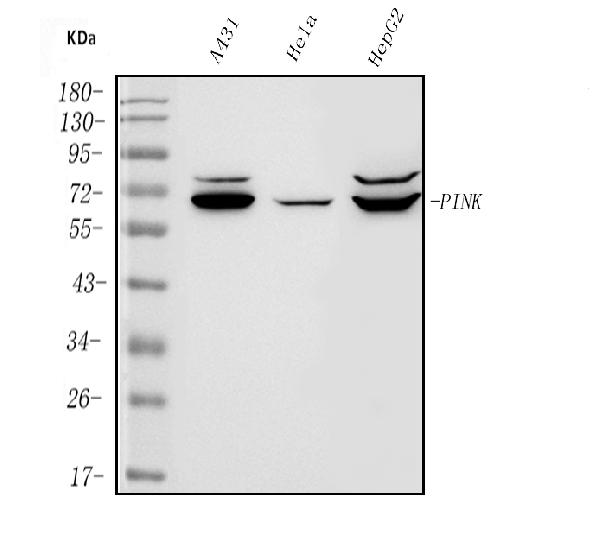
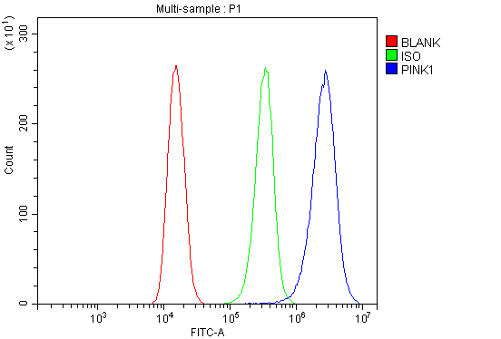 FCM/FACS (Flow Cytometry)
(Figure 3. Flow Cytometry analysis of 293T cells using anti-PINK1 antibody (AAA125528).Overlay histogram showing 293T cells stained with AAA125528 (Blue line). The cells were blocked with 10% normal goat serum. And then incubated with rabbit anti-PINK1 Antibody (AAA125528, 1μg/1x106 cells) for 30 min at 20 degree C. DyLight®488 conjugated goat anti-rabbit IgG (5-10μg/1x106 cells) was used as secondary antibody for 30 minutes at 20 degree C. Isotype control antibody (Green line) was rabbit IgG (1μg/1x106) used under the same conditions. Unlabelled sample (Red line) was also used as a control.)
FCM/FACS (Flow Cytometry)
(Figure 3. Flow Cytometry analysis of 293T cells using anti-PINK1 antibody (AAA125528).Overlay histogram showing 293T cells stained with AAA125528 (Blue line). The cells were blocked with 10% normal goat serum. And then incubated with rabbit anti-PINK1 Antibody (AAA125528, 1μg/1x106 cells) for 30 min at 20 degree C. DyLight®488 conjugated goat anti-rabbit IgG (5-10μg/1x106 cells) was used as secondary antibody for 30 minutes at 20 degree C. Isotype control antibody (Green line) was rabbit IgG (1μg/1x106) used under the same conditions. Unlabelled sample (Red line) was also used as a control.)
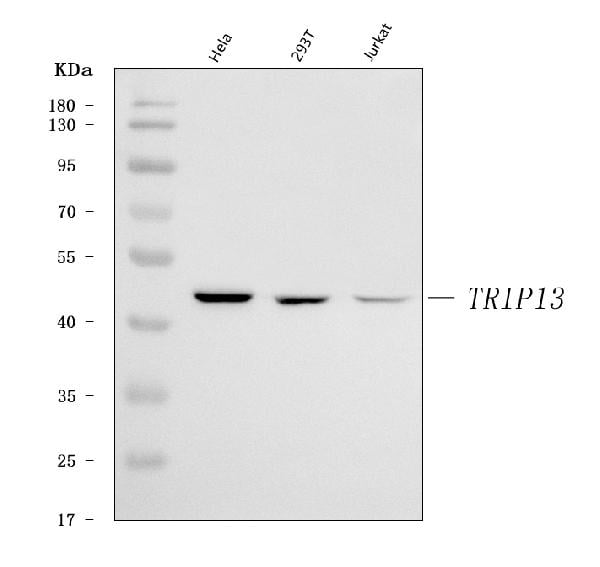
PINK1, Polyclonal Antibody (Cat# AAA125528)
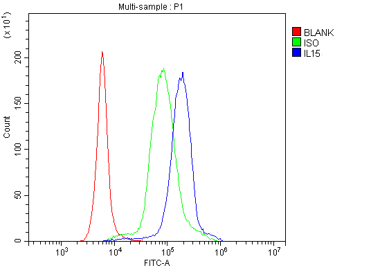 FCM/FACS (Flow Cytometry)
(Figure 3. Flow Cytometry analysis of human PBMC cells using anti-IL15 antibody (AAA125529).Overlay histogram showing human PBMC cells stained with AAA125529 (Blue line). The cells were blocked with 10% normal goat serum. And then incubated with rabbit anti-IL15 Antibody (AAA125529, 1μg/1x106 cells) for 30 min at 20 degree C. DyLight®488 conjugated goat anti-rabbit IgG (5-10μg/1x106 cells) was used as secondary antibody for 30 minutes at 20 degree C. Isotype control antibody (Green line) was rabbit IgG (1μg/1x106) used under the same conditions. Unlabelled sample (Red line) was also used as a control.)
FCM/FACS (Flow Cytometry)
(Figure 3. Flow Cytometry analysis of human PBMC cells using anti-IL15 antibody (AAA125529).Overlay histogram showing human PBMC cells stained with AAA125529 (Blue line). The cells were blocked with 10% normal goat serum. And then incubated with rabbit anti-IL15 Antibody (AAA125529, 1μg/1x106 cells) for 30 min at 20 degree C. DyLight®488 conjugated goat anti-rabbit IgG (5-10μg/1x106 cells) was used as secondary antibody for 30 minutes at 20 degree C. Isotype control antibody (Green line) was rabbit IgG (1μg/1x106) used under the same conditions. Unlabelled sample (Red line) was also used as a control.)
IL15, Polyclonal Antibody (Cat# AAA125529)
 FCM/FACS (Flow Cytometry)
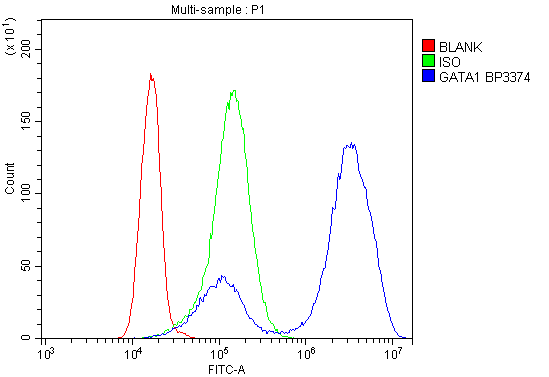
(Figure 2. Flow Cytometry analysis of K562 cells using anti- GATA1 antibody (AAA125545).Overlay histogram showing K562 cells stained with AAA125545 (Blue line). The cells were blocked with 10% normal goat serum. And then incubated with rabbit anti- GATA1 Antibody (AAA125545, 1μg/1x106 cells) for 30 min at 20 degree C. DyLight®488 conjugated goat anti-rabbit IgG (5-10μg/1x106 cells) was used as secondary antibody for 30 minutes at 20 degree C. Isotype control antibody (Green line) was rabbit IgG (1μg/1x106) used under the same conditions. Unlabelled sample (Red line) was also used as a control.)
FCM/FACS (Flow Cytometry)
(Figure 2. Flow Cytometry analysis of K562 cells using anti- GATA1 antibody (AAA125545).Overlay histogram showing K562 cells stained with AAA125545 (Blue line). The cells were blocked with 10% normal goat serum. And then incubated with rabbit anti- GATA1 Antibody (AAA125545, 1μg/1x106 cells) for 30 min at 20 degree C. DyLight®488 conjugated goat anti-rabbit IgG (5-10μg/1x106 cells) was used as secondary antibody for 30 minutes at 20 degree C. Isotype control antibody (Green line) was rabbit IgG (1μg/1x106) used under the same conditions. Unlabelled sample (Red line) was also used as a control.)
GATA1, Polyclonal Antibody (Cat# AAA125545)
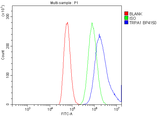 FCM/FACS (Flow Cytometry)
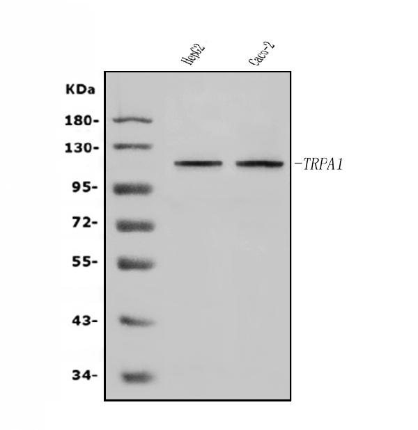
(Figure 2. Flow Cytometry analysis of A549 cells using anti-TRPA1/TSA antibody (AAA125546).Overlay histogram showing A549 cells stained with AAA125546 (Blue line). The cells were blocked with 10% normal goat serum. And then incubated with rabbit anti-TRPA1/TSA Antibody (AAA125546, 1μg/1x106 cells) for 30 min at 20 degree C. DyLight®488 conjugated goat anti-rabbit IgG (5-10μg/1x106 cells) was used as secondary antibody for 30 minutes at 20 degree C. Isotype control antibody (Green line) was rabbit IgG (1μg/1x106) used under the same conditions. Unlabelled sample (Red line) was also used as a control.)
FCM/FACS (Flow Cytometry)
(Figure 2. Flow Cytometry analysis of A549 cells using anti-TRPA1/TSA antibody (AAA125546).Overlay histogram showing A549 cells stained with AAA125546 (Blue line). The cells were blocked with 10% normal goat serum. And then incubated with rabbit anti-TRPA1/TSA Antibody (AAA125546, 1μg/1x106 cells) for 30 min at 20 degree C. DyLight®488 conjugated goat anti-rabbit IgG (5-10μg/1x106 cells) was used as secondary antibody for 30 minutes at 20 degree C. Isotype control antibody (Green line) was rabbit IgG (1μg/1x106) used under the same conditions. Unlabelled sample (Red line) was also used as a control.)
TRPA1/TSA, Polyclonal Antibody (Cat# AAA125546)
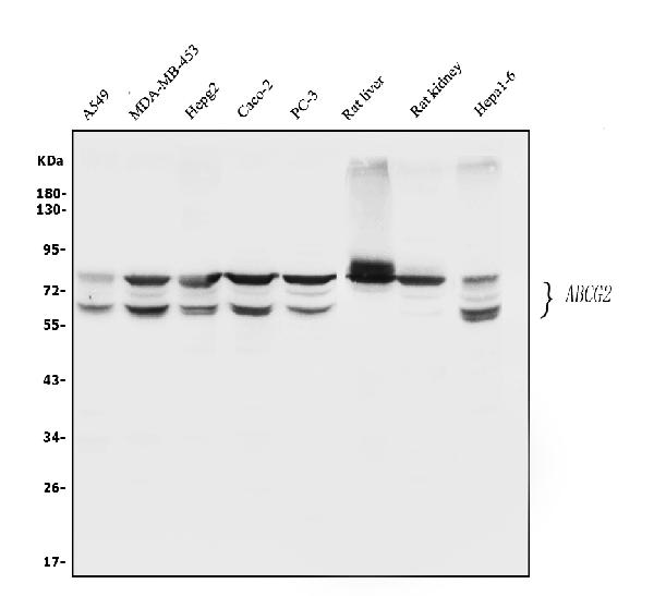
 FCM/FACS (Flow Cytometry)
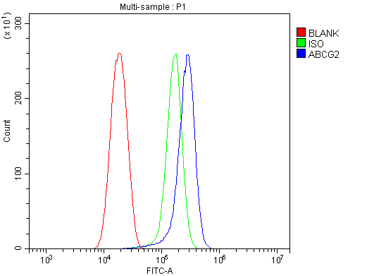
(Figure 4. Flow Cytometry analysis of SiHa cells using anti-BCRP/ABCG2 antibody (AAA125549).Overlay histogram showing SiHa cells stained with AAA125549 (Blue line). The cells were blocked with 10% normal goat serum. And then incubated with rabbit anti-BCRP/ABCG2 Antibody (AAA125549,1μg/1x106 cells) for 30 min at 20 degree C. DyLight®488 conjugated goat anti-rabbit IgG (5-10μg/1x106 cells) was used as secondary antibody for 30 minutes at 20 degree C. Isotype control antibody (Green line) was rabbit IgG (1μg/1x106) used under the same conditions. Unlabelled sample (Red line) was also used as a control.)
FCM/FACS (Flow Cytometry)
(Figure 4. Flow Cytometry analysis of SiHa cells using anti-BCRP/ABCG2 antibody (AAA125549).Overlay histogram showing SiHa cells stained with AAA125549 (Blue line). The cells were blocked with 10% normal goat serum. And then incubated with rabbit anti-BCRP/ABCG2 Antibody (AAA125549,1μg/1x106 cells) for 30 min at 20 degree C. DyLight®488 conjugated goat anti-rabbit IgG (5-10μg/1x106 cells) was used as secondary antibody for 30 minutes at 20 degree C. Isotype control antibody (Green line) was rabbit IgG (1μg/1x106) used under the same conditions. Unlabelled sample (Red line) was also used as a control.)
BCRP/ABCG2, Polyclonal Antibody (Cat# AAA125549)
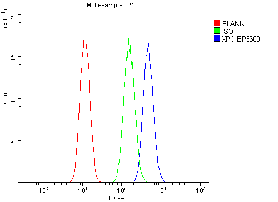
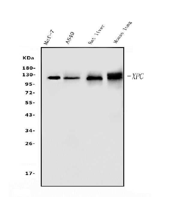
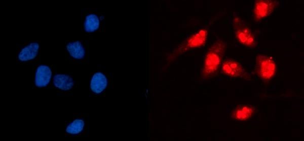
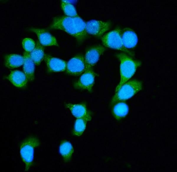
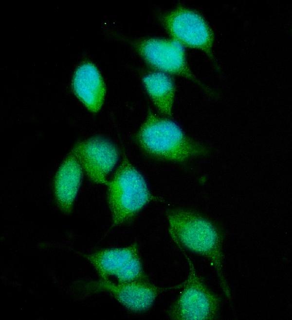 IF (Immunofluorescence)
(Figure 3. IF analysis of XPC using anti- XPC antibody (AAA125551).XPC was detected in immunocytochemical section of HELA cells. Enzyme antigen retrieval was performed using IHC enzyme antigen retrieval reagent for 15 mins. The cells were blocked with 10% goat serum. And then incubated with 5μg/mL rabbit anti-XPC Antibody (AAA125551) overnight at 4 degree C. DyLight®488 Conjugated Goat Anti-Rabbit IgG was used as secondary antibody at 1:100 dilution and incubated for 30 minutes at 37 degree C. The section was counterstained with DAPI. Visualize using a fluorescence microscope and filter sets appropriate for the label used.)
IF (Immunofluorescence)
(Figure 3. IF analysis of XPC using anti- XPC antibody (AAA125551).XPC was detected in immunocytochemical section of HELA cells. Enzyme antigen retrieval was performed using IHC enzyme antigen retrieval reagent for 15 mins. The cells were blocked with 10% goat serum. And then incubated with 5μg/mL rabbit anti-XPC Antibody (AAA125551) overnight at 4 degree C. DyLight®488 Conjugated Goat Anti-Rabbit IgG was used as secondary antibody at 1:100 dilution and incubated for 30 minutes at 37 degree C. The section was counterstained with DAPI. Visualize using a fluorescence microscope and filter sets appropriate for the label used.)
XPC, Polyclonal Antibody (Cat# AAA125551)
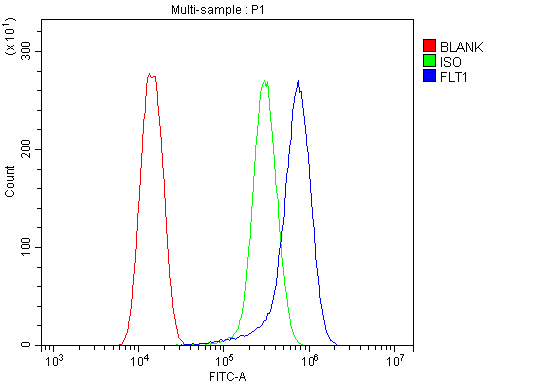 FCM/FACS (Flow Cytometry)
(Figure 3. Flow Cytometry analysis of U20S cells using anti-FLT1 antibody (AAA125557).Overlay histogram showing U20S cells stained with AAA125557 (Blue line). The cells were blocked with 10% normal goat serum. And then incubated with rabbit anti-FLT1 Antibody (AAA125557, 1μg/1x106 cells) for 30 min at 20 degree C. DyLight®488 conjugated goat anti-rabbit IgG (5-10μg/1x106 cells) was used as secondary antibody for 30 minutes at 20 degree C. Isotype control antibody (Green line) was rabbit IgG (1μg/1x106) used under the same conditions. Unlabelled sample (Red line) was also used as a control.)
FCM/FACS (Flow Cytometry)
(Figure 3. Flow Cytometry analysis of U20S cells using anti-FLT1 antibody (AAA125557).Overlay histogram showing U20S cells stained with AAA125557 (Blue line). The cells were blocked with 10% normal goat serum. And then incubated with rabbit anti-FLT1 Antibody (AAA125557, 1μg/1x106 cells) for 30 min at 20 degree C. DyLight®488 conjugated goat anti-rabbit IgG (5-10μg/1x106 cells) was used as secondary antibody for 30 minutes at 20 degree C. Isotype control antibody (Green line) was rabbit IgG (1μg/1x106) used under the same conditions. Unlabelled sample (Red line) was also used as a control.)
FLT1, Polyclonal Antibody (Cat# AAA125557)
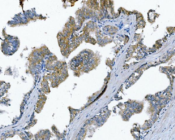
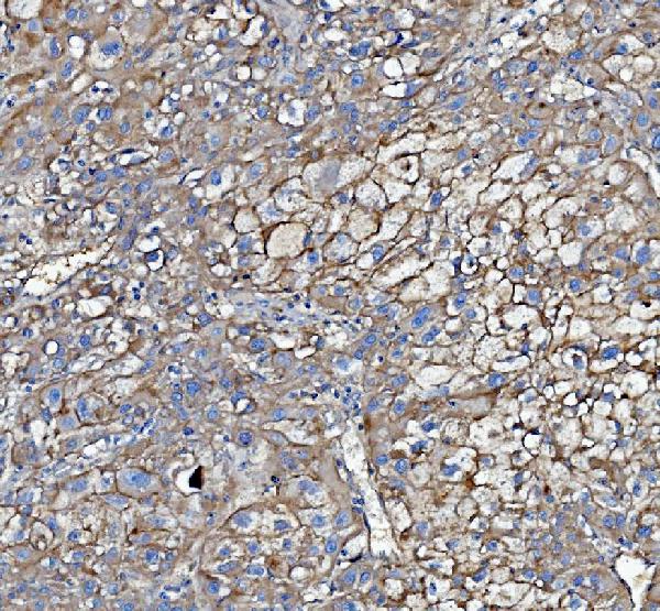
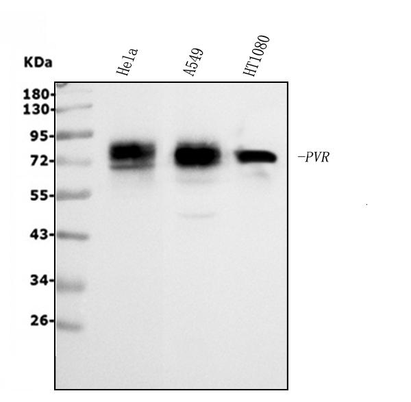
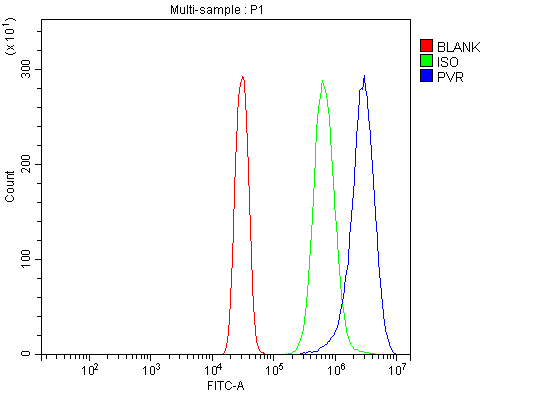 FCM/FACS (Flow Cytometry)
(Figure 5. Flow Cytometry analysis of SiHa cells using anti-Poliovirus Receptor/PVR antibody (AAA125567).Overlay histogram showing SiHa cells stained with AAA125567 (Blue line). The cells were blocked with 10% normal goat serum. And then incubated with rabbit anti-Poliovirus Receptor/PVR Antibody (AAA125567, 1μg/1x106 cells) for 30 min at 20 degree C. DyLight®488 conjugated goat anti-rabbit IgG (5-10μg/1x106 cells) was used as secondary antibody for 30 minutes at 20 degree C. Isotype control antibody (Green line) was rabbit IgG (1μg/1x106) used under the same conditions. Unlabelled sample (Red line) was also used as a control.)
FCM/FACS (Flow Cytometry)
(Figure 5. Flow Cytometry analysis of SiHa cells using anti-Poliovirus Receptor/PVR antibody (AAA125567).Overlay histogram showing SiHa cells stained with AAA125567 (Blue line). The cells were blocked with 10% normal goat serum. And then incubated with rabbit anti-Poliovirus Receptor/PVR Antibody (AAA125567, 1μg/1x106 cells) for 30 min at 20 degree C. DyLight®488 conjugated goat anti-rabbit IgG (5-10μg/1x106 cells) was used as secondary antibody for 30 minutes at 20 degree C. Isotype control antibody (Green line) was rabbit IgG (1μg/1x106) used under the same conditions. Unlabelled sample (Red line) was also used as a control.)
Poliovirus Receptor/PVR, Polyclonal Antibody (Cat# AAA125567)
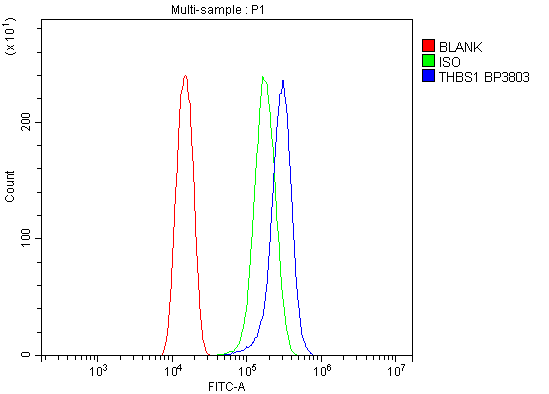 FCM/FACS (Flow Cytometry)
(Figure 2. Flow Cytometry analysis of U20S cells using anti-Thrombospondin/THBS1 antibody (AAA125568).Overlay histogram showing U20S cells stained with AAA125568 (Blue line). The cells were blocked with 10% normal goat serum. And then incubated with rabbit anti-Thrombospondin/THBS1 Antibody (AAA125568, 1μg/1x106 cells) for 30 min at 20 degree C. DyLight®488 conjugated goat anti-rabbit IgG (5-10μg/1x106 cells) was used as secondary antibody for 30 minutes at 20 degree C. Isotype control antibody (Green line) was rabbit IgG (1μg/1x106) used under the same conditions. Unlabelled sample (Red line) was also used as a control.)
FCM/FACS (Flow Cytometry)
(Figure 2. Flow Cytometry analysis of U20S cells using anti-Thrombospondin/THBS1 antibody (AAA125568).Overlay histogram showing U20S cells stained with AAA125568 (Blue line). The cells were blocked with 10% normal goat serum. And then incubated with rabbit anti-Thrombospondin/THBS1 Antibody (AAA125568, 1μg/1x106 cells) for 30 min at 20 degree C. DyLight®488 conjugated goat anti-rabbit IgG (5-10μg/1x106 cells) was used as secondary antibody for 30 minutes at 20 degree C. Isotype control antibody (Green line) was rabbit IgG (1μg/1x106) used under the same conditions. Unlabelled sample (Red line) was also used as a control.)
Thrombospondin/THBS1, Polyclonal Antibody (Cat# AAA125568)
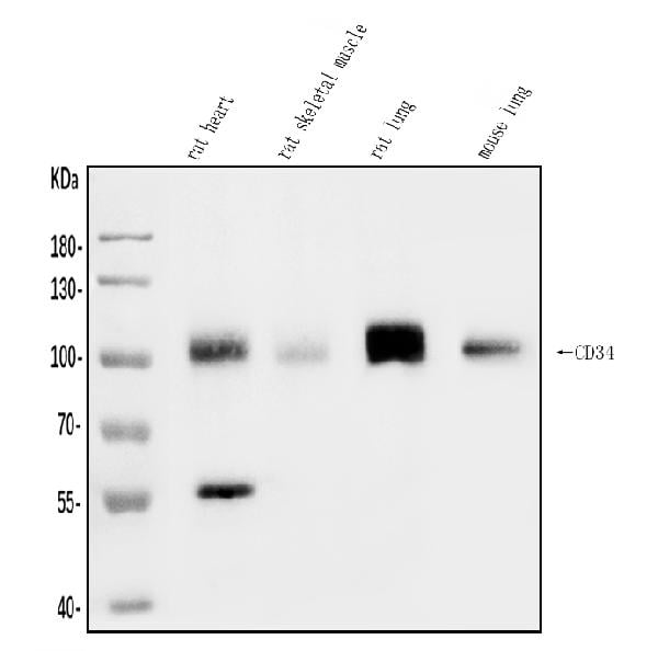
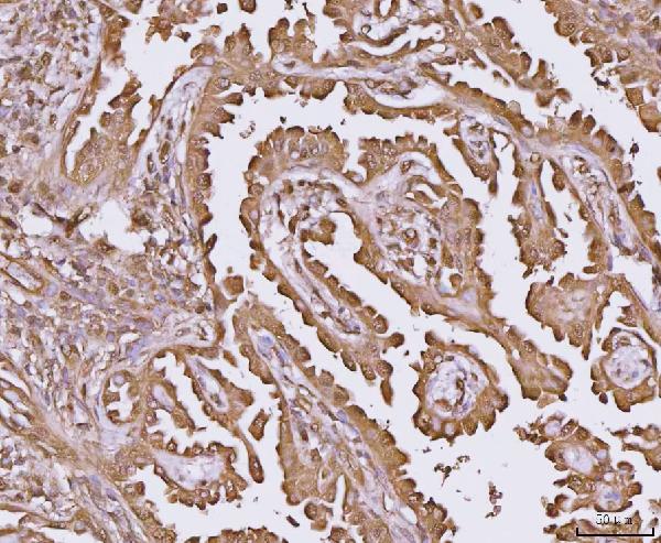
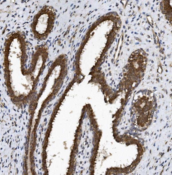
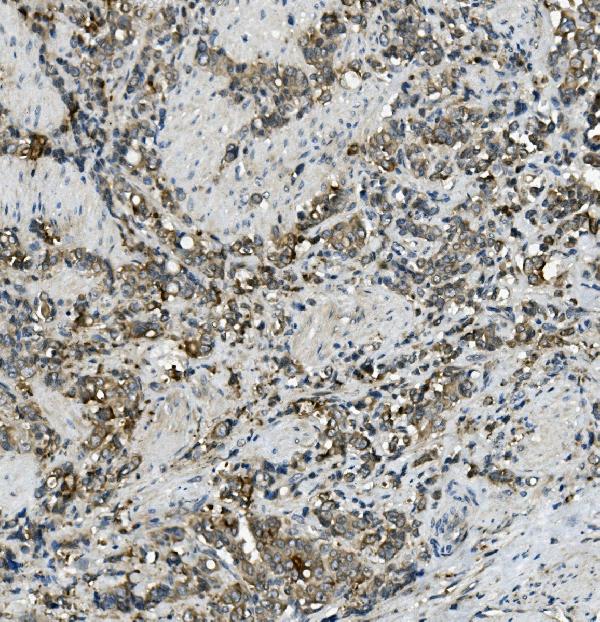
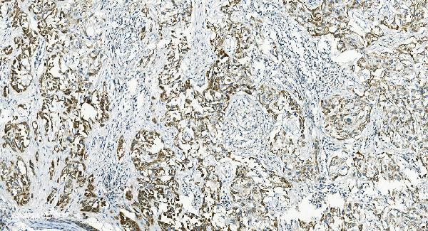
 IHC (Immunohistochemisry)
(Figure 3. IHC analysis of CD34 using anti-CD34 antibody (AAA125577).CD34 was detected in paraffin-embedded section of rat kidney tissue. Heat mediated antigen retrieval was performed in EDTA buffer (pH8. 0, epitope retrieval solution). The tissue section was blocked with 10% goat serum. The tissue section was then incubated with 2μg/ml rabbit anti-CD34 Antibody (AAA125577) overnight at 4 degree C. Biotinylated goat anti-rabbit IgG was used as secondary antibody and incubated for 30 minutes at 37 degree C. The tissue section was developed using Strepavidin-Biotin-Complex (SABC) with DAB as the chromogen.)
IHC (Immunohistochemisry)
(Figure 3. IHC analysis of CD34 using anti-CD34 antibody (AAA125577).CD34 was detected in paraffin-embedded section of rat kidney tissue. Heat mediated antigen retrieval was performed in EDTA buffer (pH8. 0, epitope retrieval solution). The tissue section was blocked with 10% goat serum. The tissue section was then incubated with 2μg/ml rabbit anti-CD34 Antibody (AAA125577) overnight at 4 degree C. Biotinylated goat anti-rabbit IgG was used as secondary antibody and incubated for 30 minutes at 37 degree C. The tissue section was developed using Strepavidin-Biotin-Complex (SABC) with DAB as the chromogen.)
CD34, Polyclonal Antibody (Cat# AAA125577)
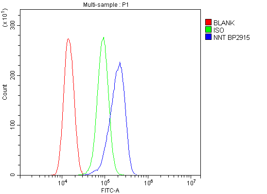 FCM/FACS (Flow Cytometry)
(Figure 3. Flow Cytometry analysis of THP-1 cells using anti-NNT antibody (AAA125578).Overlay histogram showing THP-1 cells stained with AAA125578 (Blue line). The cells were blocked with 10% normal goat serum. And then incubated with rabbit anti-NNT Antibody (AAA125578,1μg/1x106 cells) for 30 min at 20 degree C. DyLight®488 conjugated goat anti-rabbit IgG (5-10μg/1x106 cells) was used as secondary antibody for 30 minutes at 20 degree C. Isotype control antibody (Green line) was rabbit IgG (1μg/1x106) used under the same conditions. Unlabelled sample (Red line) was also used as a control.)
FCM/FACS (Flow Cytometry)
(Figure 3. Flow Cytometry analysis of THP-1 cells using anti-NNT antibody (AAA125578).Overlay histogram showing THP-1 cells stained with AAA125578 (Blue line). The cells were blocked with 10% normal goat serum. And then incubated with rabbit anti-NNT Antibody (AAA125578,1μg/1x106 cells) for 30 min at 20 degree C. DyLight®488 conjugated goat anti-rabbit IgG (5-10μg/1x106 cells) was used as secondary antibody for 30 minutes at 20 degree C. Isotype control antibody (Green line) was rabbit IgG (1μg/1x106) used under the same conditions. Unlabelled sample (Red line) was also used as a control.)
NNT, Polyclonal Antibody (Cat# AAA125578)
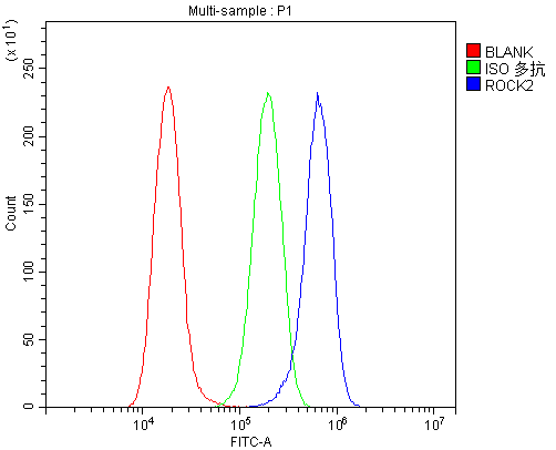
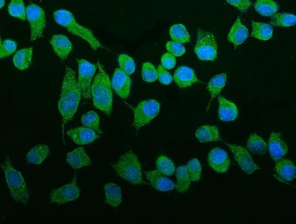 IF (Immunofluorescence)
(Figure 3. IF analysis of ROCK2 using anti- ROCK2 antibody (AAA125586).ROCK2 was detected in immunocytochemical section of Hep cells. Enzyme antigen retrieval was performed using IHC enzyme antigen retrieval reagent for 15 mins. The cells were blocked with 10% goat serum. And then incubated with 5μg/mL rabbit anti- ROCK2 Antibody (AAA125586) overnight at 4 degree C. DyLight®488 Conjugated Goat Anti-Rabbit IgG was used as secondary antibody at 1:100 dilution and incubated for 30 minutes at 37 degree C. The section was counterstained with DAPI. Visualize using a fluorescence microscope and filter sets appropriate for the label used.)
IF (Immunofluorescence)
(Figure 3. IF analysis of ROCK2 using anti- ROCK2 antibody (AAA125586).ROCK2 was detected in immunocytochemical section of Hep cells. Enzyme antigen retrieval was performed using IHC enzyme antigen retrieval reagent for 15 mins. The cells were blocked with 10% goat serum. And then incubated with 5μg/mL rabbit anti- ROCK2 Antibody (AAA125586) overnight at 4 degree C. DyLight®488 Conjugated Goat Anti-Rabbit IgG was used as secondary antibody at 1:100 dilution and incubated for 30 minutes at 37 degree C. The section was counterstained with DAPI. Visualize using a fluorescence microscope and filter sets appropriate for the label used.)
ROCK2, Polyclonal Antibody (Cat# AAA125586)
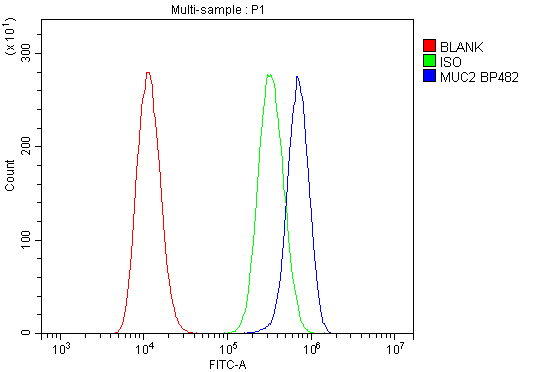 FCM/FACS (Flow Cytometry)
(Figure 3. Flow Cytometry analysis of U20S cells using anti-MUC2 antibody (AAA125594).Overlay histogram showing U20S cells stained with AAA125594 (Blue line). The cells were blocked with 10% normal goat serum. And then incubated with rabbit anti-MUC2 Antibody (AAA125594,1μg/1x106 cells) for 30 min at 20 degree C. DyLight®488 conjugated goat anti-rabbit IgG (5-10μg/1x106 cells) was used as secondary antibody for 30 minutes at 20 degree C. Isotype control antibody (Green line) was rabbit IgG (1μg/1x106) used under the same conditions. Unlabelled sample (Red line) was also used as a control.)
FCM/FACS (Flow Cytometry)
(Figure 3. Flow Cytometry analysis of U20S cells using anti-MUC2 antibody (AAA125594).Overlay histogram showing U20S cells stained with AAA125594 (Blue line). The cells were blocked with 10% normal goat serum. And then incubated with rabbit anti-MUC2 Antibody (AAA125594,1μg/1x106 cells) for 30 min at 20 degree C. DyLight®488 conjugated goat anti-rabbit IgG (5-10μg/1x106 cells) was used as secondary antibody for 30 minutes at 20 degree C. Isotype control antibody (Green line) was rabbit IgG (1μg/1x106) used under the same conditions. Unlabelled sample (Red line) was also used as a control.)
MUC2, Polyclonal Antibody (Cat# AAA125594)
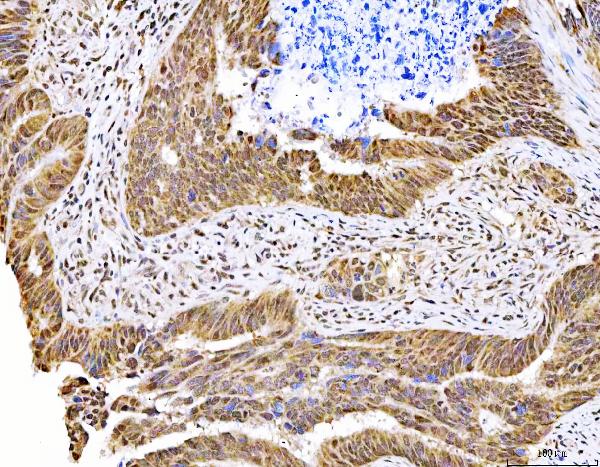
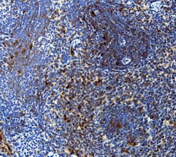
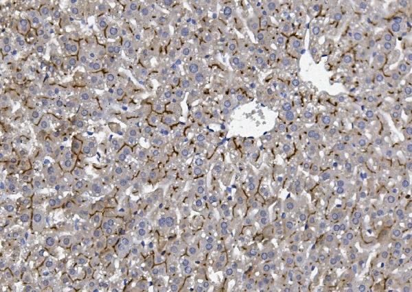
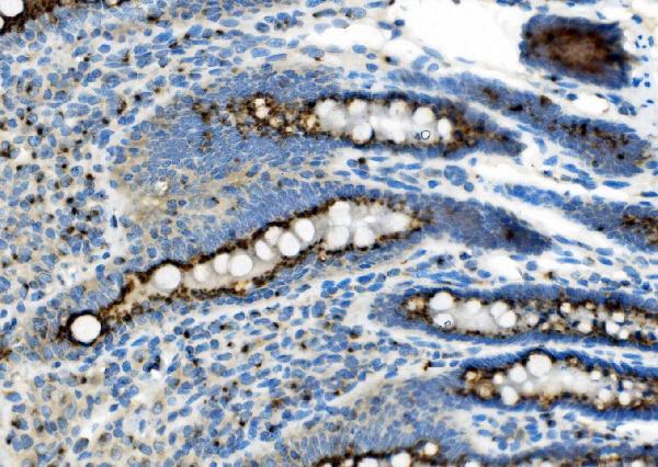
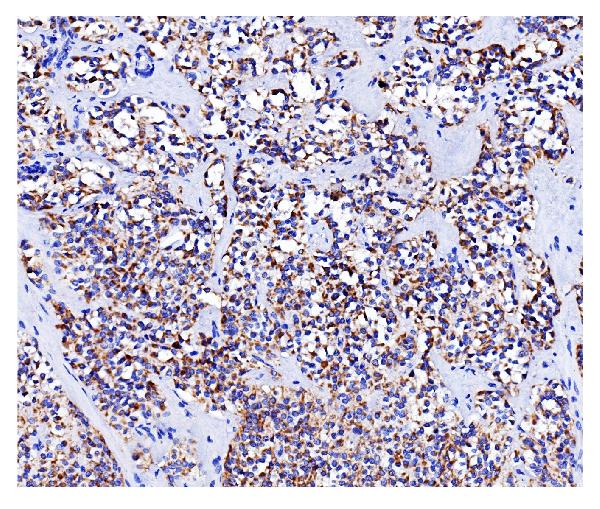
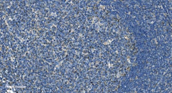
 IHC (Immunohiostchemistry)
(Figure 2. IHC analysis of CD30/TNFRSF8 using anti-CD30/TNFRSF8 antibody (AAA125595).CD30/TNFRSF8 was detected in paraffin-embedded section of human endometrial carcinoma tissue. Heat mediated antigen retrieval was performed in EDTA buffer (pH8. 0, epitope retrieval solution). The tissue section was blocked with 10% goat serum. The tissue section was then incubated with 2μg/ml rabbit anti-CD30/TNFRSF8 Antibody (AAA125595) overnight at 4 degree C. Biotinylated goat anti-rabbit IgG was used as secondary antibody and incubated for 30 minutes at 37 degree C. The tissue section was developed using Strepavidin-Biotin-Complex (SABC) with DAB as the chromogen.)
IHC (Immunohiostchemistry)
(Figure 2. IHC analysis of CD30/TNFRSF8 using anti-CD30/TNFRSF8 antibody (AAA125595).CD30/TNFRSF8 was detected in paraffin-embedded section of human endometrial carcinoma tissue. Heat mediated antigen retrieval was performed in EDTA buffer (pH8. 0, epitope retrieval solution). The tissue section was blocked with 10% goat serum. The tissue section was then incubated with 2μg/ml rabbit anti-CD30/TNFRSF8 Antibody (AAA125595) overnight at 4 degree C. Biotinylated goat anti-rabbit IgG was used as secondary antibody and incubated for 30 minutes at 37 degree C. The tissue section was developed using Strepavidin-Biotin-Complex (SABC) with DAB as the chromogen.)
CD30/TNFRSF8, Polyclonal Antibody (Cat# AAA125595)
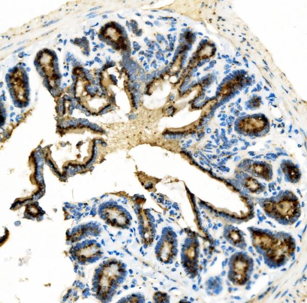
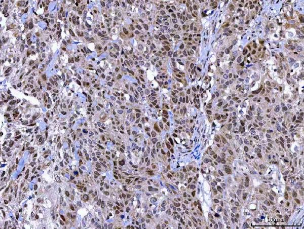
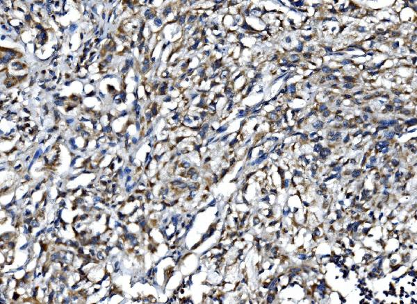
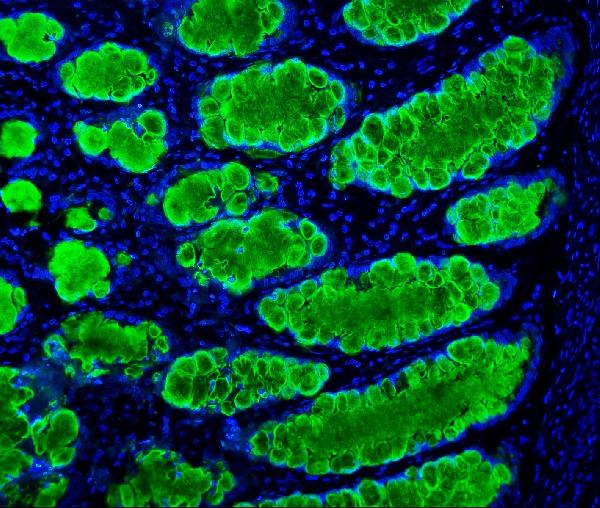
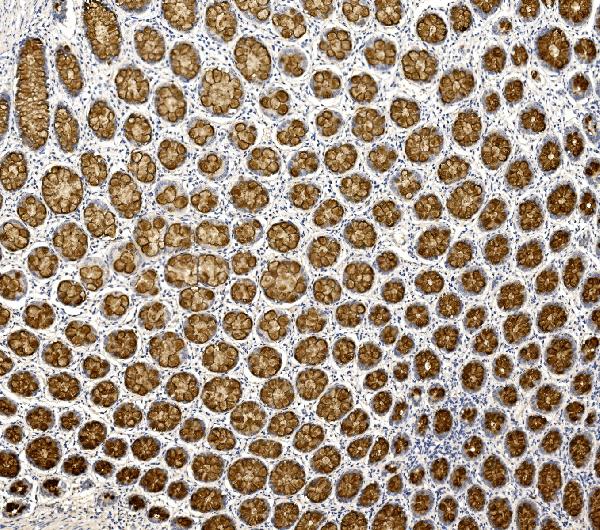
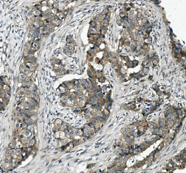
 IHC (Immunohistochemistry)
(Figure 5. IHC analysis of Golm1/GOLPH2 using anti-Golm1/GOLPH2 antibody (AAA125681).Golm1/GOLPH2 was detected in paraffin-embedded section of rat intestine tissue. Heat mediated antigen retrieval was performed in EDTA buffer (pH8. 0, epitope retrieval solution). The tissue section was blocked with 10% goat serum. The tissue section was then incubated with 2μg/ml rabbit anti-Golm1/GOLPH2 Antibody (AAA125681) overnight at 4 degree C. Biotinylated goat anti-rabbit IgG was used as secondary antibody and incubated for 30 minutes at 37 degree C. The tissue section was developed using Strepavidin-Biotin-Complex (SABC) with DAB as the chromogen.)
IHC (Immunohistochemistry)
(Figure 5. IHC analysis of Golm1/GOLPH2 using anti-Golm1/GOLPH2 antibody (AAA125681).Golm1/GOLPH2 was detected in paraffin-embedded section of rat intestine tissue. Heat mediated antigen retrieval was performed in EDTA buffer (pH8. 0, epitope retrieval solution). The tissue section was blocked with 10% goat serum. The tissue section was then incubated with 2μg/ml rabbit anti-Golm1/GOLPH2 Antibody (AAA125681) overnight at 4 degree C. Biotinylated goat anti-rabbit IgG was used as secondary antibody and incubated for 30 minutes at 37 degree C. The tissue section was developed using Strepavidin-Biotin-Complex (SABC) with DAB as the chromogen.)
Golm1/GOLPH2, Polyclonal Antibody (Cat# AAA125681)
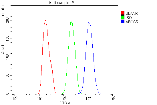 FCM/FACS (Flow Cytometry)
(Figure 3. Flow Cytometry analysis of PC-3 cells using anti-MRP5/ABCC5 antibody (AAA125683).Overlay histogram showing PC-3 cells stained with AAA125683 (Blue line). The cells were blocked with 10% normal goat serum. And then incubated with rabbit anti-MRP5/ABCC5 Antibody (AAA125683, 1μg/1x106 cells) for 30 min at 20 degree C. DyLight®488 conjugated goat anti-rabbit IgG (5-10μg/1x106 cells) was used as secondary antibody for 30 minutes at 20 degree C. Isotype control antibody (Green line) was rabbit IgG (1μg/1x106) used under the same conditions. Unlabelled sample (Red line) was also used as a control.)
FCM/FACS (Flow Cytometry)
(Figure 3. Flow Cytometry analysis of PC-3 cells using anti-MRP5/ABCC5 antibody (AAA125683).Overlay histogram showing PC-3 cells stained with AAA125683 (Blue line). The cells were blocked with 10% normal goat serum. And then incubated with rabbit anti-MRP5/ABCC5 Antibody (AAA125683, 1μg/1x106 cells) for 30 min at 20 degree C. DyLight®488 conjugated goat anti-rabbit IgG (5-10μg/1x106 cells) was used as secondary antibody for 30 minutes at 20 degree C. Isotype control antibody (Green line) was rabbit IgG (1μg/1x106) used under the same conditions. Unlabelled sample (Red line) was also used as a control.)
MRP5/ABCC5, Polyclonal Antibody (Cat# AAA125683)
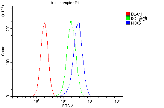 FCM/FACS (Flow Cytometry)
(Figure 5. Flow Cytometry analysis of PC-3 cells using anti-NOX5 antibody (AAA125684).Overlay histogram showing PC-3 cells stained with AAA125684 (Blue line). The cells were blocked with 10% normal goat serum. And then incubated with rabbit anti-NOX5 Antibody (AAA125684,1μg/1x106 cells) for 30 min at 20 degree C. DyLight®488 conjugated goat anti-rabbit IgG (5-10μg/1x106 cells) was used as secondary antibody for 30 minutes at 20 degree C. Isotype control antibody (Green line) was rabbit IgG (1μg/1x106) used under the same conditions. Unlabelled sample (Red line) was also used as a control.)
FCM/FACS (Flow Cytometry)
(Figure 5. Flow Cytometry analysis of PC-3 cells using anti-NOX5 antibody (AAA125684).Overlay histogram showing PC-3 cells stained with AAA125684 (Blue line). The cells were blocked with 10% normal goat serum. And then incubated with rabbit anti-NOX5 Antibody (AAA125684,1μg/1x106 cells) for 30 min at 20 degree C. DyLight®488 conjugated goat anti-rabbit IgG (5-10μg/1x106 cells) was used as secondary antibody for 30 minutes at 20 degree C. Isotype control antibody (Green line) was rabbit IgG (1μg/1x106) used under the same conditions. Unlabelled sample (Red line) was also used as a control.)
NOX5, Polyclonal Antibody (Cat# AAA125684)
CD39/ENTPD1, Polyclonal Antibody (Cat# AAA125685)
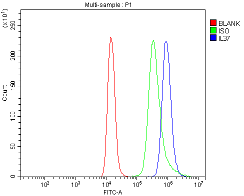
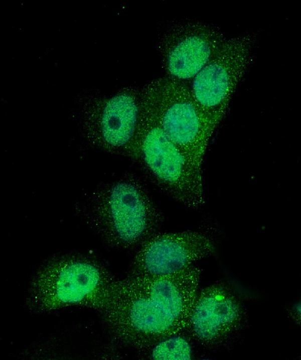
 IF (Immunofluorescence)
(Figure 4. IF analysis of IL37 using anti- IL37 antibody (AAA125697).IL37 was detected in immunocytochemical section of A431 cells. Enzyme antigen retrieval was performed using IHC enzyme antigen retrieval reagent for 15 mins. The cells were blocked with 10% goat serum. And then incubated with 5μg/mL rabbit anti- IL37 Antibody (AAA125697) overnight at 4 degree C. DyLight®488 Conjugated Goat Anti-Rabbit IgG was used as secondary antibody at 1:100 dilution and incubated for 30 minutes at 37 degree C. The section was counterstained with DAPI. Visualize using a fluorescence microscope and filter sets appropriate for the label used.)
IF (Immunofluorescence)
(Figure 4. IF analysis of IL37 using anti- IL37 antibody (AAA125697).IL37 was detected in immunocytochemical section of A431 cells. Enzyme antigen retrieval was performed using IHC enzyme antigen retrieval reagent for 15 mins. The cells were blocked with 10% goat serum. And then incubated with 5μg/mL rabbit anti- IL37 Antibody (AAA125697) overnight at 4 degree C. DyLight®488 Conjugated Goat Anti-Rabbit IgG was used as secondary antibody at 1:100 dilution and incubated for 30 minutes at 37 degree C. The section was counterstained with DAPI. Visualize using a fluorescence microscope and filter sets appropriate for the label used.)
IL37, Polyclonal Antibody (Cat# AAA125697)
What are Polyclonal Antibodies?
Polyclonal antibodies are antibodies that come from multiple B cell clones of a host animal. The typical hosts used for the majority of polyclonal antibody production are rabbits, goats, sheep, and donkeys. These polyclonal antibodies, once having identified their target, will bind to different epitopes located at different regions or sequences on the same protein/antigen. This ability to bind multiple epitopes is what makes polyclonal antibodies highly sensitive, as explained in our detailed guide on polyclonal antibodies and why they matter.
As a result, they are ideal at locating and binding to the target, even if the target is in very low concentrations (due to many different antibodies being able to bind to the same target molecule, which allows for significant amplification of a downstream signal).
Polyclonal antibodies are typically produced by injecting an antigen into a host animal, which causes the animal’s immune system to attack the foreign antigen by mass generating antibodies against it. After a period of time, serum is collected from the animal and purified using physicochemical fractionation, class-specific affinity purification, and/or antigen-affinity purification.
Key Uses of Polyclonal Antibodies
- Western Blotting: This method is used to find specific proteins in biological samples after separating them by size.
- Immunohistochemistry: IHC helps visualize the location of proteins in tissue sections using various staining techniques.
- ELISA: (Enzyme-Linked Immunosorbent Assay) is typically used to identify specific protein quantities in a sample. ELISAs can be either “Quantitative” or “Qualitative”.
- Flow Cytometry: technique that identifies and measures the specific protein on the surface or inside the cells in a fluid suspension.
- Immunoprecipitation: IP isolates and studies a specific protein from a complex mixture using antibodies.
Why Buy Polyclonal Antibodies from AAA Biotech?
1. Ideal for Various Applications
Our antibodies are generally going to be validated for use in multiple types of assays, including ELISA, Western Blotting, Immunohistochemistry, Immunoprecipitation, amongst others. They are ideal for a wide range of research applications.
2. Rigorous Quality Control
All of the antibodies in our catalog undergo strict quality testing to ensure specificity, sensitivity, and consistent performance. We are confident in the ability of our antibodies to provide you with accurate results.
3. Wide Assortment of Antibodies
Antibodies in our catalog can be found for both common and exotic species, and these antibodies are also available in both conjugated and recombinant forms to suit many diverse experimental needs.
4. Highly Purified
Our antibodies are available in purified forms with over 85% purity, as confirmed by SDS-PAGE. They are also available with tags such as His, Flag, GST, or MBP. We cater to customers worldwide.
FAQ
1. How are polyclonal antibodies produced?
Traditionally, polyclonal antibodies are produced by injecting an antigen into a host animal (such as a rabbit or goat), which then triggers an immune response from the host animal. The animal’s B cells produce antibodies that will recognize different parts of the injected antigen. These antibodies are then collected from the animal’s blood and purified for use.
2. How do polyclonal antibodies differ from monoclonal antibodies?
Polyclonal antibodies are a mix of antibodies that bind to different locations (epitopes) of the same antigen, while monoclonal antibodies are identical and bind to just one specific epitope. This makes polyclonal antibodies more versatile and better at detecting proteins that may be present in low quantities or in altered/modified forms.
3. How should I store polyclonal antibodies?
Polyclonal antibodies should be stored at 4°C for short-term use (up to a few weeks) and at -20°C or -80°C for long-term storage. Avoid repeated freeze-thaw cycles by dividing them into small aliquots. Always check the datasheet for specific storage instructions.