Filters
▼Clonality
▼Type
▼Reactivity
▼Gene Name
▼Isotype
▼Host
▼Application
▼Clone
▼Polyclonal Antibodies
At AAA Biotech also known as AAA Bio or AAABio, we provide a broad range of purified polyclonal antibodies (pAbs) that are able to all be browsed online through our website. Due to their high specificity and strong binding affinity, these antibodies are ideal for wide swathes of research and experimental applications.
Our polyclonal antibodies can easily support your work, whether you use them for Western Blotting, Immunocytochemistry (with or without Immunofluorescence used in conjunction), Immunohistochemistry, Immunoprecipitation, and ELISA tests. We highly encourage you to browse our range of pAbs and choose the one that best suits your experimental model.
Viewing 9200-9250 of 96812 product results
 FCM/FACS (Flow Cytometry)
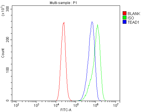
(Figure 3. Flow Cytometry analysis of U87 cells using anti-TEF1/TEAD-1/TEAD1 antibody (AAA125689).Overlay histogram showing U87 cells stained with AAA125689 (Blue line). The cells were blocked with 10% normal goat serum. And then incubated with rabbit anti-TEF1/TEAD-1/TEAD1 Antibody (AAA125689, 1μg/1x106 cells) for 30 min at 20 degree C. DyLight®488 conjugated goat anti-rabbit IgG (5-10μg/1x106 cells) was used as secondary antibody for 30 minutes at 20 degree C. Isotype control antibody (Green line) was rabbit IgG (1μg/1x106) used under the same conditions. Unlabelled sample (Red line) was also used as a control.)
FCM/FACS (Flow Cytometry)
(Figure 3. Flow Cytometry analysis of U87 cells using anti-TEF1/TEAD-1/TEAD1 antibody (AAA125689).Overlay histogram showing U87 cells stained with AAA125689 (Blue line). The cells were blocked with 10% normal goat serum. And then incubated with rabbit anti-TEF1/TEAD-1/TEAD1 Antibody (AAA125689, 1μg/1x106 cells) for 30 min at 20 degree C. DyLight®488 conjugated goat anti-rabbit IgG (5-10μg/1x106 cells) was used as secondary antibody for 30 minutes at 20 degree C. Isotype control antibody (Green line) was rabbit IgG (1μg/1x106) used under the same conditions. Unlabelled sample (Red line) was also used as a control.)
TEF1/TEAD-1/TEAD1, Polyclonal Antibody (Cat# AAA125689)
 FCM/FACS (Flow Cytometry)
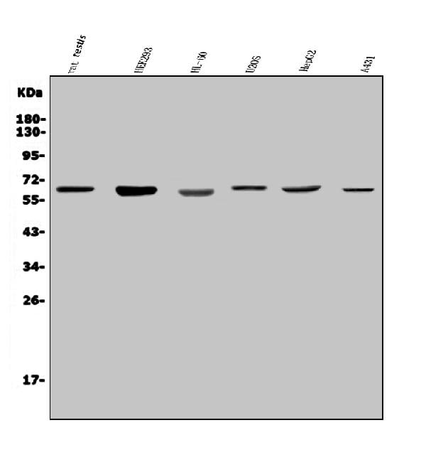
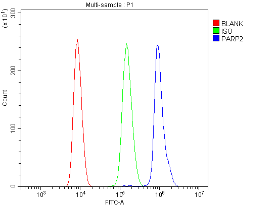
(Figure 3. Flow Cytometry analysis of HL-60 cells using anti-PARP2 antibody (AAA125690).Overlay histogram showing HL-60 cells stained with AAA125690 (Blue line). The cells were blocked with 10% normal goat serum. And then incubated with rabbit anti-PARP2 Antibody (AAA125690, 1μg/1x106 cells) for 30 min at 20 degree C. DyLight®488 conjugated goat anti-rabbit IgG (5-10μg/1x106 cells) was used as secondary antibody for 30 minutes at 20 degree C. Isotype control antibody (Green line) was rabbit IgG (1μg/1x106) used under the same conditions. Unlabelled sample (Red line) was also used as a control.)
FCM/FACS (Flow Cytometry)
(Figure 3. Flow Cytometry analysis of HL-60 cells using anti-PARP2 antibody (AAA125690).Overlay histogram showing HL-60 cells stained with AAA125690 (Blue line). The cells were blocked with 10% normal goat serum. And then incubated with rabbit anti-PARP2 Antibody (AAA125690, 1μg/1x106 cells) for 30 min at 20 degree C. DyLight®488 conjugated goat anti-rabbit IgG (5-10μg/1x106 cells) was used as secondary antibody for 30 minutes at 20 degree C. Isotype control antibody (Green line) was rabbit IgG (1μg/1x106) used under the same conditions. Unlabelled sample (Red line) was also used as a control.)
PARP2, Polyclonal Antibody (Cat# AAA125690)
 FCM/FACS (Flow Cytometry)
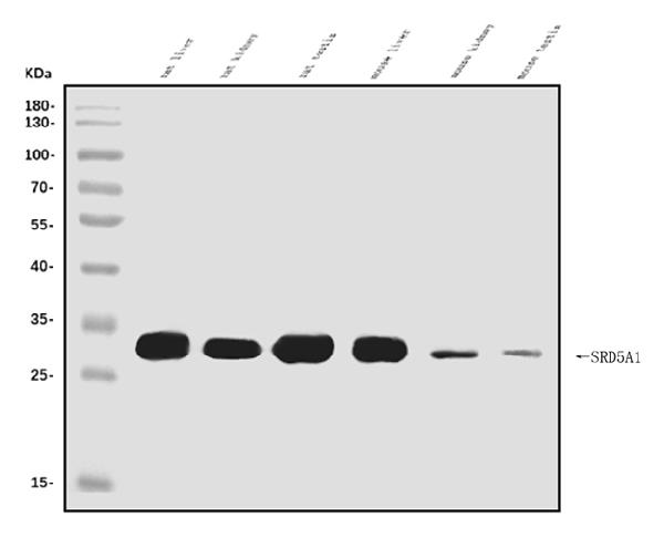
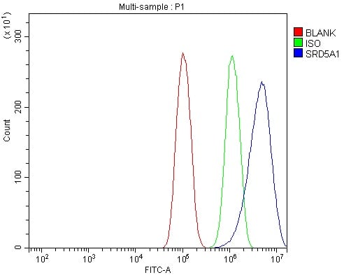
(Figure 2. Flow Cytometry analysis of PC-3 cells using anti-SRD5A1 antibody (AAA125694).Overlay histogram showing PC-3 cells stained with AAA125694 (Blue line). The cells were blocked with 10% normal goat serum. And then incubated with rabbit anti-SRD5A1 Antibody (AAA125694, 1μg/1x106 cells) for 30 min at 20 degree C. DyLight®488 conjugated goat anti-rabbit IgG (5-10μg/1x106 cells) was used as secondary antibody for 30 minutes at 20 degree C. Isotype control antibody (Green line) was rabbit IgG (1μg/1x106) used under the same conditions. Unlabelled sample (Red line) was also used as a control.)
FCM/FACS (Flow Cytometry)
(Figure 2. Flow Cytometry analysis of PC-3 cells using anti-SRD5A1 antibody (AAA125694).Overlay histogram showing PC-3 cells stained with AAA125694 (Blue line). The cells were blocked with 10% normal goat serum. And then incubated with rabbit anti-SRD5A1 Antibody (AAA125694, 1μg/1x106 cells) for 30 min at 20 degree C. DyLight®488 conjugated goat anti-rabbit IgG (5-10μg/1x106 cells) was used as secondary antibody for 30 minutes at 20 degree C. Isotype control antibody (Green line) was rabbit IgG (1μg/1x106) used under the same conditions. Unlabelled sample (Red line) was also used as a control.)
SRD5A1, Polyclonal Antibody (Cat# AAA125694)
 FCM/FACS (Flow Cytometry)
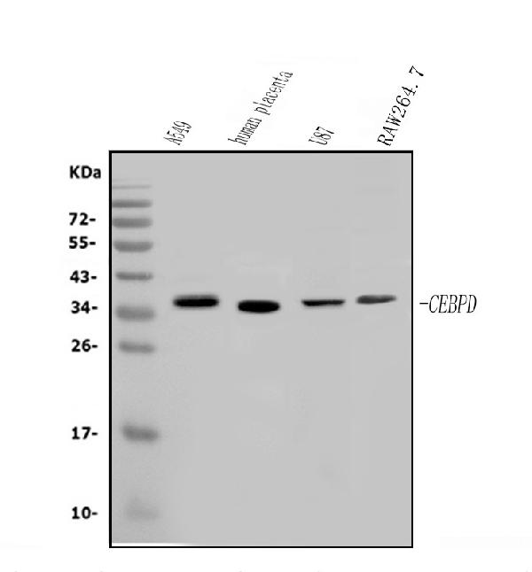
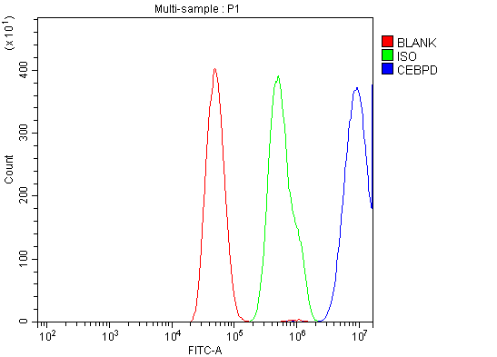
(Figure 2. Flow Cytometry analysis of SiHa cells using anti-CEBP Delta/CEBPD antibody (AAA125696).Overlay histogram showing SiHa cells stained with AAA125696 (Blue line). The cells were blocked with 10% normal goat serum. And then incubated with rabbit anti-CEBP Delta/CEBPD Antibody (AAA125696, 1μg/1x106 cells) for 30 min at 20 degree C. DyLight®488 conjugated goat anti-rabbit IgG (5-10μg/1x106 cells) was used as secondary antibody for 30 minutes at 20 degree C. Isotype control antibody (Green line) was rabbit IgG (1μg/1x106) used under the same conditions. Unlabelled sample (Red line) was also used as a control.)
FCM/FACS (Flow Cytometry)
(Figure 2. Flow Cytometry analysis of SiHa cells using anti-CEBP Delta/CEBPD antibody (AAA125696).Overlay histogram showing SiHa cells stained with AAA125696 (Blue line). The cells were blocked with 10% normal goat serum. And then incubated with rabbit anti-CEBP Delta/CEBPD Antibody (AAA125696, 1μg/1x106 cells) for 30 min at 20 degree C. DyLight®488 conjugated goat anti-rabbit IgG (5-10μg/1x106 cells) was used as secondary antibody for 30 minutes at 20 degree C. Isotype control antibody (Green line) was rabbit IgG (1μg/1x106) used under the same conditions. Unlabelled sample (Red line) was also used as a control.)
CEBP Delta/CEBPD, Polyclonal Antibody (Cat# AAA125696)
 FCM/FACS (Flow Cytometry)
(Figure 4. Flow Cytometry analysis of HEPA1-6 cells using anti-CD20/MS4A1 antibody (AAA125704).Overlay histogram showing HEPA1-6 cells stained with AAA125704 (Blue line). The cells were blocked with 10% normal goat serum. And then incubated with rabbit anti-CD20/MS4A1 Antibody (AAA125704, 1μg/1x106 cells) for 30 min at 20 degree C. DyLight®488 conjugated goat anti-rabbit IgG (5-10μg/1x106 cells) was used as secondary antibody for 30 minutes at 20 degree C. Isotype control antibody (Green line) was rabbit IgG (1μg/1x106) used under the same conditions. Unlabelled sample (Red line) was also used as a control.)
FCM/FACS (Flow Cytometry)
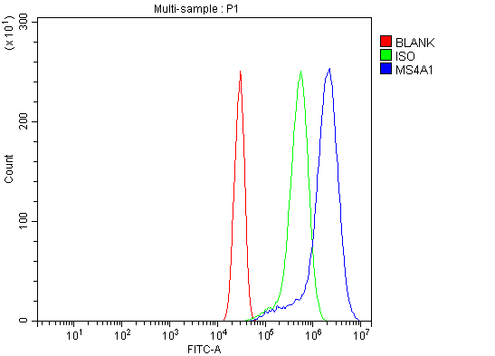
(Figure 4. Flow Cytometry analysis of HEPA1-6 cells using anti-CD20/MS4A1 antibody (AAA125704).Overlay histogram showing HEPA1-6 cells stained with AAA125704 (Blue line). The cells were blocked with 10% normal goat serum. And then incubated with rabbit anti-CD20/MS4A1 Antibody (AAA125704, 1μg/1x106 cells) for 30 min at 20 degree C. DyLight®488 conjugated goat anti-rabbit IgG (5-10μg/1x106 cells) was used as secondary antibody for 30 minutes at 20 degree C. Isotype control antibody (Green line) was rabbit IgG (1μg/1x106) used under the same conditions. Unlabelled sample (Red line) was also used as a control.)
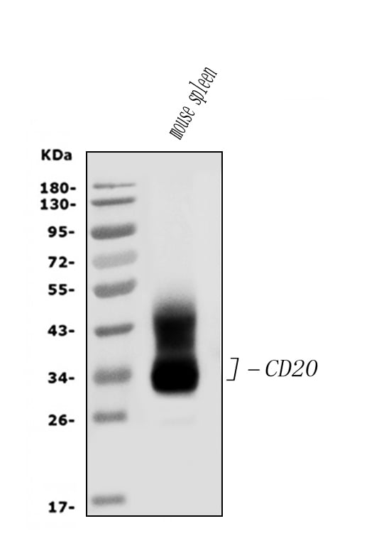
CD20/MS4A1, Polyclonal Antibody (Cat# AAA125704)
 FCM/FACS (Flow Cytometry)
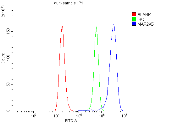
(Figure 4. Flow Cytometry analysis of 293T cells using anti-MEK5/MAP2K5 antibody (AAA125708).Overlay histogram showing 293T cells stained with AAA125708 (Blue line). The cells were blocked with 10% normal goat serum. And then incubated with rabbit anti-MEK5/MAP2K5 Antibody (AAA125708, 1μg/1x106 cells) for 30 min at 20 degree C. DyLight®488 conjugated goat anti-rabbit IgG (5-10μg/1x106 cells) was used as secondary antibody for 30 minutes at 20 degree C. Isotype control antibody (Green line) was rabbit IgG (1μg/1x106) used under the same conditions. Unlabelled sample (Red line) was also used as a control.)
FCM/FACS (Flow Cytometry)
(Figure 4. Flow Cytometry analysis of 293T cells using anti-MEK5/MAP2K5 antibody (AAA125708).Overlay histogram showing 293T cells stained with AAA125708 (Blue line). The cells were blocked with 10% normal goat serum. And then incubated with rabbit anti-MEK5/MAP2K5 Antibody (AAA125708, 1μg/1x106 cells) for 30 min at 20 degree C. DyLight®488 conjugated goat anti-rabbit IgG (5-10μg/1x106 cells) was used as secondary antibody for 30 minutes at 20 degree C. Isotype control antibody (Green line) was rabbit IgG (1μg/1x106) used under the same conditions. Unlabelled sample (Red line) was also used as a control.)
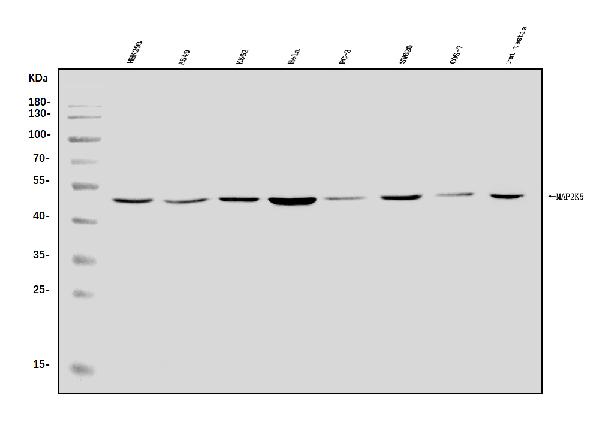
MEK5/MAP2K5, Polyclonal Antibody (Cat# AAA125708)
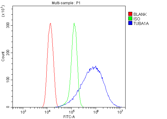 FCM/FACS (Flow Cytometry)
(Figure 5. Flow Cytometry analysis of Hela cells using anti-Tubulin alpha antibody (AAA125709).Overlay histogram showing Hela cells stained with AAA125709 (Blue line). The cells were blocked with 10% normal goat serum. And then incubated with rabbit anti-Tubulin alpha Antibody (AAA125709,1μg/1x106 cells) for 30 min at 20 degree C. DyLight®488 conjugated goat anti-rabbit IgG (5-10μg/1x106 cells) was used as secondary antibody for 30 minutes at 20 degree C. Isotype control antibody (Green line) was rabbit IgG (1μg/1x106) used under the same conditions. Unlabelled sample (Red line) was also used as a control.)
FCM/FACS (Flow Cytometry)
(Figure 5. Flow Cytometry analysis of Hela cells using anti-Tubulin alpha antibody (AAA125709).Overlay histogram showing Hela cells stained with AAA125709 (Blue line). The cells were blocked with 10% normal goat serum. And then incubated with rabbit anti-Tubulin alpha Antibody (AAA125709,1μg/1x106 cells) for 30 min at 20 degree C. DyLight®488 conjugated goat anti-rabbit IgG (5-10μg/1x106 cells) was used as secondary antibody for 30 minutes at 20 degree C. Isotype control antibody (Green line) was rabbit IgG (1μg/1x106) used under the same conditions. Unlabelled sample (Red line) was also used as a control.)
Tubulin alpha, Polyclonal Antibody (Cat# AAA125709)
 FCM/FACS (Flow Cytometry)
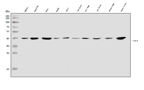
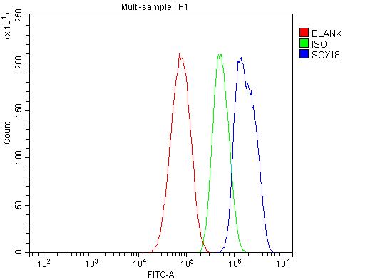
(Figure 3. Flow Cytometry analysis of HepG2 cells using anti-SOX18 antibody (AAA125710).Overlay histogram showing HepG2 cells stained with AAA125710 (Blue line). The cells were blocked with 10% normal goat serum. And then incubated with rabbit anti-SOX18 Antibody (AAA125710, 1μg/1x106 cells) for 30 min at 20 degree C. DyLight®488 conjugated goat anti-rabbit IgG (5-10μg/1x106 cells) was used as secondary antibody for 30 minutes at 20 degree C. Isotype control antibody (Green line) was rabbit IgG (1μg/1x106) used under the same conditions. Unlabelled sample (Red line) was also used as a control.)
FCM/FACS (Flow Cytometry)
(Figure 3. Flow Cytometry analysis of HepG2 cells using anti-SOX18 antibody (AAA125710).Overlay histogram showing HepG2 cells stained with AAA125710 (Blue line). The cells were blocked with 10% normal goat serum. And then incubated with rabbit anti-SOX18 Antibody (AAA125710, 1μg/1x106 cells) for 30 min at 20 degree C. DyLight®488 conjugated goat anti-rabbit IgG (5-10μg/1x106 cells) was used as secondary antibody for 30 minutes at 20 degree C. Isotype control antibody (Green line) was rabbit IgG (1μg/1x106) used under the same conditions. Unlabelled sample (Red line) was also used as a control.)
SOX18, Polyclonal Antibody (Cat# AAA125710)
 FCM/FACS (Flow Cytometry)
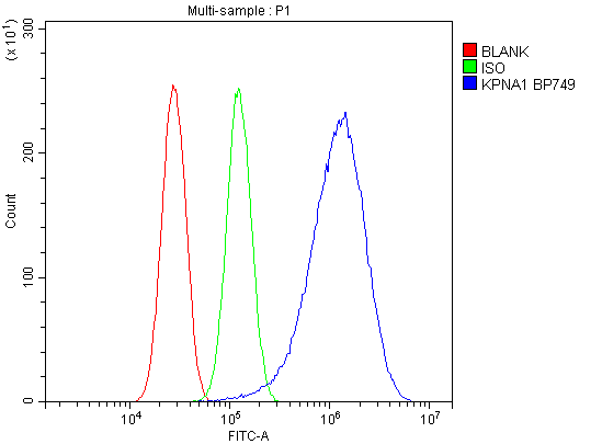
(Figure 3. Flow Cytometry analysis of U87 cells using anti-SRP1/KPNA1 antibody (AAA125711).Overlay histogram showing U87 cells stained with AAA125711 (Blue line). The cells were blocked with 10% normal goat serum. And then incubated with rabbit anti-SRP1/KPNA1 Antibody (AAA125711, 1μg/1x106 cells) for 30 min at 20 degree C. DyLight®488 conjugated goat anti-rabbit IgG (5-10μg/1x106 cells) was used as secondary antibody for 30 minutes at 20 degree C. Isotype control antibody (Green line) was rabbit IgG (1μg/1x106) used under the same conditions. Unlabelled sample (Red line) was also used as a control.)
FCM/FACS (Flow Cytometry)
(Figure 3. Flow Cytometry analysis of U87 cells using anti-SRP1/KPNA1 antibody (AAA125711).Overlay histogram showing U87 cells stained with AAA125711 (Blue line). The cells were blocked with 10% normal goat serum. And then incubated with rabbit anti-SRP1/KPNA1 Antibody (AAA125711, 1μg/1x106 cells) for 30 min at 20 degree C. DyLight®488 conjugated goat anti-rabbit IgG (5-10μg/1x106 cells) was used as secondary antibody for 30 minutes at 20 degree C. Isotype control antibody (Green line) was rabbit IgG (1μg/1x106) used under the same conditions. Unlabelled sample (Red line) was also used as a control.)
SRP1/KPNA1, Polyclonal Antibody (Cat# AAA125711)
 FCM/FACS (Flow Cytometry)
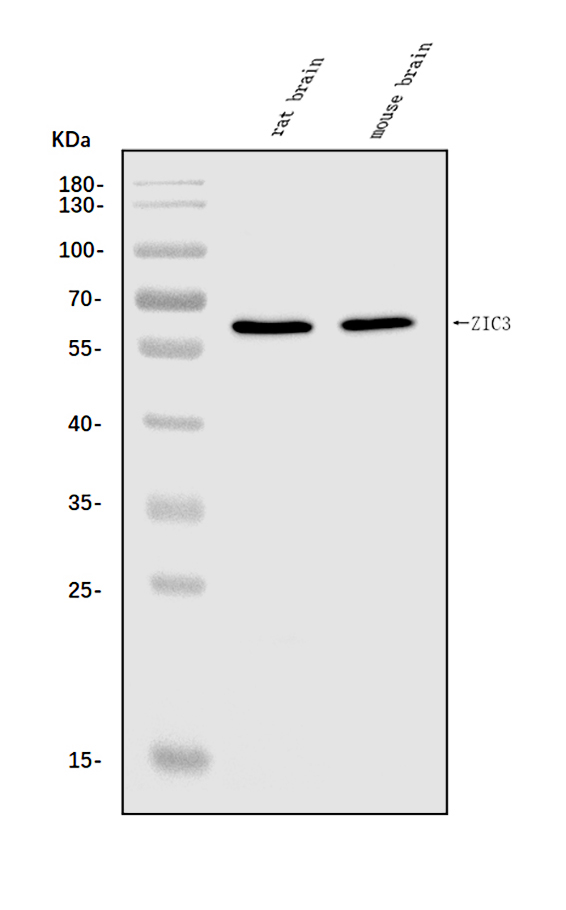
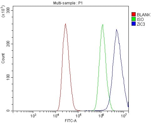
(Figure 2. Flow Cytometry analysis of MCF-7 cells using anti-ZIC3 antibody (AAA125713).Overlay histogram showing MCF-7 cells stained with AAA125713 (Blue line). The cells were blocked with 10% normal goat serum. And then incubated with rabbit anti-ZIC3 Antibody (AAA125713, 1μg/1x106 cells) for 30 min at 20 degree C. DyLight®488 conjugated goat anti-rabbit IgG (5-10μg/1x106 cells) was used as secondary antibody for 30 minutes at 20 degree C. Isotype control antibody (Green line) was rabbit IgG (1μg/1x106) used under the same conditions. Unlabelled sample (Red line) was also used as a control.)
FCM/FACS (Flow Cytometry)
(Figure 2. Flow Cytometry analysis of MCF-7 cells using anti-ZIC3 antibody (AAA125713).Overlay histogram showing MCF-7 cells stained with AAA125713 (Blue line). The cells were blocked with 10% normal goat serum. And then incubated with rabbit anti-ZIC3 Antibody (AAA125713, 1μg/1x106 cells) for 30 min at 20 degree C. DyLight®488 conjugated goat anti-rabbit IgG (5-10μg/1x106 cells) was used as secondary antibody for 30 minutes at 20 degree C. Isotype control antibody (Green line) was rabbit IgG (1μg/1x106) used under the same conditions. Unlabelled sample (Red line) was also used as a control.)
ZIC3, Polyclonal Antibody (Cat# AAA125713)
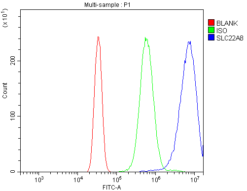 FCM/FACS (Flow Cytometry)
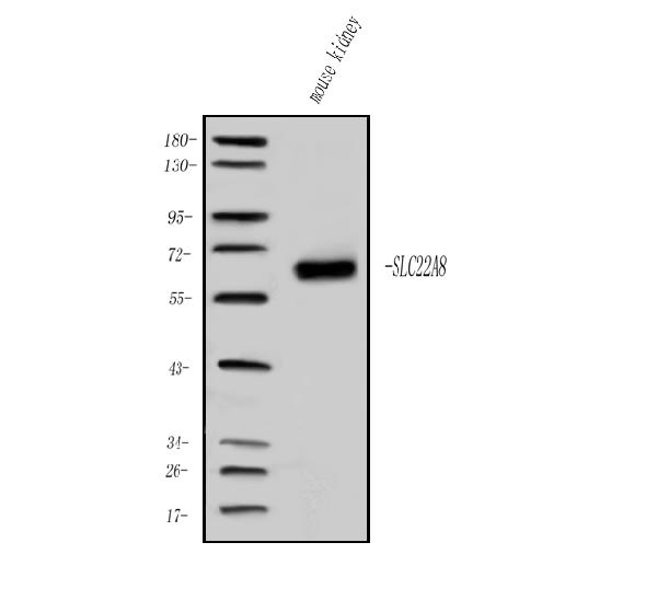
(Figure 2. Flow Cytometry analysis of Neuro-2a cells using anti-OAT3/Slc22a8 antibody (AAA125715).Overlay histogram showing Neuro-2a cells stained with AAA125715 (Blue line). The cells were blocked with 10% normal goat serum. And then incubated with rabbit anti-OAT3/Slc22a8 Antibody (AAA125715, 1μg/1x106 cells) for 30 min at 20 degree C. DyLight®488 conjugated goat anti-rabbit IgG (5-10μg/1x106 cells) was used as secondary antibody for 30 minutes at 20 degree C. Isotype control antibody (Green line) was rabbit IgG (1μg/1x106) used under the same conditions. Unlabelled sample (Red line) was also used as a control.)
FCM/FACS (Flow Cytometry)
(Figure 2. Flow Cytometry analysis of Neuro-2a cells using anti-OAT3/Slc22a8 antibody (AAA125715).Overlay histogram showing Neuro-2a cells stained with AAA125715 (Blue line). The cells were blocked with 10% normal goat serum. And then incubated with rabbit anti-OAT3/Slc22a8 Antibody (AAA125715, 1μg/1x106 cells) for 30 min at 20 degree C. DyLight®488 conjugated goat anti-rabbit IgG (5-10μg/1x106 cells) was used as secondary antibody for 30 minutes at 20 degree C. Isotype control antibody (Green line) was rabbit IgG (1μg/1x106) used under the same conditions. Unlabelled sample (Red line) was also used as a control.)
OAT3/Slc22a8, Polyclonal Antibody (Cat# AAA125715)
 FCM/FACS (Flow Cytometry)
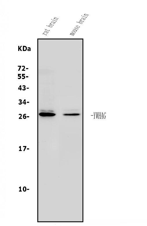
(Figure 2. Flow Cytometry analysis of A549 cells using anti-14-3-3 Gamma/YWHAG antibody (AAA125716).Overlay histogram showing A549 cells stained with AAA125716 (Blue line). The cells were blocked with 10% normal goat serum. And then incubated with rabbit anti-14-3-3 Gamma/YWHAG Antibody (AAA125716, 1μg/1x106 cells) for 30 min at 20 degree C. DyLight®488 conjugated goat anti-rabbit IgG (5-10μg/1x106 cells) was used as secondary antibody for 30 minutes at 20 degree C. Isotype control antibody (Green line) was rabbit IgG (1μg/1x106) used under the same conditions. Unlabelled sample (Red line) was also used as a control.)
FCM/FACS (Flow Cytometry)
(Figure 2. Flow Cytometry analysis of A549 cells using anti-14-3-3 Gamma/YWHAG antibody (AAA125716).Overlay histogram showing A549 cells stained with AAA125716 (Blue line). The cells were blocked with 10% normal goat serum. And then incubated with rabbit anti-14-3-3 Gamma/YWHAG Antibody (AAA125716, 1μg/1x106 cells) for 30 min at 20 degree C. DyLight®488 conjugated goat anti-rabbit IgG (5-10μg/1x106 cells) was used as secondary antibody for 30 minutes at 20 degree C. Isotype control antibody (Green line) was rabbit IgG (1μg/1x106) used under the same conditions. Unlabelled sample (Red line) was also used as a control.)
14-3-3 gamma/YWHAG, Polyclonal Antibody (Cat# AAA125716)
 FCM/FACS (Flow Cytometry)
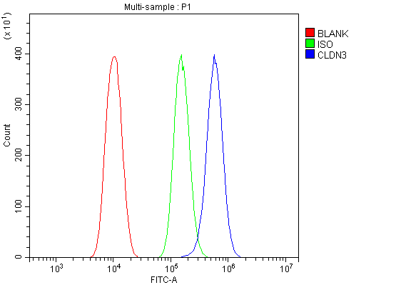
(Figure 4. Flow Cytometry analysis of PC-3 cells using anti-Claudin 3/CLDN3 antibody (AAA125722).Overlay histogram showing PC-3 cells stained with AAA125722 (Blue line). The cells were blocked with 10% normal goat serum. And then incubated with rabbit anti-Claudin 3/CLDN3 Antibody (AAA125722,1μg/1x106 cells) for 30 min at 20 degree C. DyLight®488 conjugated goat anti-rabbit IgG (5-10μg/1x106 cells) was used as secondary antibody for 30 minutes at 20 degree C. Isotype control antibody (Green line) was rabbit IgG (1μg/1x106) used under the same conditions. Unlabelled sample (Red line) was also used as a control.)
FCM/FACS (Flow Cytometry)
(Figure 4. Flow Cytometry analysis of PC-3 cells using anti-Claudin 3/CLDN3 antibody (AAA125722).Overlay histogram showing PC-3 cells stained with AAA125722 (Blue line). The cells were blocked with 10% normal goat serum. And then incubated with rabbit anti-Claudin 3/CLDN3 Antibody (AAA125722,1μg/1x106 cells) for 30 min at 20 degree C. DyLight®488 conjugated goat anti-rabbit IgG (5-10μg/1x106 cells) was used as secondary antibody for 30 minutes at 20 degree C. Isotype control antibody (Green line) was rabbit IgG (1μg/1x106) used under the same conditions. Unlabelled sample (Red line) was also used as a control.)
Claudin 3/CLDN3, Polyclonal Antibody (Cat# AAA125722)
 FCM/FACS (Flow Cytometry)
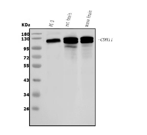
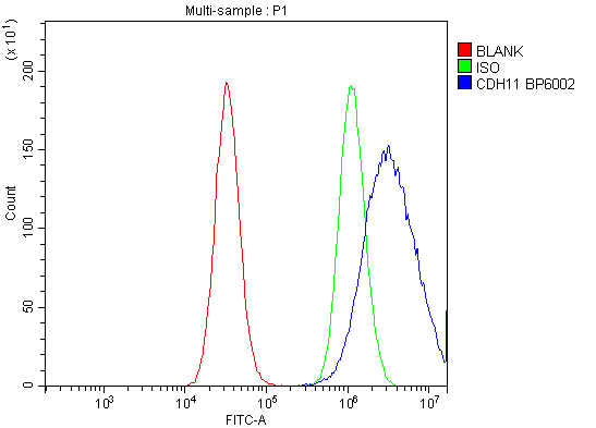
(Figure 4. Flow Cytometry analysis of PC-3 cells using anti-OB Cadherin/CDH11 antibody (AAA125724).Overlay histogram showing PC-3 cells stained with AAA125724 (Blue line). The cells were blocked with 10% normal goat serum. And then incubated with rabbit anti-OB Cadherin/CDH11 Antibody (AAA125724, 1μg/1x106 cells) for 30 min at 20 degree C. DyLight®488 conjugated goat anti-rabbit IgG (5-10μg/1x106 cells) was used as secondary antibody for 30 minutes at 20 degree C. Isotype control antibody (Green line) was rabbit IgG (1μg/1x106) used under the same conditions. Unlabelled sample (Red line) was also used as a control.)
FCM/FACS (Flow Cytometry)
(Figure 4. Flow Cytometry analysis of PC-3 cells using anti-OB Cadherin/CDH11 antibody (AAA125724).Overlay histogram showing PC-3 cells stained with AAA125724 (Blue line). The cells were blocked with 10% normal goat serum. And then incubated with rabbit anti-OB Cadherin/CDH11 Antibody (AAA125724, 1μg/1x106 cells) for 30 min at 20 degree C. DyLight®488 conjugated goat anti-rabbit IgG (5-10μg/1x106 cells) was used as secondary antibody for 30 minutes at 20 degree C. Isotype control antibody (Green line) was rabbit IgG (1μg/1x106) used under the same conditions. Unlabelled sample (Red line) was also used as a control.)
OB Cadherin/CDH11, Polyclonal Antibody (Cat# AAA125724)
 FCM/FACS (Flow Cytometry)
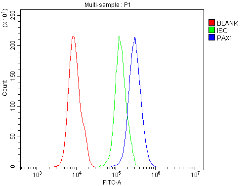
(Figure 2. Flow Cytometry analysis of 293T cells using anti-PAX1 antibody (AAA125727).Overlay histogram showing 293T cells stained with AAA125727 (Blue line). The cells were blocked with 10% normal goat serum. And then incubated with rabbit anti-PAX1 Antibody (AAA125727, 1μg/1x106 cells) for 30 min at 20 degree C. DyLight®488 conjugated goat anti-rabbit IgG (5-10μg/1x106 cells) was used as secondary antibody for 30 minutes at 20 degree C. Isotype control antibody (Green line) was rabbit IgG (1μg/1x106) used under the same conditions. Unlabelled sample (Red line) was also used as a control.)
FCM/FACS (Flow Cytometry)
(Figure 2. Flow Cytometry analysis of 293T cells using anti-PAX1 antibody (AAA125727).Overlay histogram showing 293T cells stained with AAA125727 (Blue line). The cells were blocked with 10% normal goat serum. And then incubated with rabbit anti-PAX1 Antibody (AAA125727, 1μg/1x106 cells) for 30 min at 20 degree C. DyLight®488 conjugated goat anti-rabbit IgG (5-10μg/1x106 cells) was used as secondary antibody for 30 minutes at 20 degree C. Isotype control antibody (Green line) was rabbit IgG (1μg/1x106) used under the same conditions. Unlabelled sample (Red line) was also used as a control.)
PAX1, Polyclonal Antibody (Cat# AAA125727)
 FCM/FACS (Flow Cytometry)
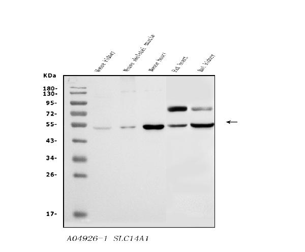
(Figure 2. Flow Cytometry analysis of HEPA1-6 cells using anti-SLC14A1/UTE antibody (AAA125734).Overlay histogram showing HEPA1-6 cells stained with AAA125734 (Blue line). The cells were blocked with 10% normal goat serum. And then incubated with rabbit anti-SLC14A1/UTE Antibody (AAA125734, 1μg/1x106 cells) for 30 min at 20 degree C. DyLight®488 conjugated goat anti-rabbit IgG (5-10μg/1x106 cells) was used as secondary antibody for 30 minutes at 20 degree C. Isotype control antibody (Green line) was rabbit IgG (1μg/1x106) used under the same conditions. Unlabelled sample (Red line) was also used as a control.)
FCM/FACS (Flow Cytometry)
(Figure 2. Flow Cytometry analysis of HEPA1-6 cells using anti-SLC14A1/UTE antibody (AAA125734).Overlay histogram showing HEPA1-6 cells stained with AAA125734 (Blue line). The cells were blocked with 10% normal goat serum. And then incubated with rabbit anti-SLC14A1/UTE Antibody (AAA125734, 1μg/1x106 cells) for 30 min at 20 degree C. DyLight®488 conjugated goat anti-rabbit IgG (5-10μg/1x106 cells) was used as secondary antibody for 30 minutes at 20 degree C. Isotype control antibody (Green line) was rabbit IgG (1μg/1x106) used under the same conditions. Unlabelled sample (Red line) was also used as a control.)
SLC14A1/UTE, Polyclonal Antibody (Cat# AAA125734)
 FCM/FACS (Flow Cytometry)
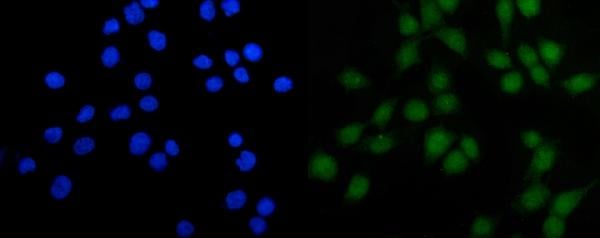
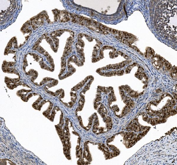
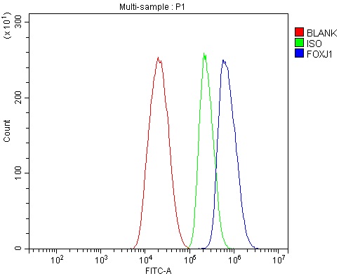
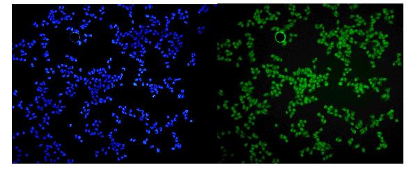
(Figure 4. Flow Cytometry analysis of HepG2 cells using anti-FOXJ1 antibody (AAA125736).Overlay histogram showing HepG2 cells stained with AAA125736 (Blue line). The cells were blocked with 10% normal goat serum. And then incubated with rabbit anti-FOXJ1 Antibody (AAA125736, 1μg/1x106 cells) for 30 min at 20 degree C. DyLight®488 conjugated goat anti-rabbit IgG (5-10μg/1x106 cells) was used as secondary antibody for 30 minutes at 20 degree C. Isotype control antibody (Green line) was rabbit IgG (1μg/1x106) used under the same conditions. Unlabelled sample (Red line) was also used as a control.)
FCM/FACS (Flow Cytometry)
(Figure 4. Flow Cytometry analysis of HepG2 cells using anti-FOXJ1 antibody (AAA125736).Overlay histogram showing HepG2 cells stained with AAA125736 (Blue line). The cells were blocked with 10% normal goat serum. And then incubated with rabbit anti-FOXJ1 Antibody (AAA125736, 1μg/1x106 cells) for 30 min at 20 degree C. DyLight®488 conjugated goat anti-rabbit IgG (5-10μg/1x106 cells) was used as secondary antibody for 30 minutes at 20 degree C. Isotype control antibody (Green line) was rabbit IgG (1μg/1x106) used under the same conditions. Unlabelled sample (Red line) was also used as a control.)
FOXJ1, Polyclonal Antibody (Cat# AAA125736)
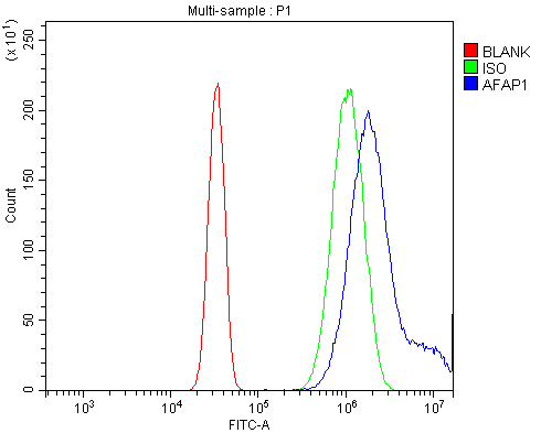
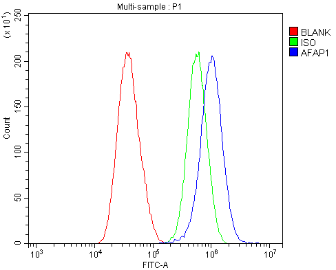
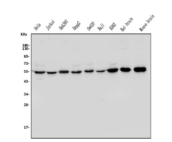
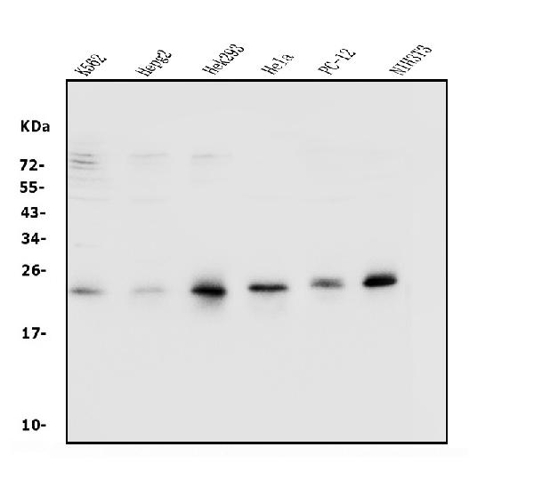
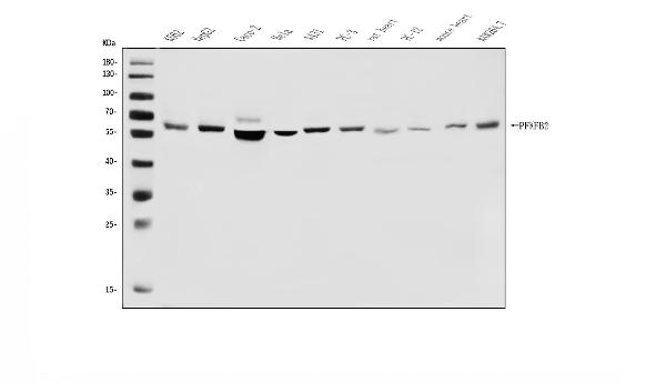
 WB (Western Blot)
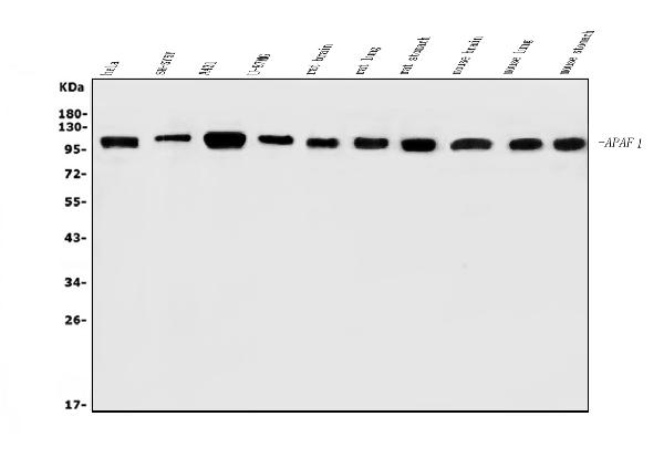
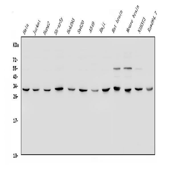
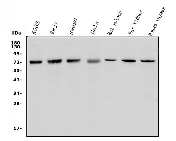
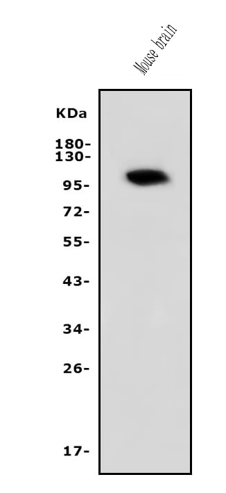
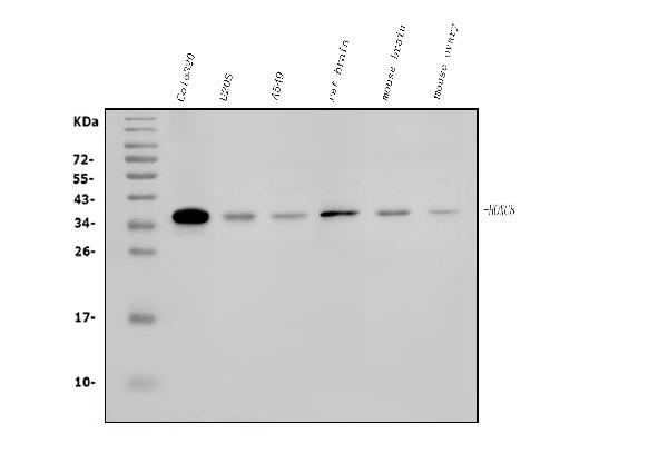
(Figure 1. Western blot analysis of AFAP/AFAP1 using anti-AFAP/AFAP1 antibody (AAA125742).Electrophoresis was performed on a 5-20% SDS-PAGE gel at 70V (Stacking gel) / 90V (Resolving gel) for 2-3 hours. The sample well of each lane was loaded with 50ug of sample under reducing conditions.Lane 1: human Hela whole cell lysatesLane 2: human SH-SY5Y whole cell lysatesLane 3: human A431 whole cell lysatesLane 4: human U-87MG whole cell lysatesLane 5: rat brain tissue lysatesLane 6: rat lung tissue lysatesLane 7: rat stomach tissue lysatesLane 8: mouse brain tissue lysatesLane 9: mouse lung tissue lysatesLane 10: mouse stomach tissue lysates.After Electrophoresis, proteins were transferred to a Nitrocellulose membrane at 150mA for 50-90 minutes. Blocked the membrane with 5% Non-fat Milk/ TBS for 1. 5 hour at RT. The membrane was incubated with rabbit anti-AFAP/AFAP1 antigen affinity purified polyclonal antibody (Catalog # AAA125742) at 0. 25 μg/mL overnight at 4 degree C, then washed with TBS-0. 1%Tween 3 times with 5 minutes each and probed with a goat anti-rabbit IgG-HRP secondary antibody at a dilution of 1:5000 for 1. 5 hour at RT. The signal is developed using an Enhanced Chemiluminescent detection (ECL) kit with Tanon 5200 system. A specific band was detected for AFAP/AFAP1 at approximately 120KD. The expected band size for AFAP/AFAP1 is at 120KD.)
WB (Western Blot)
(Figure 1. Western blot analysis of AFAP/AFAP1 using anti-AFAP/AFAP1 antibody (AAA125742).Electrophoresis was performed on a 5-20% SDS-PAGE gel at 70V (Stacking gel) / 90V (Resolving gel) for 2-3 hours. The sample well of each lane was loaded with 50ug of sample under reducing conditions.Lane 1: human Hela whole cell lysatesLane 2: human SH-SY5Y whole cell lysatesLane 3: human A431 whole cell lysatesLane 4: human U-87MG whole cell lysatesLane 5: rat brain tissue lysatesLane 6: rat lung tissue lysatesLane 7: rat stomach tissue lysatesLane 8: mouse brain tissue lysatesLane 9: mouse lung tissue lysatesLane 10: mouse stomach tissue lysates.After Electrophoresis, proteins were transferred to a Nitrocellulose membrane at 150mA for 50-90 minutes. Blocked the membrane with 5% Non-fat Milk/ TBS for 1. 5 hour at RT. The membrane was incubated with rabbit anti-AFAP/AFAP1 antigen affinity purified polyclonal antibody (Catalog # AAA125742) at 0. 25 μg/mL overnight at 4 degree C, then washed with TBS-0. 1%Tween 3 times with 5 minutes each and probed with a goat anti-rabbit IgG-HRP secondary antibody at a dilution of 1:5000 for 1. 5 hour at RT. The signal is developed using an Enhanced Chemiluminescent detection (ECL) kit with Tanon 5200 system. A specific band was detected for AFAP/AFAP1 at approximately 120KD. The expected band size for AFAP/AFAP1 is at 120KD.)
AFAP/AFAP1, Polyclonal Antibody (Cat# AAA125742)
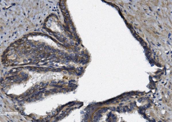
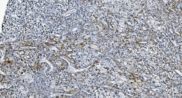
 IHC (Immunohistochemistry)
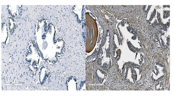
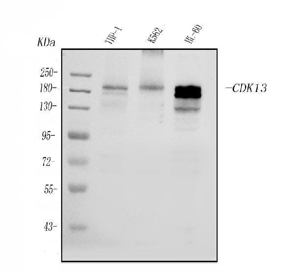
(Figure 4. IHC analysis of CDK13 using anti-CDK13 antibody (AAA125745).CDK13 was detected in paraffin-embedded section of human esophageal squamous carcinoma tissue. Heat mediated antigen retrieval was performed in EDTA buffer (pH8. 0, epitope retrieval solution). The tissue section was blocked with 10% goat serum. The tissue section was then incubated with 2μg/ml rabbit anti-CDK13 Antibody (AAA125745) overnight at 4 degree C. Biotinylated goat anti-rabbit IgG was used as secondary antibody and incubated for 30 minutes at 37 degree C. The tissue section was developed using Strepavidin-Biotin-Complex (SABC) with DAB as the chromogen.)
IHC (Immunohistochemistry)
(Figure 4. IHC analysis of CDK13 using anti-CDK13 antibody (AAA125745).CDK13 was detected in paraffin-embedded section of human esophageal squamous carcinoma tissue. Heat mediated antigen retrieval was performed in EDTA buffer (pH8. 0, epitope retrieval solution). The tissue section was blocked with 10% goat serum. The tissue section was then incubated with 2μg/ml rabbit anti-CDK13 Antibody (AAA125745) overnight at 4 degree C. Biotinylated goat anti-rabbit IgG was used as secondary antibody and incubated for 30 minutes at 37 degree C. The tissue section was developed using Strepavidin-Biotin-Complex (SABC) with DAB as the chromogen.)
CDK13, Polyclonal Antibody (Cat# AAA125745)
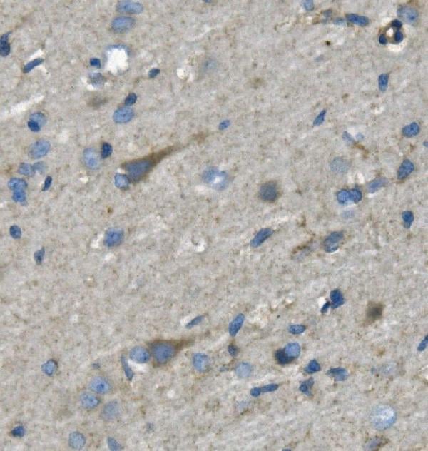
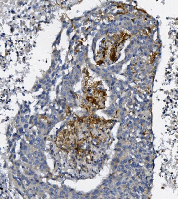
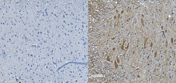 IHC (Immunohistochemistry)
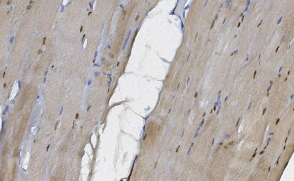
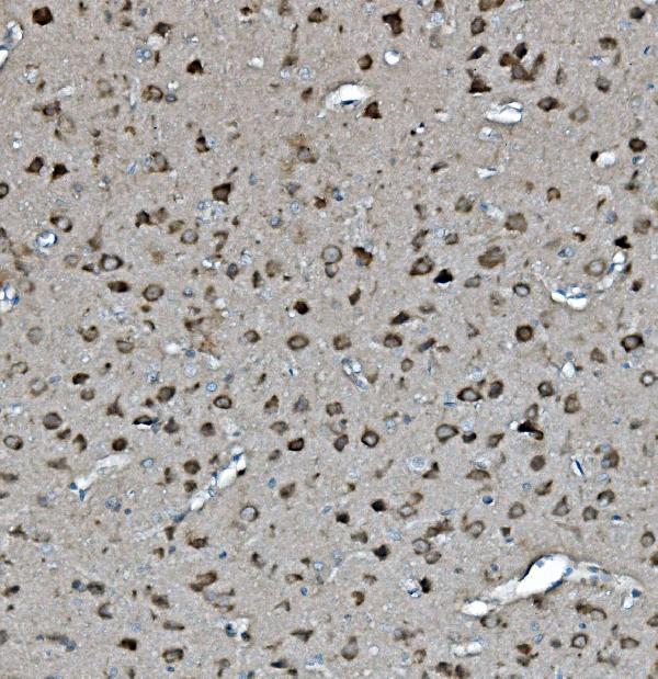
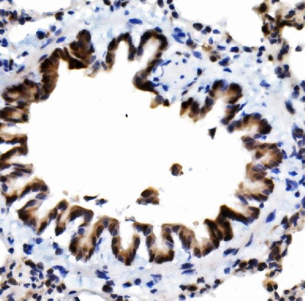
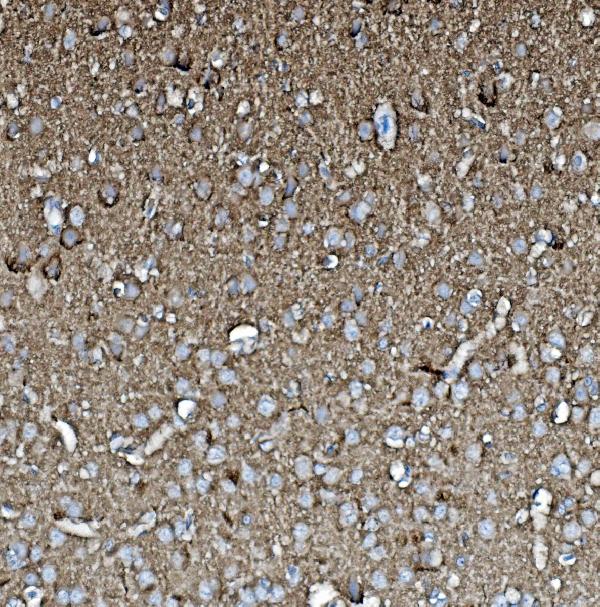
(Figure 4. IHC analysis of Nefh using anti-Nefh antibody (AAA125747).Nefh was detected in paraffin-embedded section of mouse spinal cord tissue. Heat mediated antigen retrieval was performed in EDTA buffer (pH8. 0, epitope retrieval solution). The tissue section was blocked with 10% goat serum. The tissue section was then incubated with 2μg/ml rabbit anti-Nefh Antibody (AAA125747) overnight at 4 degree C. Biotinylated goat anti-rabbit IgG was used as secondary antibody and incubated for 30 minutes at 37 degree C. The tissue section was developed using Strepavidin-Biotin-Complex (SABC) with DAB as the chromogen.)
IHC (Immunohistochemistry)
(Figure 4. IHC analysis of Nefh using anti-Nefh antibody (AAA125747).Nefh was detected in paraffin-embedded section of mouse spinal cord tissue. Heat mediated antigen retrieval was performed in EDTA buffer (pH8. 0, epitope retrieval solution). The tissue section was blocked with 10% goat serum. The tissue section was then incubated with 2μg/ml rabbit anti-Nefh Antibody (AAA125747) overnight at 4 degree C. Biotinylated goat anti-rabbit IgG was used as secondary antibody and incubated for 30 minutes at 37 degree C. The tissue section was developed using Strepavidin-Biotin-Complex (SABC) with DAB as the chromogen.)
Nefh, Polyclonal Antibody (Cat# AAA125747)

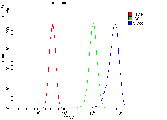 FCM/FACS (Flow Cytometry)
(Figure 2. Flow Cytometry analysis of HELA cells using anti-N WASP/WASL antibody (AAA125749).Overlay histogram showing HELA cells stained with AAA125749 (Blue line). The cells were blocked with 10% normal goat serum. And then incubated with rabbit anti-N WASP/WASL Antibody (AAA125749, 1μg/1x106 cells) for 30 min at 20 degree C. DyLight®488 conjugated goat anti-rabbit IgG (5-10μg/1x106 cells) was used as secondary antibody for 30 minutes at 20 degree C. Isotype control antibody (Green line) was rabbit IgG (1μg/1x106) used under the same conditions. Unlabelled sample (Red line) was also used as a control.)
FCM/FACS (Flow Cytometry)
(Figure 2. Flow Cytometry analysis of HELA cells using anti-N WASP/WASL antibody (AAA125749).Overlay histogram showing HELA cells stained with AAA125749 (Blue line). The cells were blocked with 10% normal goat serum. And then incubated with rabbit anti-N WASP/WASL Antibody (AAA125749, 1μg/1x106 cells) for 30 min at 20 degree C. DyLight®488 conjugated goat anti-rabbit IgG (5-10μg/1x106 cells) was used as secondary antibody for 30 minutes at 20 degree C. Isotype control antibody (Green line) was rabbit IgG (1μg/1x106) used under the same conditions. Unlabelled sample (Red line) was also used as a control.)
N WASP/WASL, Polyclonal Antibody (Cat# AAA125749)
 FCM/FACS (Flow Cytometry)
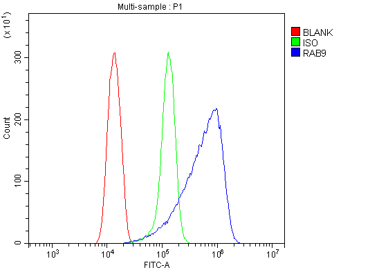
(Figure 3. Flow Cytometry analysis of Hela cells using anti-Rab9/RAB9A antibody (AAA125757).Overlay histogram showing Hela cells stained with AAA125757 (Blue line). The cells were blocked with 10% normal goat serum. And then incubated with rabbit anti-Rab9/RAB9A Antibody (AAA125757,1μg/1x106 cells) for 30 min at 20 degree C. DyLight®488 conjugated goat anti-rabbit IgG (5-10μg/1x106 cells) was used as secondary antibody for 30 minutes at 20 degree C. Isotype control antibody (Green line) was rabbit IgG (1μg/1x106) used under the same conditions. Unlabelled sample (Red line) was also used as a control.)
FCM/FACS (Flow Cytometry)
(Figure 3. Flow Cytometry analysis of Hela cells using anti-Rab9/RAB9A antibody (AAA125757).Overlay histogram showing Hela cells stained with AAA125757 (Blue line). The cells were blocked with 10% normal goat serum. And then incubated with rabbit anti-Rab9/RAB9A Antibody (AAA125757,1μg/1x106 cells) for 30 min at 20 degree C. DyLight®488 conjugated goat anti-rabbit IgG (5-10μg/1x106 cells) was used as secondary antibody for 30 minutes at 20 degree C. Isotype control antibody (Green line) was rabbit IgG (1μg/1x106) used under the same conditions. Unlabelled sample (Red line) was also used as a control.)
Rab9/RAB9A, Polyclonal Antibody (Cat# AAA125757)
 FCM/FACS (Flow Cytometry)
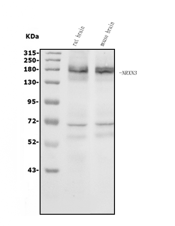
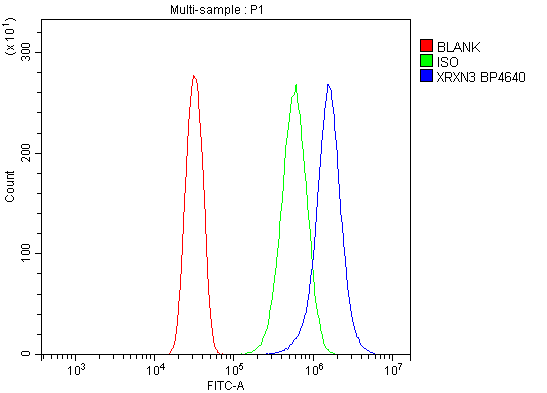
(Figure 2. Flow Cytometry analysis of U20S cells using anti-NRXN3 antibody (AAA125758).Overlay histogram showing U20S cells stained with AAA125758 (Blue line). The cells were blocked with 10% normal goat serum. And then incubated with rabbit anti-NRXN3 Antibody (AAA125758, 1μg/1x106 cells) for 30 min at 20 degree C. DyLight®488 conjugated goat anti-rabbit IgG (5-10μg/1x106 cells) was used as secondary antibody for 30 minutes at 20 degree C. Isotype control antibody (Green line) was rabbit IgG (1μg/1x106) used under the same conditions. Unlabelled sample (Red line) was also used as a control.)
FCM/FACS (Flow Cytometry)
(Figure 2. Flow Cytometry analysis of U20S cells using anti-NRXN3 antibody (AAA125758).Overlay histogram showing U20S cells stained with AAA125758 (Blue line). The cells were blocked with 10% normal goat serum. And then incubated with rabbit anti-NRXN3 Antibody (AAA125758, 1μg/1x106 cells) for 30 min at 20 degree C. DyLight®488 conjugated goat anti-rabbit IgG (5-10μg/1x106 cells) was used as secondary antibody for 30 minutes at 20 degree C. Isotype control antibody (Green line) was rabbit IgG (1μg/1x106) used under the same conditions. Unlabelled sample (Red line) was also used as a control.)
NRXN3, Polyclonal Antibody (Cat# AAA125758)


 FCM/FACS (Flow Cytometry)
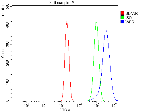
(Figure 4. Flow Cytometry analysis of U20S cells using anti-WFS1 antibody (AAA125604).Overlay histogram showing U20S cells stained with AAA125604 (Blue line). The cells were blocked with 10% normal goat serum. And then incubated with rabbit anti-WFS1 Antibody (AAA125604, 1μg/1x106 cells) for 30 min at 20 degree C. DyLight®488 conjugated goat anti-rabbit IgG (5-10μg/1x106 cells) was used as secondary antibody for 30 minutes at 20 degree C. Isotype control antibody (Green line) was rabbit IgG (1μg/1x106) used under the same conditions. Unlabelled sample (Red line) was also used as a control.)
FCM/FACS (Flow Cytometry)
(Figure 4. Flow Cytometry analysis of U20S cells using anti-WFS1 antibody (AAA125604).Overlay histogram showing U20S cells stained with AAA125604 (Blue line). The cells were blocked with 10% normal goat serum. And then incubated with rabbit anti-WFS1 Antibody (AAA125604, 1μg/1x106 cells) for 30 min at 20 degree C. DyLight®488 conjugated goat anti-rabbit IgG (5-10μg/1x106 cells) was used as secondary antibody for 30 minutes at 20 degree C. Isotype control antibody (Green line) was rabbit IgG (1μg/1x106) used under the same conditions. Unlabelled sample (Red line) was also used as a control.)
WFS1, Polyclonal Antibody (Cat# AAA125604)
 FCM/FACS (Flow Cytometry)
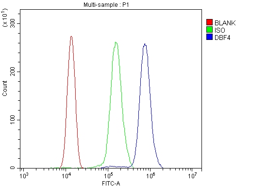
(Figure 3. Flow Cytometry analysis of THP-1 cells using anti-DBF4 antibody (AAA125605).Overlay histogram showing THP-1 cells stained with AAA125605 (Blue line). The cells were blocked with 10% normal goat serum. And then incubated with rabbit anti-DBF4 Antibody (AAA125605, 1μg/1x106 cells) for 30 min at 20 degree C. DyLight®488 conjugated goat anti-rabbit IgG (5-10μg/1x106 cells) was used as secondary antibody for 30 minutes at 20 degree C. Isotype control antibody (Green line) was rabbit IgG (1μg/1x106) used under the same conditions. Unlabelled sample (Red line) was also used as a control.)
FCM/FACS (Flow Cytometry)
(Figure 3. Flow Cytometry analysis of THP-1 cells using anti-DBF4 antibody (AAA125605).Overlay histogram showing THP-1 cells stained with AAA125605 (Blue line). The cells were blocked with 10% normal goat serum. And then incubated with rabbit anti-DBF4 Antibody (AAA125605, 1μg/1x106 cells) for 30 min at 20 degree C. DyLight®488 conjugated goat anti-rabbit IgG (5-10μg/1x106 cells) was used as secondary antibody for 30 minutes at 20 degree C. Isotype control antibody (Green line) was rabbit IgG (1μg/1x106) used under the same conditions. Unlabelled sample (Red line) was also used as a control.)
DBF4, Polyclonal Antibody (Cat# AAA125605)
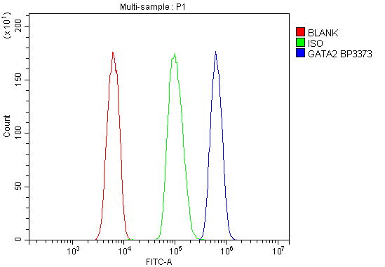 FCM/FACS (Flow Cytometry)
(Figure 3. Flow Cytometry analysis of HL-60 cells using anti- GATA2 antibody (AAA125607).Overlay histogram showing HL-60 cells stained withAAA125607 (Blue line). The cells were blocked with 10% normal goat serum. And then incubated with rabbit anti- GATA2 Antibody (AAA125607, 1μg/1x106 cells) for 30 min at 20 degree C. DyLight®488 conjugated goat anti-rabbit IgG (5-10μg/1x106 cells) was used as secondary antibody for 30 minutes at 20 degree C. Isotype control antibody (Green line) was rabbit IgG (1μg/1x106) used under the same conditions. Unlabelled sample (Red line) was also used as a control.)
FCM/FACS (Flow Cytometry)
(Figure 3. Flow Cytometry analysis of HL-60 cells using anti- GATA2 antibody (AAA125607).Overlay histogram showing HL-60 cells stained withAAA125607 (Blue line). The cells were blocked with 10% normal goat serum. And then incubated with rabbit anti- GATA2 Antibody (AAA125607, 1μg/1x106 cells) for 30 min at 20 degree C. DyLight®488 conjugated goat anti-rabbit IgG (5-10μg/1x106 cells) was used as secondary antibody for 30 minutes at 20 degree C. Isotype control antibody (Green line) was rabbit IgG (1μg/1x106) used under the same conditions. Unlabelled sample (Red line) was also used as a control.)
GATA2, Polyclonal Antibody (Cat# AAA125607)
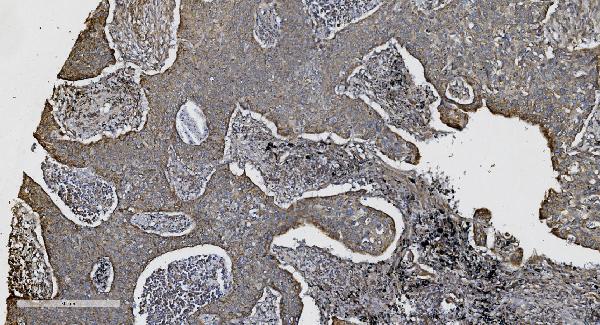
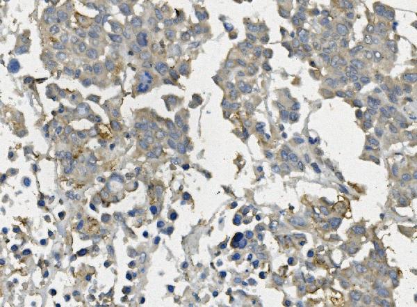
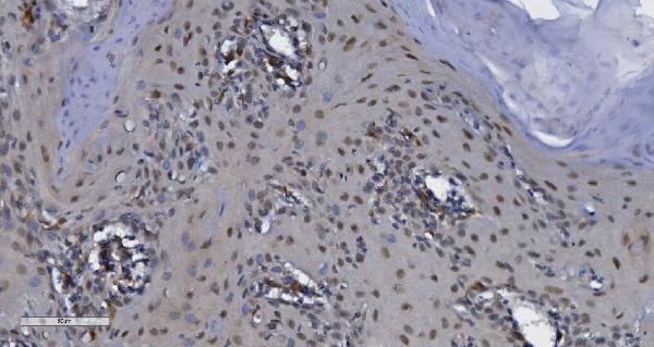
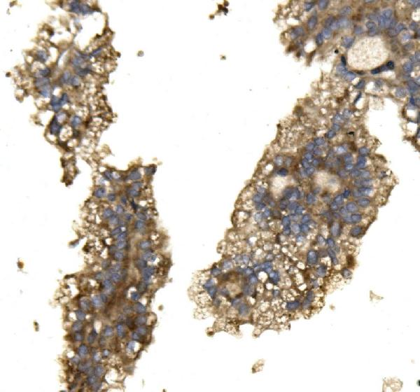
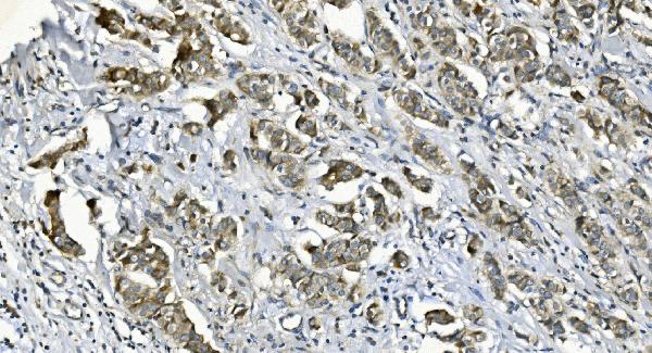
 IHC (Immunohistochemistry)
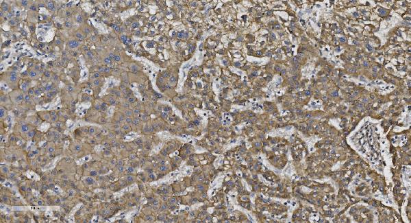
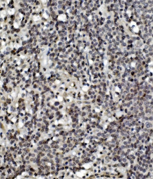
(Figure 5. IHC analysis of N Cadherin/CDH2 using anti-N Cadherin/CDH2 antibody (AAA125611).N Cadherin/CDH2 was detected in paraffin-embedded section of human pancreatic cancer tissue. Heat mediated antigen retrieval was performed in EDTA buffer (pH8. 0, epitope retrieval solution). The tissue section was blocked with 10% goat serum. The tissue section was then incubated with 2μg/ml rabbit anti-N Cadherin/CDH2 Antibody (AAA125611) overnight at 4 degree C. Biotinylated goat anti-rabbit IgG was used as secondary antibody and incubated for 30 minutes at 37 degree C. The tissue section was developed using Strepavidin-Biotin-Complex (SABC) with DAB as the chromogen.)
IHC (Immunohistochemistry)
(Figure 5. IHC analysis of N Cadherin/CDH2 using anti-N Cadherin/CDH2 antibody (AAA125611).N Cadherin/CDH2 was detected in paraffin-embedded section of human pancreatic cancer tissue. Heat mediated antigen retrieval was performed in EDTA buffer (pH8. 0, epitope retrieval solution). The tissue section was blocked with 10% goat serum. The tissue section was then incubated with 2μg/ml rabbit anti-N Cadherin/CDH2 Antibody (AAA125611) overnight at 4 degree C. Biotinylated goat anti-rabbit IgG was used as secondary antibody and incubated for 30 minutes at 37 degree C. The tissue section was developed using Strepavidin-Biotin-Complex (SABC) with DAB as the chromogen.)
N Cadherin/CDH2, Polyclonal Antibody (Cat# AAA125611)
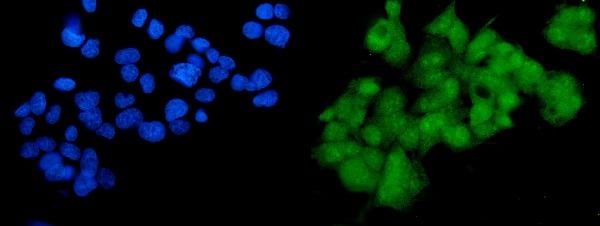
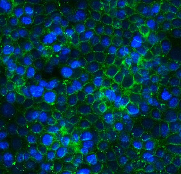
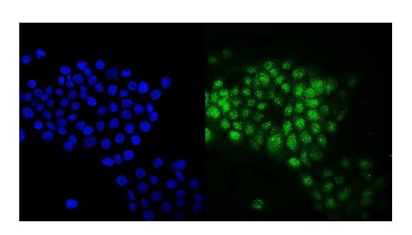
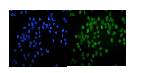
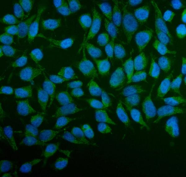
 IF (Immunofluorescence)
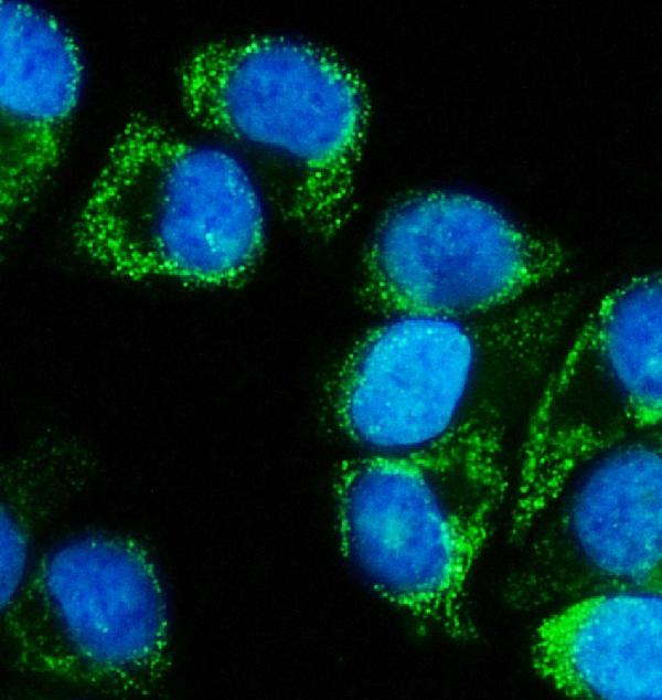
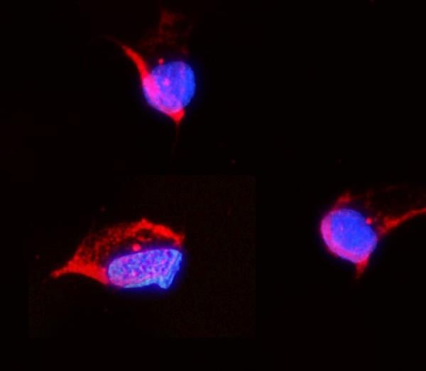
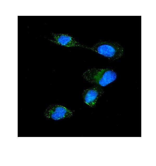
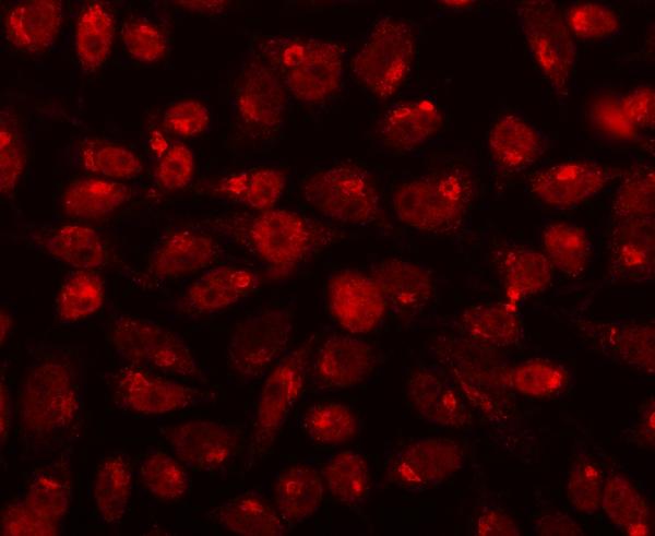
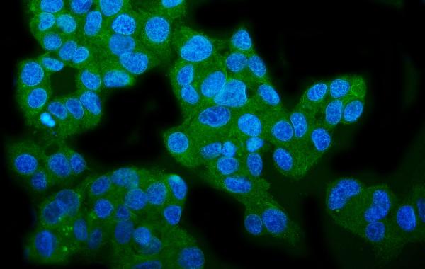
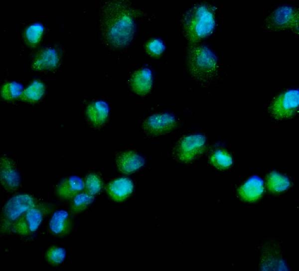
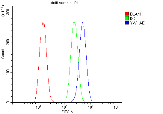
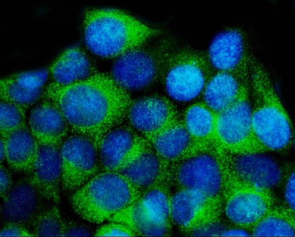
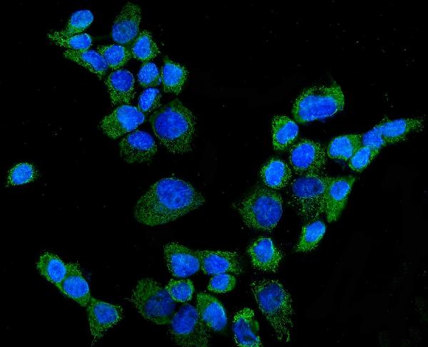
(Figure 4. IF analysis of YWHAE using anti- YWHAE antibody (AAA125617).YWHAE was detected in immunocytochemical section of MCF-7 cells. Enzyme antigen retrieval was performed using IHC enzyme antigen retrieval reagent for 15 mins. The cells were blocked with 10% goat serum. And then incubated with 5μg/mL rabbit anti- YWHAE Antibody (AAA125617) overnight at 4 degree C. DyLight®488 Conjugated Goat Anti-Rabbit IgG was used as secondary antibody at 1:100 dilution and incubated for 30 minutes at 37 degree C. The section was counterstained with DAPI. Visualize using a fluorescence microscope and filter sets appropriate for the label used.)
IF (Immunofluorescence)
(Figure 4. IF analysis of YWHAE using anti- YWHAE antibody (AAA125617).YWHAE was detected in immunocytochemical section of MCF-7 cells. Enzyme antigen retrieval was performed using IHC enzyme antigen retrieval reagent for 15 mins. The cells were blocked with 10% goat serum. And then incubated with 5μg/mL rabbit anti- YWHAE Antibody (AAA125617) overnight at 4 degree C. DyLight®488 Conjugated Goat Anti-Rabbit IgG was used as secondary antibody at 1:100 dilution and incubated for 30 minutes at 37 degree C. The section was counterstained with DAPI. Visualize using a fluorescence microscope and filter sets appropriate for the label used.)
YWHAE, Polyclonal Antibody (Cat# AAA125617)
 FCM/FACS (Flow Cytometry)
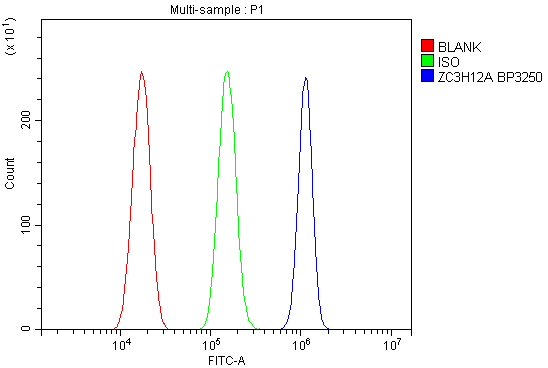
(Figure 5. Flow Cytometry analysis of U87 cells using anti-MCPIP1/ZC3H12A antibody (AAA125618).Overlay histogram showing U87 cells stained with AAA125618 (Blue line). The cells were blocked with 10% normal goat serum. And then incubated with rabbit anti-MCPIP1/ZC3H12A Antibody (AAA125618, 1μg/1x106 cells) for 30 min at 20 degree C. DyLight®488 conjugated goat anti-rabbit IgG (5-10μg/1x106 cells) was used as secondary antibody for 30 minutes at 20 degree C. Isotype control antibody (Green line) was rabbit IgG (1μg/1x106) used under the same conditions. Unlabelled sample (Red line) was also used as a control.)
FCM/FACS (Flow Cytometry)
(Figure 5. Flow Cytometry analysis of U87 cells using anti-MCPIP1/ZC3H12A antibody (AAA125618).Overlay histogram showing U87 cells stained with AAA125618 (Blue line). The cells were blocked with 10% normal goat serum. And then incubated with rabbit anti-MCPIP1/ZC3H12A Antibody (AAA125618, 1μg/1x106 cells) for 30 min at 20 degree C. DyLight®488 conjugated goat anti-rabbit IgG (5-10μg/1x106 cells) was used as secondary antibody for 30 minutes at 20 degree C. Isotype control antibody (Green line) was rabbit IgG (1μg/1x106) used under the same conditions. Unlabelled sample (Red line) was also used as a control.)
MCPIP1/ZC3H12A, Polyclonal Antibody (Cat# AAA125618)
 FCM/FACS (Flow Cytometry)
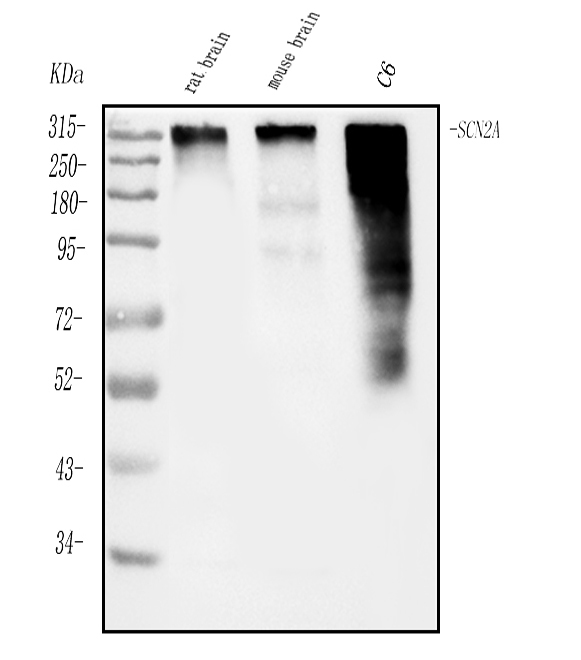
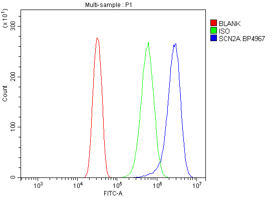
(Figure 2. Flow Cytometry analysis of U20S cells using anti-SCN2A antibody (AAA125619).Overlay histogram showing U20S cells stained with AAA125619 (Blue line). The cells were blocked with 10% normal goat serum. And then incubated with rabbit anti-SCN2A Antibody (AAA125619, 1μg/1x106 cells) for 30 min at 20 degree C. DyLight®488 conjugated goat anti-rabbit IgG (5-10μg/1x106 cells) was used as secondary antibody for 30 minutes at 20 degree C. Isotype control antibody (Green line) was rabbit IgG (1μg/1x106) used under the same conditions. Unlabelled sample (Red line) was also used as a control.)
FCM/FACS (Flow Cytometry)
(Figure 2. Flow Cytometry analysis of U20S cells using anti-SCN2A antibody (AAA125619).Overlay histogram showing U20S cells stained with AAA125619 (Blue line). The cells were blocked with 10% normal goat serum. And then incubated with rabbit anti-SCN2A Antibody (AAA125619, 1μg/1x106 cells) for 30 min at 20 degree C. DyLight®488 conjugated goat anti-rabbit IgG (5-10μg/1x106 cells) was used as secondary antibody for 30 minutes at 20 degree C. Isotype control antibody (Green line) was rabbit IgG (1μg/1x106) used under the same conditions. Unlabelled sample (Red line) was also used as a control.)
SCN2A, Polyclonal Antibody (Cat# AAA125619)
 FCM/FACS (Flow Cytometry)
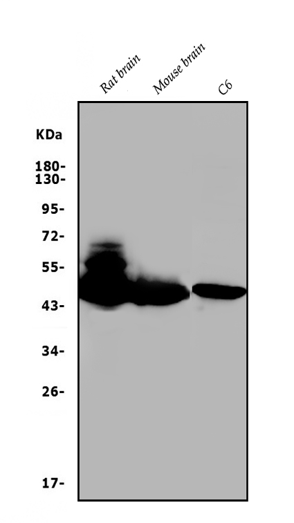
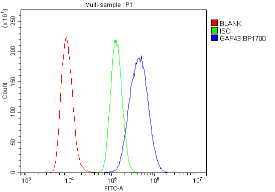
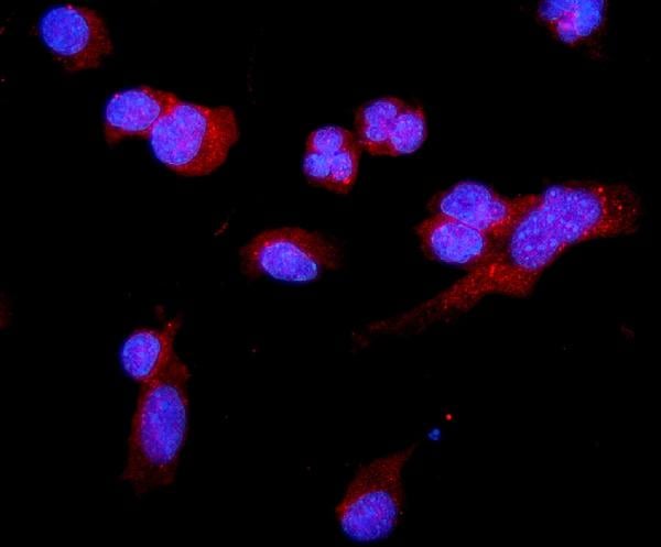
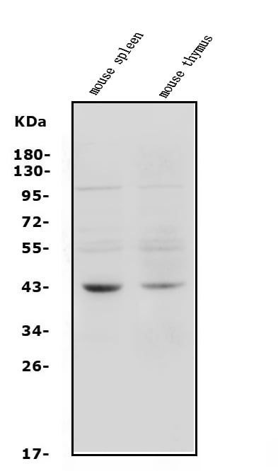
(Figure 3. Flow Cytometry analysis of Raji cells using anti-GAP43 antibody (AAA125626).Overlay histogram showing Raji cells stained with AAA125626 (Blue line). The cells were blocked with 10% normal goat serum. And then incubated with rabbit anti-GAP43 Antibody (AAA125626,1μg/1x106 cells) for 30 min at 20 degree C. DyLight®488 conjugated goat anti-rabbit IgG (5-10μg/1x106 cells) was used as secondary antibody for 30 minutes at 20 degree C. Isotype control antibody (Green line) was rabbit IgG (1μg/1x106) used under the same conditions. Unlabelled sample (Red line) was also used as a control.)
FCM/FACS (Flow Cytometry)
(Figure 3. Flow Cytometry analysis of Raji cells using anti-GAP43 antibody (AAA125626).Overlay histogram showing Raji cells stained with AAA125626 (Blue line). The cells were blocked with 10% normal goat serum. And then incubated with rabbit anti-GAP43 Antibody (AAA125626,1μg/1x106 cells) for 30 min at 20 degree C. DyLight®488 conjugated goat anti-rabbit IgG (5-10μg/1x106 cells) was used as secondary antibody for 30 minutes at 20 degree C. Isotype control antibody (Green line) was rabbit IgG (1μg/1x106) used under the same conditions. Unlabelled sample (Red line) was also used as a control.)
GAP43, Polyclonal Antibody (Cat# AAA125626)
 FCM/FACS (Flow Cytometry)
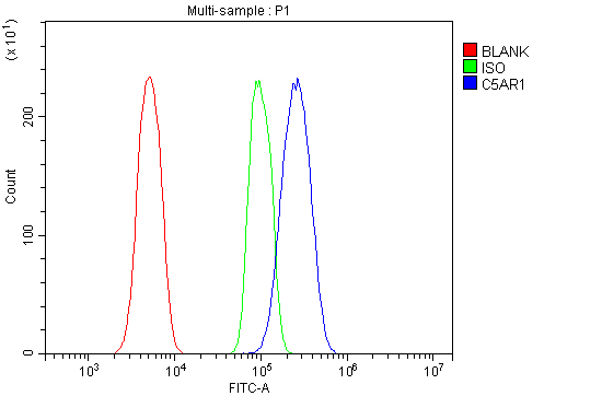
(Figure 3. Flow Cytometry analysis of RAW264. 7 cells using anti-C5a-R/C5ar1 antibody (AAA125628).Overlay histogram showing RAW264. 7 cells stained with AAA125628 (Blue line). The cells were blocked with 10% normal goat serum. And then incubated with rabbit anti-C5a-R/C5ar1 Antibody (AAA125628,1μg/1x106 cells) for 30 min at 20 degree C. DyLight®488 conjugated goat anti-rabbit IgG (5-10μg/1x106 cells) was used as secondary antibody for 30 minutes at 20 degree C. Isotype control antibody (Green line) was rabbit IgG (1μg/1x106) used under the same conditions. Unlabelled sample (Red line) was also used as a control.)
FCM/FACS (Flow Cytometry)
(Figure 3. Flow Cytometry analysis of RAW264. 7 cells using anti-C5a-R/C5ar1 antibody (AAA125628).Overlay histogram showing RAW264. 7 cells stained with AAA125628 (Blue line). The cells were blocked with 10% normal goat serum. And then incubated with rabbit anti-C5a-R/C5ar1 Antibody (AAA125628,1μg/1x106 cells) for 30 min at 20 degree C. DyLight®488 conjugated goat anti-rabbit IgG (5-10μg/1x106 cells) was used as secondary antibody for 30 minutes at 20 degree C. Isotype control antibody (Green line) was rabbit IgG (1μg/1x106) used under the same conditions. Unlabelled sample (Red line) was also used as a control.)
C5a-R/C5ar1, Polyclonal Antibody (Cat# AAA125628)
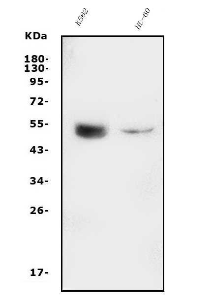
 WB (Western Blot)
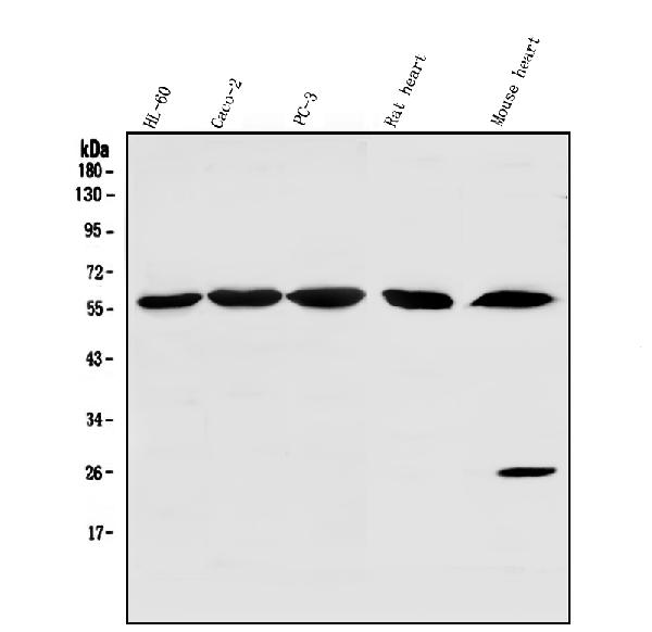
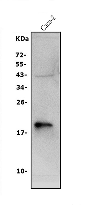
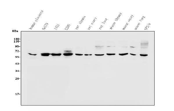
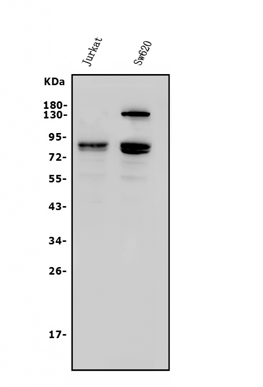
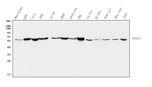
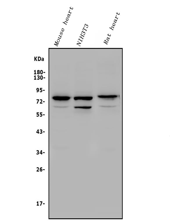
(Figure 2. Western blot analysis of CYP27A1 using anti-CYP27A1 antibody (AAA125635).Electrophoresis was performed on a 5-20% SDS-PAGE gel at 70V (Stacking gel) / 90V (Resolving gel) for 2-3 hours. The sample well of each lane was loaded with 50ug of sample under reducing conditions.Lane 1: human placenta tissue lysatesLane 2: human HEPG2 whole cell lysatesLane 3: human CACO-2 whole cell lysatesLane 4: human SW620 whole cell lysatesLane 5: human SGC-7901 whole cell lysatesLane 6: human HEK293 whole cell lysatesLane 7: monkey liver tissue lysatesLane 8: human U20S whole cell lysatesLane 9: rat liver tissue lysatesLane 10: rat heart tissue lysatesLane 11: mouse liver tissue lysatesLane 12: mouse heart tissue lysatesLane 13: mouse NIH/3T3 whole cell lysates.After Electrophoresis, proteins were transferred to a Nitrocellulose membrane at 150mA for 50-90 minutes. Blocked the membrane with 5% Non-fat Milk/ TBS for 1. 5 hour at RT. The membrane was incubated with rabbit anti-CYP27A1 antigen affinity purified polyclonal antibody (Catalog # AAA125635) at 0. 25 μg/mL overnight at 4 degree C, then washed with TBS-0. 1%Tween 3 times with 5 minutes each and probed with a goat anti-rabbit IgG-HRP secondary antibody at a dilution of 1:5000 for 1. 5 hour at RT. The signal is developed using an Enhanced Chemiluminescent detection (ECL) kit with Tanon 5200 system. A specific band was detected for CYP27A1 at approximately 60KD. The expected band size for CYP27A1 is at 60KD.)
WB (Western Blot)
(Figure 2. Western blot analysis of CYP27A1 using anti-CYP27A1 antibody (AAA125635).Electrophoresis was performed on a 5-20% SDS-PAGE gel at 70V (Stacking gel) / 90V (Resolving gel) for 2-3 hours. The sample well of each lane was loaded with 50ug of sample under reducing conditions.Lane 1: human placenta tissue lysatesLane 2: human HEPG2 whole cell lysatesLane 3: human CACO-2 whole cell lysatesLane 4: human SW620 whole cell lysatesLane 5: human SGC-7901 whole cell lysatesLane 6: human HEK293 whole cell lysatesLane 7: monkey liver tissue lysatesLane 8: human U20S whole cell lysatesLane 9: rat liver tissue lysatesLane 10: rat heart tissue lysatesLane 11: mouse liver tissue lysatesLane 12: mouse heart tissue lysatesLane 13: mouse NIH/3T3 whole cell lysates.After Electrophoresis, proteins were transferred to a Nitrocellulose membrane at 150mA for 50-90 minutes. Blocked the membrane with 5% Non-fat Milk/ TBS for 1. 5 hour at RT. The membrane was incubated with rabbit anti-CYP27A1 antigen affinity purified polyclonal antibody (Catalog # AAA125635) at 0. 25 μg/mL overnight at 4 degree C, then washed with TBS-0. 1%Tween 3 times with 5 minutes each and probed with a goat anti-rabbit IgG-HRP secondary antibody at a dilution of 1:5000 for 1. 5 hour at RT. The signal is developed using an Enhanced Chemiluminescent detection (ECL) kit with Tanon 5200 system. A specific band was detected for CYP27A1 at approximately 60KD. The expected band size for CYP27A1 is at 60KD.)
CYP27A1, Polyclonal Antibody (Cat# AAA125635)
 FCM/FACS (Flow Cytometry)
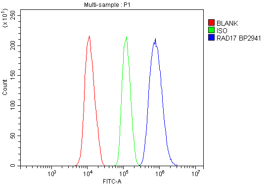
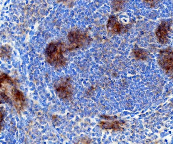
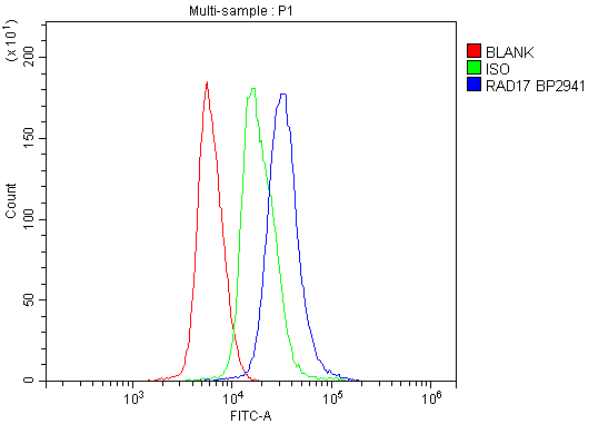
(Figure 5. Flow Cytometry analysis of mouse spleen tissues using anti-Rad17 antibody (AAA125638).Overlay histogram showing mouse spleen tissues stained with AAA125638 (Blue line). The tissues were blocked with 10% normal goat serum. And then incubated with rabbit anti-Rad17 Antibody (AAA125638,1μg/1x106 cells) for 30 min at 20 degree C. DyLight®488 conjugated goat anti-rabbit IgG (5-10μg/1x106 cells) was used as secondary antibody for 30 minutes at 20 degree C. Isotype control antibody (Green line) was rabbit IgG (1μg/1x106) used under the same conditions. Unlabelled sample (Red line) was also used as a control.)
FCM/FACS (Flow Cytometry)
(Figure 5. Flow Cytometry analysis of mouse spleen tissues using anti-Rad17 antibody (AAA125638).Overlay histogram showing mouse spleen tissues stained with AAA125638 (Blue line). The tissues were blocked with 10% normal goat serum. And then incubated with rabbit anti-Rad17 Antibody (AAA125638,1μg/1x106 cells) for 30 min at 20 degree C. DyLight®488 conjugated goat anti-rabbit IgG (5-10μg/1x106 cells) was used as secondary antibody for 30 minutes at 20 degree C. Isotype control antibody (Green line) was rabbit IgG (1μg/1x106) used under the same conditions. Unlabelled sample (Red line) was also used as a control.)
Rad17, Polyclonal Antibody (Cat# AAA125638)

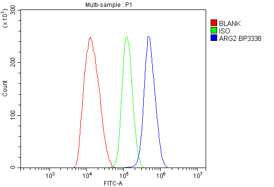 FCM/FACS (Flow Cytometry)
(Figure 5. Flow Cytometry analysis of HEPG2 cells using anti-ARG2 antibody (AAA125644).Overlay histogram showing HEPG2 cells stained with AAA125644(Blue line). The cells were blocked with 10% normal goat serum. And then incubated with rabbit anti-ARG2 Antibody (AAA125644, 1μg/1x106 cells) for 30 min at 20 degree C. DyLight®488 conjugated goat anti-rabbit IgG (5-10μg/1x106 cells) was used as secondary antibody for 30 minutes at 20 degree C. Isotype control antibody (Green line) was rabbit IgG (1μg/1x106) used under the same conditions. Unlabelled sample (Red line) was also used as a control.)
FCM/FACS (Flow Cytometry)
(Figure 5. Flow Cytometry analysis of HEPG2 cells using anti-ARG2 antibody (AAA125644).Overlay histogram showing HEPG2 cells stained with AAA125644(Blue line). The cells were blocked with 10% normal goat serum. And then incubated with rabbit anti-ARG2 Antibody (AAA125644, 1μg/1x106 cells) for 30 min at 20 degree C. DyLight®488 conjugated goat anti-rabbit IgG (5-10μg/1x106 cells) was used as secondary antibody for 30 minutes at 20 degree C. Isotype control antibody (Green line) was rabbit IgG (1μg/1x106) used under the same conditions. Unlabelled sample (Red line) was also used as a control.)
ARG2, Polyclonal Antibody (Cat# AAA125644)

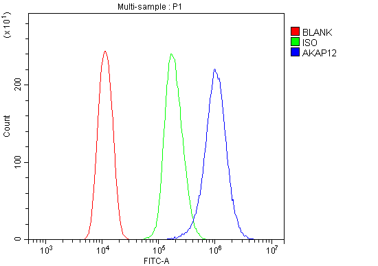
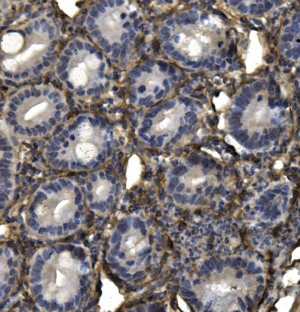
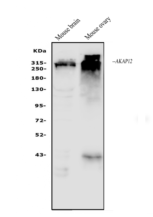
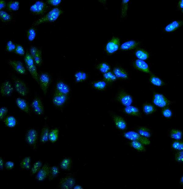
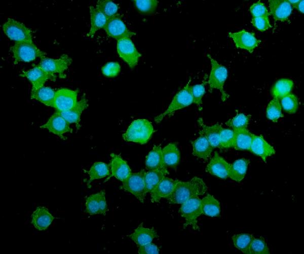 IF (Immunofluorescence)
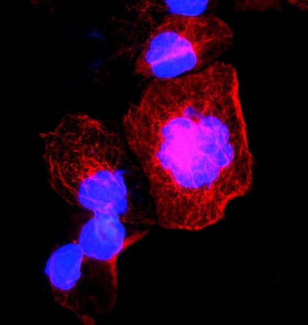
(Figure 5. IF analysis of AKAP12 using anti- AKAP12 antibody (AAA125648).AKAP12 was detected in immunocytochemical section of HEPA1-6 cells. Enzyme antigen retrieval was performed using IHC enzyme antigen retrieval reagent for 15 mins. The cells were blocked with 10% goat serum. And then incubated with 5μg/mL rabbit anti- AKAP12 Antibody (AAA125648) overnight at 4 degree C. DyLight®488 Conjugated Goat Anti-Rabbit IgG was used as secondary antibody at 1:100 dilution and incubated for 30 minutes at 37 degree C. The section was counterstained with DAPI. Visualize using a fluorescence microscope and filter sets appropriate for the label used.)
IF (Immunofluorescence)
(Figure 5. IF analysis of AKAP12 using anti- AKAP12 antibody (AAA125648).AKAP12 was detected in immunocytochemical section of HEPA1-6 cells. Enzyme antigen retrieval was performed using IHC enzyme antigen retrieval reagent for 15 mins. The cells were blocked with 10% goat serum. And then incubated with 5μg/mL rabbit anti- AKAP12 Antibody (AAA125648) overnight at 4 degree C. DyLight®488 Conjugated Goat Anti-Rabbit IgG was used as secondary antibody at 1:100 dilution and incubated for 30 minutes at 37 degree C. The section was counterstained with DAPI. Visualize using a fluorescence microscope and filter sets appropriate for the label used.)
AKAP12, Polyclonal Antibody (Cat# AAA125648)
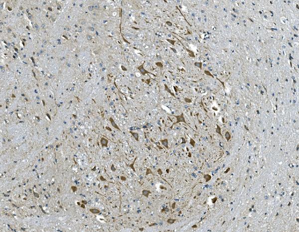
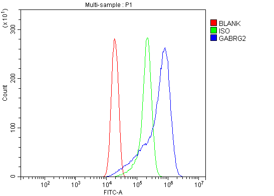 FCM/FACS (Flow Cytometry)
(Figure 3. Flow Cytometry analysis of THP-1 cells using anti-GABA A Receptor gamma 2/GABRG2 antibody (AAA125651).Overlay histogram showing THP-1 cells stained with AAA125651 (Blue line). The cells were blocked with 10% normal goat serum. And then incubated with rabbit anti-GABA A Receptor gamma 2/GABRG2 Antibody (AAA125651, 1μg/1x106 cells) for 30 min at 20 degree C. DyLight®488 conjugated goat anti-rabbit IgG (5-10μg/1x106 cells) was used as secondary antibody for 30 minutes at 20 degree C. Isotype control antibody (Green line) was rabbit IgG (1μg/1x106) used under the same conditions. Unlabelled sample (Red line) was also used as a control.)
FCM/FACS (Flow Cytometry)
(Figure 3. Flow Cytometry analysis of THP-1 cells using anti-GABA A Receptor gamma 2/GABRG2 antibody (AAA125651).Overlay histogram showing THP-1 cells stained with AAA125651 (Blue line). The cells were blocked with 10% normal goat serum. And then incubated with rabbit anti-GABA A Receptor gamma 2/GABRG2 Antibody (AAA125651, 1μg/1x106 cells) for 30 min at 20 degree C. DyLight®488 conjugated goat anti-rabbit IgG (5-10μg/1x106 cells) was used as secondary antibody for 30 minutes at 20 degree C. Isotype control antibody (Green line) was rabbit IgG (1μg/1x106) used under the same conditions. Unlabelled sample (Red line) was also used as a control.)
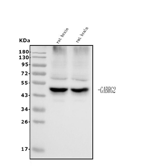
GABA A Receptor gamma 2/GABRG2, Polyclonal Antibody (Cat# AAA125651)
 FCM/FACS (Flow Cytometry)
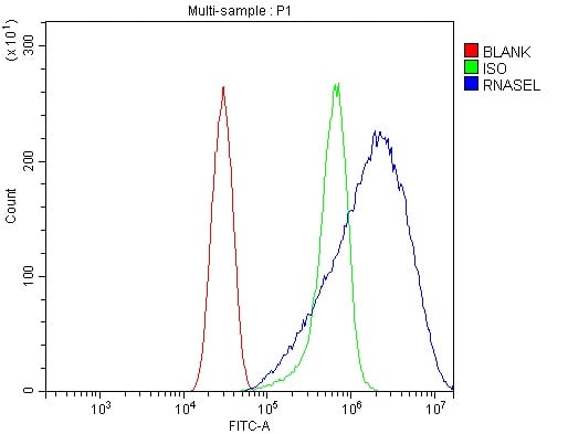
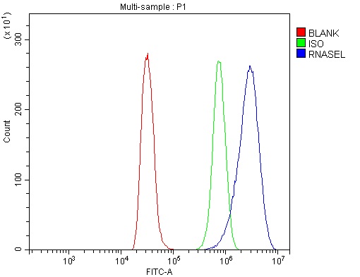
(Figure 3. Flow Cytometry analysis of NRK cells using anti-RNase L/Rnasel antibody (AAA125662).Overlay histogram showing NRK cells stained with AAA125662 (Blue line). The cells were blocked with 10% normal goat serum. And then incubated with rabbit anti-RNase L/Rnasel Antibody (AAA125662, 1μg/1x106 cells) for 30 min at 20 degree C. DyLight®488 conjugated goat anti-rabbit IgG (5-10μg/1x106 cells) was used as secondary antibody for 30 minutes at 20 degree C. Isotype control antibody (Green line) was rabbit IgG (1μg/1x106) used under the same conditions. Unlabelled sample (Red line) was also used as a control.)
FCM/FACS (Flow Cytometry)
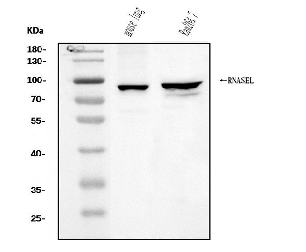
(Figure 3. Flow Cytometry analysis of NRK cells using anti-RNase L/Rnasel antibody (AAA125662).Overlay histogram showing NRK cells stained with AAA125662 (Blue line). The cells were blocked with 10% normal goat serum. And then incubated with rabbit anti-RNase L/Rnasel Antibody (AAA125662, 1μg/1x106 cells) for 30 min at 20 degree C. DyLight®488 conjugated goat anti-rabbit IgG (5-10μg/1x106 cells) was used as secondary antibody for 30 minutes at 20 degree C. Isotype control antibody (Green line) was rabbit IgG (1μg/1x106) used under the same conditions. Unlabelled sample (Red line) was also used as a control.)
RNase L/Rnasel, Polyclonal Antibody (Cat# AAA125662)
 FCM/FACS (Flow Cytometry)
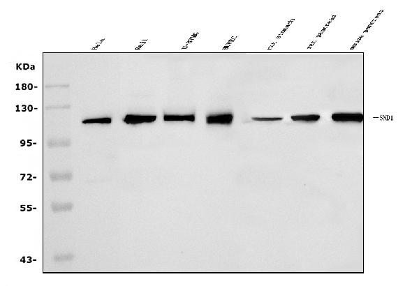
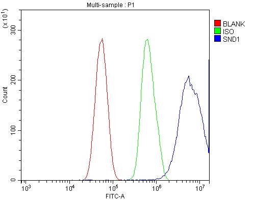
(Figure 5. Flow Cytometry analysis of U87 cells using anti-SND1 antibody (AAA125665).Overlay histogram showing U87 cells stained with AAA125665 (Blue line). The cells were blocked with 10% normal goat serum. And then incubated with rabbit anti-SND1 Antibody (AAA125665, 1μg/1x106 cells) for 30 min at 20 degree C. DyLight®488 conjugated goat anti-rabbit IgG (5-10μg/1x106 cells) was used as secondary antibody for 30 minutes at 20 degree C. Isotype control antibody (Green line) was rabbit IgG (1μg/1x106) used under the same conditions. Unlabelled sample (Red line) was also used as a control.)
FCM/FACS (Flow Cytometry)
(Figure 5. Flow Cytometry analysis of U87 cells using anti-SND1 antibody (AAA125665).Overlay histogram showing U87 cells stained with AAA125665 (Blue line). The cells were blocked with 10% normal goat serum. And then incubated with rabbit anti-SND1 Antibody (AAA125665, 1μg/1x106 cells) for 30 min at 20 degree C. DyLight®488 conjugated goat anti-rabbit IgG (5-10μg/1x106 cells) was used as secondary antibody for 30 minutes at 20 degree C. Isotype control antibody (Green line) was rabbit IgG (1μg/1x106) used under the same conditions. Unlabelled sample (Red line) was also used as a control.)
SND1, Polyclonal Antibody (Cat# AAA125665)

 FCM/FACS (Flow Cytometry)
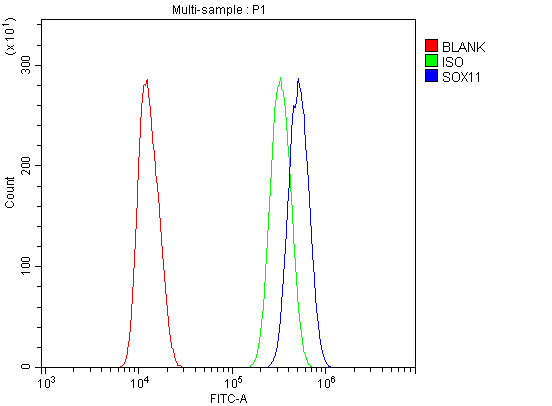
(Figure 3. Flow Cytometry analysis of U20S cells using anti-SOX11 antibody (AAA125666).Overlay histogram showing U20S cells stained with AAA125666 (Blue line). The cells were blocked with 10% normal goat serum. And then incubated with rabbit anti-SOX11 Antibody (AAA125666, 1μg/1x106 cells) for 30 min at 20 degree C. DyLight®488 conjugated goat anti-rabbit IgG (5-10μg/1x106 cells) was used as secondary antibody for 30 minutes at 20 degree C. Isotype control antibody (Green line) was rabbit IgG (1μg/1x106) used under the same conditions. Unlabelled sample (Red line) was also used as a control.)
FCM/FACS (Flow Cytometry)
(Figure 3. Flow Cytometry analysis of U20S cells using anti-SOX11 antibody (AAA125666).Overlay histogram showing U20S cells stained with AAA125666 (Blue line). The cells were blocked with 10% normal goat serum. And then incubated with rabbit anti-SOX11 Antibody (AAA125666, 1μg/1x106 cells) for 30 min at 20 degree C. DyLight®488 conjugated goat anti-rabbit IgG (5-10μg/1x106 cells) was used as secondary antibody for 30 minutes at 20 degree C. Isotype control antibody (Green line) was rabbit IgG (1μg/1x106) used under the same conditions. Unlabelled sample (Red line) was also used as a control.)
SOX11, Polyclonal Antibody (Cat# AAA125666)
 FCM/FACS (Flow Cytometry)
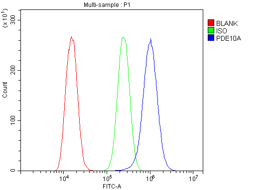
(Figure 2. Flow Cytometry analysis of A549 cells using anti-PDE10A antibody (AAA125667).Overlay histogram showing A549 cells stained with AAA125667 (Blue line). The cells were blocked with 10% normal goat serum. And then incubated with rabbit anti-PDE10A Antibody (AAA125667, 1μg/1x106 cells) for 30 min at 20 degree C. DyLight®488 conjugated goat anti-rabbit IgG (5-10μg/1x106 cells) was used as secondary antibody for 30 minutes at 20 degree C. Isotype control antibody (Green line) was rabbit IgG (1μg/1x106) used under the same conditions. Unlabelled sample (Red line) was also used as a control.)
FCM/FACS (Flow Cytometry)
(Figure 2. Flow Cytometry analysis of A549 cells using anti-PDE10A antibody (AAA125667).Overlay histogram showing A549 cells stained with AAA125667 (Blue line). The cells were blocked with 10% normal goat serum. And then incubated with rabbit anti-PDE10A Antibody (AAA125667, 1μg/1x106 cells) for 30 min at 20 degree C. DyLight®488 conjugated goat anti-rabbit IgG (5-10μg/1x106 cells) was used as secondary antibody for 30 minutes at 20 degree C. Isotype control antibody (Green line) was rabbit IgG (1μg/1x106) used under the same conditions. Unlabelled sample (Red line) was also used as a control.)
PDE10A, Polyclonal Antibody (Cat# AAA125667)
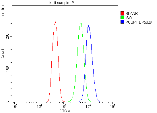
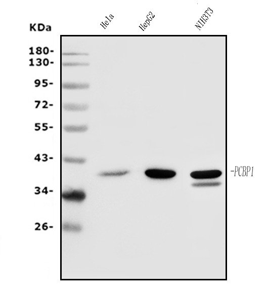
 FCM/FACS (Flow Cytometry)
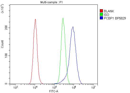
(Figure 4. Flow Cytometry analysis of RH35 cells using anti-PCBP1 antibody (AAA125668).Overlay histogram showing RH35 cells stained with AAA125668 (Blue line). The cells were blocked with 10% normal goat serum. And then incubated with rabbit anti-PCBP1 Antibody (AAA125668, 1μg/1x106 cells) for 30 min at 20 degree C. DyLight®488 conjugated goat anti-rabbit IgG (5-10μg/1x106 cells) was used as secondary antibody for 30 minutes at 20 degree C. Isotype control antibody (Green line) was rabbit IgG (1μg/1x106) used under the same conditions. Unlabelled sample (Red line) was also used as a control.)
FCM/FACS (Flow Cytometry)
(Figure 4. Flow Cytometry analysis of RH35 cells using anti-PCBP1 antibody (AAA125668).Overlay histogram showing RH35 cells stained with AAA125668 (Blue line). The cells were blocked with 10% normal goat serum. And then incubated with rabbit anti-PCBP1 Antibody (AAA125668, 1μg/1x106 cells) for 30 min at 20 degree C. DyLight®488 conjugated goat anti-rabbit IgG (5-10μg/1x106 cells) was used as secondary antibody for 30 minutes at 20 degree C. Isotype control antibody (Green line) was rabbit IgG (1μg/1x106) used under the same conditions. Unlabelled sample (Red line) was also used as a control.)
PCBP1, Polyclonal Antibody (Cat# AAA125668)
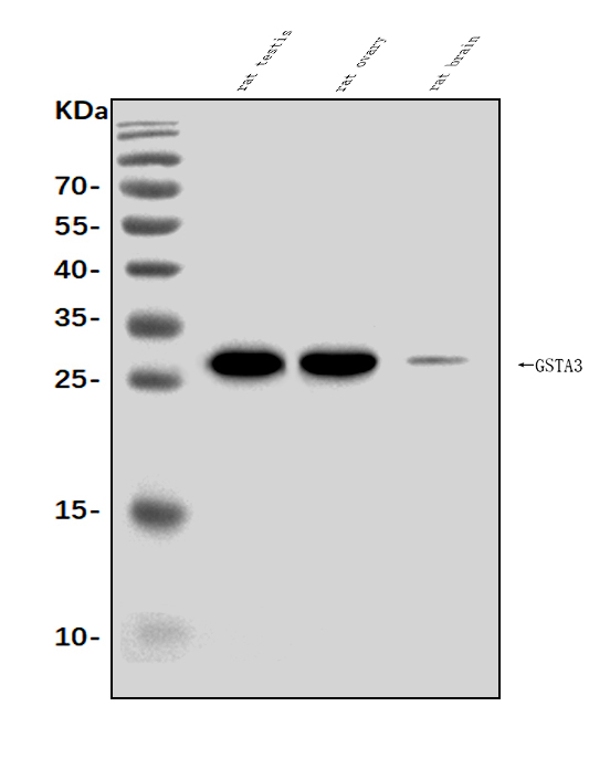
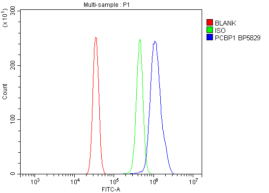
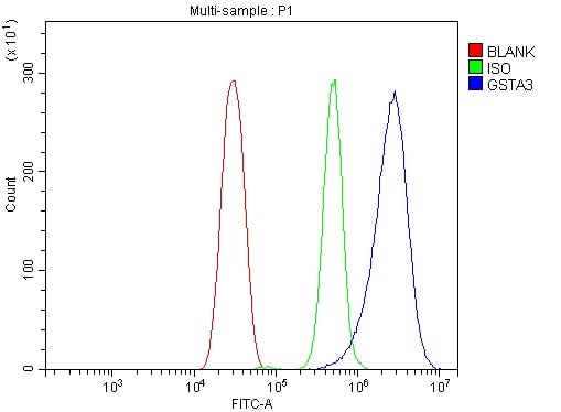 FCM/FACS (Flow Cytometry)
(Figure 2. Flow Cytometry analysis of HEPA1-6 cells using anti-Gsta3 antibody (AAA125760).Overlay histogram showing HEPA1-6 cells stained with AAA125760 (Blue line). The cells were blocked with 10% normal goat serum. And then incubated with rabbit anti-Gsta3 Antibody (AAA125760, 1μg/1x106 cells) for 30 min at 20 degree C. DyLight®488 conjugated goat anti-rabbit IgG (5-10μg/1x106 cells) was used as secondary antibody for 30 minutes at 20 degree C. Isotype control antibody (Green line) was rabbit IgG (1μg/1x106) used under the same conditions. Unlabelled sample (Red line) was also used as a control.)
FCM/FACS (Flow Cytometry)
(Figure 2. Flow Cytometry analysis of HEPA1-6 cells using anti-Gsta3 antibody (AAA125760).Overlay histogram showing HEPA1-6 cells stained with AAA125760 (Blue line). The cells were blocked with 10% normal goat serum. And then incubated with rabbit anti-Gsta3 Antibody (AAA125760, 1μg/1x106 cells) for 30 min at 20 degree C. DyLight®488 conjugated goat anti-rabbit IgG (5-10μg/1x106 cells) was used as secondary antibody for 30 minutes at 20 degree C. Isotype control antibody (Green line) was rabbit IgG (1μg/1x106) used under the same conditions. Unlabelled sample (Red line) was also used as a control.)
Gsta3, Polyclonal Antibody (Cat# AAA125760)
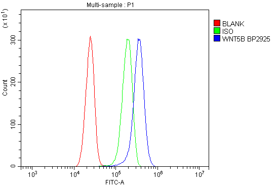 FCM/FACS (Flow Cytometry)
(Figure 2. Flow Cytometry analysis of U87 cells using anti-Wnt5b antibody (AAA125761).Overlay histogram showing U87 cells stained with AAA125761 (Blue line). The cells were blocked with 10% normal goat serum. And then incubated with rabbit anti-Wnt5b Antibody (AAA125761,1μg/1x106 cells) for 30 min at 20 degree C. DyLight®488 conjugated goat anti-rabbit IgG (5-10μg/1x106 cells) was used as secondary antibody for 30 minutes at 20 degree C. Isotype control antibody (Green line) was rabbit IgG (1μg/1x106) used under the same conditions. Unlabelled sample (Red line) was also used as a control.)
FCM/FACS (Flow Cytometry)
(Figure 2. Flow Cytometry analysis of U87 cells using anti-Wnt5b antibody (AAA125761).Overlay histogram showing U87 cells stained with AAA125761 (Blue line). The cells were blocked with 10% normal goat serum. And then incubated with rabbit anti-Wnt5b Antibody (AAA125761,1μg/1x106 cells) for 30 min at 20 degree C. DyLight®488 conjugated goat anti-rabbit IgG (5-10μg/1x106 cells) was used as secondary antibody for 30 minutes at 20 degree C. Isotype control antibody (Green line) was rabbit IgG (1μg/1x106) used under the same conditions. Unlabelled sample (Red line) was also used as a control.)
Wnt5b, Polyclonal Antibody (Cat# AAA125761)
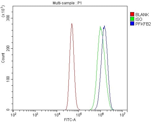
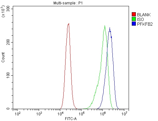 FCM/FACS (Flow Cytometry)
(Figure 4. Flow Cytometry analysis of CACO-2 cells using anti-PFKFB2 antibody (AAA125765).Overlay histogram showing CACO-2 cells stained with AAA125765 (Blue line). The cells were blocked with 10% normal goat serum. And then incubated with rabbit anti-PFKFB2 Antibody (AAA125765, 1μg/1x106 cells) for 30 min at 20 degree C. DyLight®488 conjugated goat anti-rabbit IgG (5-10μg/1x106 cells) was used as secondary antibody for 30 minutes at 20 degree C. Isotype control antibody (Green line) was rabbit IgG (1μg/1x106) used under the same conditions. Unlabelled sample (Red line) was also used as a control.)
FCM/FACS (Flow Cytometry)
(Figure 4. Flow Cytometry analysis of CACO-2 cells using anti-PFKFB2 antibody (AAA125765).Overlay histogram showing CACO-2 cells stained with AAA125765 (Blue line). The cells were blocked with 10% normal goat serum. And then incubated with rabbit anti-PFKFB2 Antibody (AAA125765, 1μg/1x106 cells) for 30 min at 20 degree C. DyLight®488 conjugated goat anti-rabbit IgG (5-10μg/1x106 cells) was used as secondary antibody for 30 minutes at 20 degree C. Isotype control antibody (Green line) was rabbit IgG (1μg/1x106) used under the same conditions. Unlabelled sample (Red line) was also used as a control.)
PFKFB2, Polyclonal Antibody (Cat# AAA125765)
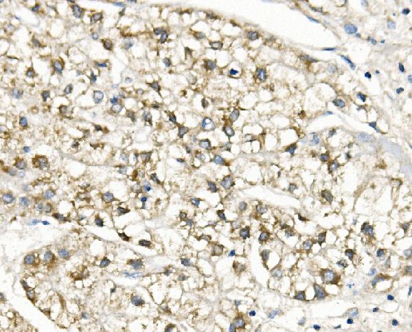
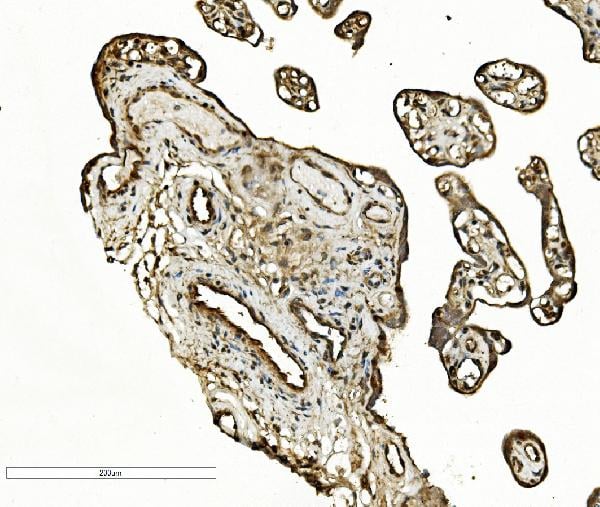
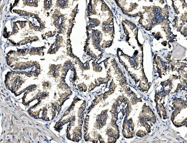
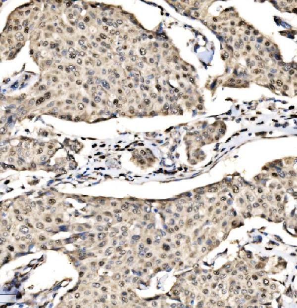
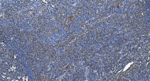
 IHC (Immunohistochemisry)
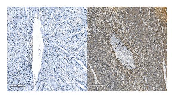
(Figure 3. IHC analysis of Slc25a1 using anti-Slc25a1 antibody (AAA125766).Slc25a1 was detected in paraffin-embedded section of human mammary cancer tissue. Heat mediated antigen retrieval was performed in EDTA buffer (pH8. 0, epitope retrieval solution). The tissue section was blocked with 10% goat serum. The tissue section was then incubated with 2μg/ml rabbit anti-Slc25a1 Antibody (AAA125766) overnight at 4 degree C. Biotinylated goat anti-rabbit IgG was used as secondary antibody and incubated for 30 minutes at 37 degree C. The tissue section was developed using Strepavidin-Biotin-Complex (SABC) with DAB as the chromogen.)
IHC (Immunohistochemisry)
(Figure 3. IHC analysis of Slc25a1 using anti-Slc25a1 antibody (AAA125766).Slc25a1 was detected in paraffin-embedded section of human mammary cancer tissue. Heat mediated antigen retrieval was performed in EDTA buffer (pH8. 0, epitope retrieval solution). The tissue section was blocked with 10% goat serum. The tissue section was then incubated with 2μg/ml rabbit anti-Slc25a1 Antibody (AAA125766) overnight at 4 degree C. Biotinylated goat anti-rabbit IgG was used as secondary antibody and incubated for 30 minutes at 37 degree C. The tissue section was developed using Strepavidin-Biotin-Complex (SABC) with DAB as the chromogen.)
Slc25a1, Polyclonal Antibody (Cat# AAA125766)
 FCM/FACS (Flow Cytometry)
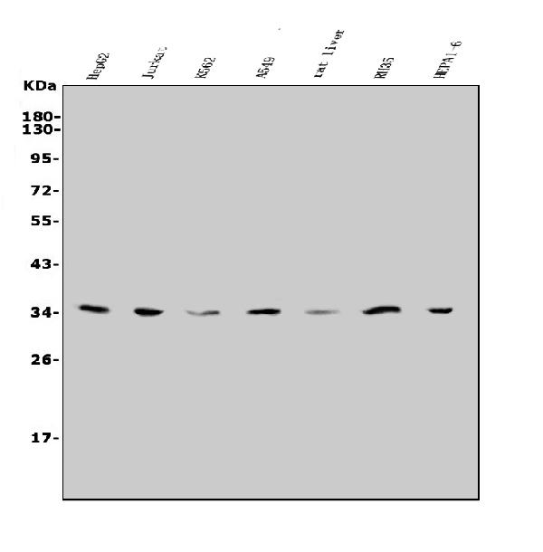
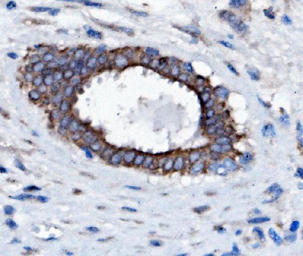
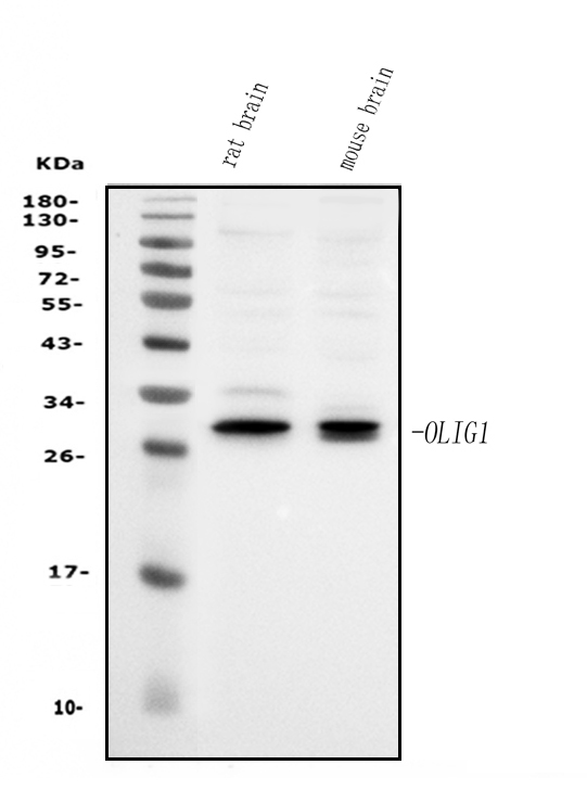
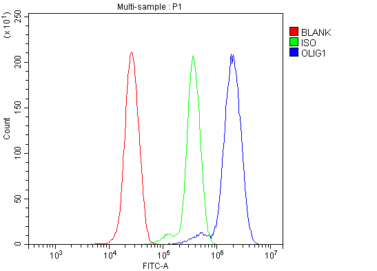
(Figure 2. Flow Cytometry analysis of THP-1 cells using anti-Olig1 antibody (AAA125768).Overlay histogram showing THP-1 cells stained with AAA125768 (Blue line). The cells were blocked with 10% normal goat serum. And then incubated with rabbit anti-Olig1 Antibody (AAA125768, 1μg/1x106 cells) for 30 min at 20 degree C. DyLight®488 conjugated goat anti-rabbit IgG (5-10μg/1x106 cells) was used as secondary antibody for 30 minutes at 20 degree C. Isotype control antibody (Green line) was rabbit IgG (1μg/1x106) used under the same conditions. Unlabelled sample (Red line) was also used as a control.)
FCM/FACS (Flow Cytometry)
(Figure 2. Flow Cytometry analysis of THP-1 cells using anti-Olig1 antibody (AAA125768).Overlay histogram showing THP-1 cells stained with AAA125768 (Blue line). The cells were blocked with 10% normal goat serum. And then incubated with rabbit anti-Olig1 Antibody (AAA125768, 1μg/1x106 cells) for 30 min at 20 degree C. DyLight®488 conjugated goat anti-rabbit IgG (5-10μg/1x106 cells) was used as secondary antibody for 30 minutes at 20 degree C. Isotype control antibody (Green line) was rabbit IgG (1μg/1x106) used under the same conditions. Unlabelled sample (Red line) was also used as a control.)
Olig1, Polyclonal Antibody (Cat# AAA125768)
 FCM/FACS (Flow Cytometry)
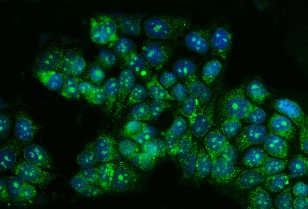
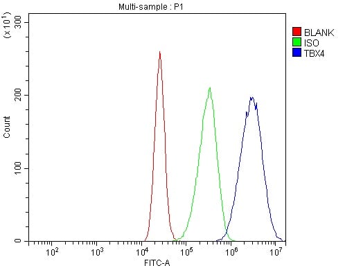
(Figure 3. Flow Cytometry analysis of CACO-2 cells using anti-TBX4 antibody (AAA125773).Overlay histogram showing CACO-2 cells stained with AAA125773 (Blue line). The cells were blocked with 10% normal goat serum. And then incubated with rabbit anti-TBX4 Antibody (AAA125773, 1μg/1x106 cells) for 30 min at 20 degree C. DyLight®488 conjugated goat anti-rabbit IgG (5-10μg/1x106 cells) was used as secondary antibody for 30 minutes at 20 degree C. Isotype control antibody (Green line) was rabbit IgG (1μg/1x106) used under the same conditions. Unlabelled sample (Red line) was also used as a control.)
FCM/FACS (Flow Cytometry)
(Figure 3. Flow Cytometry analysis of CACO-2 cells using anti-TBX4 antibody (AAA125773).Overlay histogram showing CACO-2 cells stained with AAA125773 (Blue line). The cells were blocked with 10% normal goat serum. And then incubated with rabbit anti-TBX4 Antibody (AAA125773, 1μg/1x106 cells) for 30 min at 20 degree C. DyLight®488 conjugated goat anti-rabbit IgG (5-10μg/1x106 cells) was used as secondary antibody for 30 minutes at 20 degree C. Isotype control antibody (Green line) was rabbit IgG (1μg/1x106) used under the same conditions. Unlabelled sample (Red line) was also used as a control.)
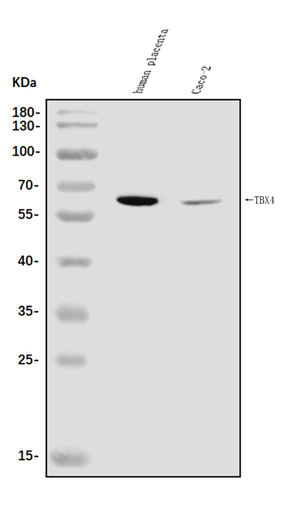
TBX4, Polyclonal Antibody (Cat# AAA125773)
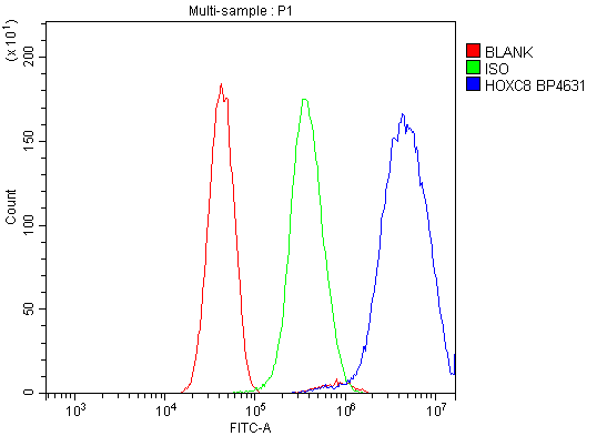 FCM/FACS (Flow Cytometry)
(Figure 2. Flow Cytometry analysis of U251 cells using anti-HOXC8 antibody (AAA125774).Overlay histogram showing U251 cells stained with AAA125774 (Blue line). The cells were blocked with 10% normal goat serum. And then incubated with rabbit anti-HOXC8 Antibody (AAA125774, 1μg/1x106 cells) for 30 min at 20 degree C. DyLight®488 conjugated goat anti-rabbit IgG (5-10μg/1x106 cells) was used as secondary antibody for 30 minutes at 20 degree C. Isotype control antibody (Green line) was rabbit IgG (1μg/1x106) used under the same conditions. Unlabelled sample (Red line) was also used as a control.)
FCM/FACS (Flow Cytometry)
(Figure 2. Flow Cytometry analysis of U251 cells using anti-HOXC8 antibody (AAA125774).Overlay histogram showing U251 cells stained with AAA125774 (Blue line). The cells were blocked with 10% normal goat serum. And then incubated with rabbit anti-HOXC8 Antibody (AAA125774, 1μg/1x106 cells) for 30 min at 20 degree C. DyLight®488 conjugated goat anti-rabbit IgG (5-10μg/1x106 cells) was used as secondary antibody for 30 minutes at 20 degree C. Isotype control antibody (Green line) was rabbit IgG (1μg/1x106) used under the same conditions. Unlabelled sample (Red line) was also used as a control.)
HOXC8, Polyclonal Antibody (Cat# AAA125774)
 FCM/FACS (Flow Cytometry)
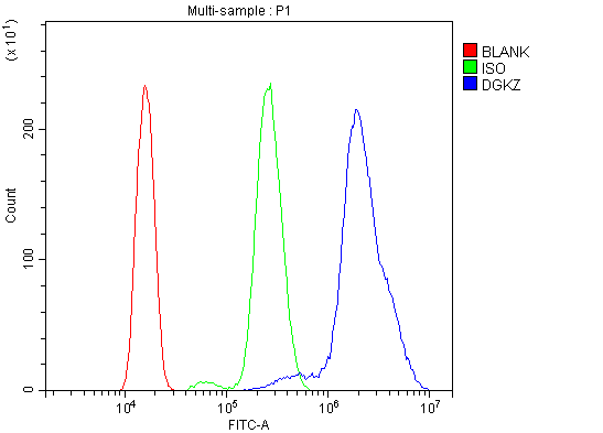
(Figure 5. Flow Cytometry analysis of HL-60 cells using anti-DGKZ/DGK-zeta antibody (AAA125777).Overlay histogram showing HL-60 cells stained with AAA125777 (Blue line). The cells were blocked with 10% normal goat serum. And then incubated with rabbit anti-DGKZ/DGK-zeta Antibody (AAA125777, 1μg/1x106 cells) for 30 min at 20 degree C. DyLight®488 conjugated goat anti-rabbit IgG (5-10μg/1x106 cells) was used as secondary antibody for 30 minutes at 20 degree C. Isotype control antibody (Green line) was rabbit IgG (1μg/1x106) used under the same conditions. Unlabelled sample (Red line) was also used as a control.)
FCM/FACS (Flow Cytometry)
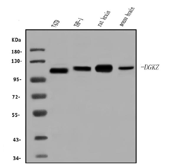
(Figure 5. Flow Cytometry analysis of HL-60 cells using anti-DGKZ/DGK-zeta antibody (AAA125777).Overlay histogram showing HL-60 cells stained with AAA125777 (Blue line). The cells were blocked with 10% normal goat serum. And then incubated with rabbit anti-DGKZ/DGK-zeta Antibody (AAA125777, 1μg/1x106 cells) for 30 min at 20 degree C. DyLight®488 conjugated goat anti-rabbit IgG (5-10μg/1x106 cells) was used as secondary antibody for 30 minutes at 20 degree C. Isotype control antibody (Green line) was rabbit IgG (1μg/1x106) used under the same conditions. Unlabelled sample (Red line) was also used as a control.)
DGKZ/DGK-zeta, Polyclonal Antibody (Cat# AAA125777)
What are Polyclonal Antibodies?
Polyclonal antibodies are antibodies that come from multiple B cell clones of a host animal. The typical hosts used for the majority of polyclonal antibody production are rabbits, goats, sheep, and donkeys. These polyclonal antibodies, once having identified their target, will bind to different epitopes located at different regions or sequences on the same protein/antigen. This ability to bind multiple epitopes is what makes polyclonal antibodies highly sensitive, as explained in our detailed guide on polyclonal antibodies and why they matter.
As a result, they are ideal at locating and binding to the target, even if the target is in very low concentrations (due to many different antibodies being able to bind to the same target molecule, which allows for significant amplification of a downstream signal).
Polyclonal antibodies are typically produced by injecting an antigen into a host animal, which causes the animal’s immune system to attack the foreign antigen by mass generating antibodies against it. After a period of time, serum is collected from the animal and purified using physicochemical fractionation, class-specific affinity purification, and/or antigen-affinity purification.
Key Uses of Polyclonal Antibodies
- Western Blotting: This method is used to find specific proteins in biological samples after separating them by size.
- Immunohistochemistry: IHC helps visualize the location of proteins in tissue sections using various staining techniques.
- ELISA: (Enzyme-Linked Immunosorbent Assay) is typically used to identify specific protein quantities in a sample. ELISAs can be either “Quantitative” or “Qualitative”.
- Flow Cytometry: technique that identifies and measures the specific protein on the surface or inside the cells in a fluid suspension.
- Immunoprecipitation: IP isolates and studies a specific protein from a complex mixture using antibodies.
Why Buy Polyclonal Antibodies from AAA Biotech?
1. Ideal for Various Applications
Our antibodies are generally going to be validated for use in multiple types of assays, including ELISA, Western Blotting, Immunohistochemistry, Immunoprecipitation, amongst others. They are ideal for a wide range of research applications.
2. Rigorous Quality Control
All of the antibodies in our catalog undergo strict quality testing to ensure specificity, sensitivity, and consistent performance. We are confident in the ability of our antibodies to provide you with accurate results.
3. Wide Assortment of Antibodies
Antibodies in our catalog can be found for both common and exotic species, and these antibodies are also available in both conjugated and recombinant forms to suit many diverse experimental needs.
4. Highly Purified
Our antibodies are available in purified forms with over 85% purity, as confirmed by SDS-PAGE. They are also available with tags such as His, Flag, GST, or MBP. We cater to customers worldwide.
FAQ
1. How are polyclonal antibodies produced?
Traditionally, polyclonal antibodies are produced by injecting an antigen into a host animal (such as a rabbit or goat), which then triggers an immune response from the host animal. The animal’s B cells produce antibodies that will recognize different parts of the injected antigen. These antibodies are then collected from the animal’s blood and purified for use.
2. How do polyclonal antibodies differ from monoclonal antibodies?
Polyclonal antibodies are a mix of antibodies that bind to different locations (epitopes) of the same antigen, while monoclonal antibodies are identical and bind to just one specific epitope. This makes polyclonal antibodies more versatile and better at detecting proteins that may be present in low quantities or in altered/modified forms.
3. How should I store polyclonal antibodies?
Polyclonal antibodies should be stored at 4°C for short-term use (up to a few weeks) and at -20°C or -80°C for long-term storage. Avoid repeated freeze-thaw cycles by dividing them into small aliquots. Always check the datasheet for specific storage instructions.