 FCM/FACS (Flow Cytometry)
(Figure 5. Flow Cytometry analysis of HepG2 cells using anti-APOA1 antibody (AAA46595).Overlay histogram showing HepG2 cells stained with AAA46595 (Blue line).The cells were blocked with 10% normal goat serum. And then incubated with rabbit anti-APOA1 Antibody (AAA46595,1ug/1x10^6 cells) for 30 min at 20 degree C. DyLight®488 conjugated goat anti-rabbit IgG (5-10ug/1x10^6 cells) was used as secondary antibody for 30 minutes at 20 degree C. Isotype control antibody (Green line) was rabbit IgG (1ug/1x106) used under the same conditions. Unlabelled sample (Red line) was also used as a control.)
FCM/FACS (Flow Cytometry)
(Figure 5. Flow Cytometry analysis of HepG2 cells using anti-APOA1 antibody (AAA46595).Overlay histogram showing HepG2 cells stained with AAA46595 (Blue line).The cells were blocked with 10% normal goat serum. And then incubated with rabbit anti-APOA1 Antibody (AAA46595,1ug/1x10^6 cells) for 30 min at 20 degree C. DyLight®488 conjugated goat anti-rabbit IgG (5-10ug/1x10^6 cells) was used as secondary antibody for 30 minutes at 20 degree C. Isotype control antibody (Green line) was rabbit IgG (1ug/1x106) used under the same conditions. Unlabelled sample (Red line) was also used as a control.)
Rabbit Apolipoprotein A I Polyclonal Antibody | anti-APOA1 antibody
Anti-Apolipoprotein A I Antibody
No cross reactivity with other proteins.
No cross reactivity with other proteins.
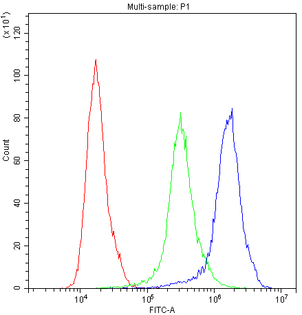
FCM/FACS (Flow Cytometry)
(Figure 5. Flow Cytometry analysis of HepG2 cells using anti-APOA1 antibody (AAA46595).Overlay histogram showing HepG2 cells stained with AAA46595 (Blue line).The cells were blocked with 10% normal goat serum. And then incubated with rabbit anti-APOA1 Antibody (AAA46595,1ug/1x10^6 cells) for 30 min at 20 degree C. DyLight®488 conjugated goat anti-rabbit IgG (5-10ug/1x10^6 cells) was used as secondary antibody for 30 minutes at 20 degree C. Isotype control antibody (Green line) was rabbit IgG (1ug/1x106) used under the same conditions. Unlabelled sample (Red line) was also used as a control.)
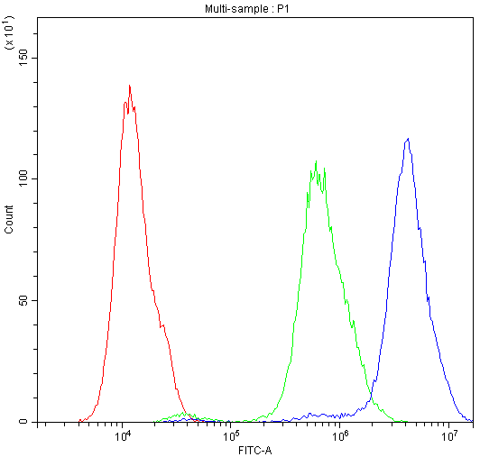
FCM/FACS (Flow Cytometry)
(Figure 4. Flow Cytometry analysis of CACO-2 cells using anti-APOA1 antibody (AAA46595).Overlay histogram showing CACO-2 cells stained with AAA46595 (Blue line).The cells were blocked with 10% normal goat serum. And then incubated with rabbit anti-APOA1 Antibody (AAA46595,1ug/1x10^6 cells) for 30 min at 20 degree C. DyLight®488 conjugated goat anti-rabbit IgG (5-10ug/1x10^6 cells) was used as secondary antibody for 30 minutes at 20 degree C. Isotype control antibody (Green line) was rabbit IgG (1ug/1x106) used under the same conditions. Unlabelled sample (Red line) was also used as a control.)
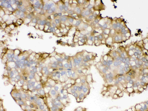
IHC (Immunohistochemisry)
(APOA1 was detected in paraffin-embedded sections of human renal cancer tissues using rabbit anti- APOA1 Antigen Affinity purified polyclonal antibody at 1ug/mL. The immunohistochemical section was developed using SABC method.)
IHC (Immunohistochemisry)
(APOA1 was detected in paraffin-embedded sections of human renal cancer tissues using rabbit anti- APOA1 Antigen Affinity purified polyclonal antibody at 1ug/mL. The immunohistochemical section was developed using SABC method.)
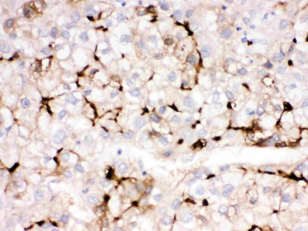
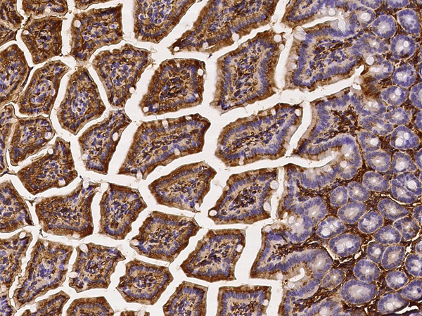
IHC (Immunohiostchemistry)
(APOA1 was detected in paraffin-embedded sections of human liver cancer tissues using rabbit anti- APOA1 Antigen Affinity purified polyclonal antibody at 1ug/mL. The immunohistochemical section was developed using SABC method.)
IHC (Immunohiostchemistry)
(APOA1 was detected in paraffin-embedded sections of human liver cancer tissues using rabbit anti- APOA1 Antigen Affinity purified polyclonal antibody at 1ug/mL. The immunohistochemical section was developed using SABC method.)
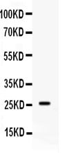
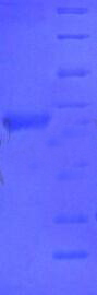
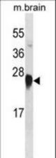
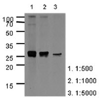
WB (Western Blot)
(Western blot analysis of APOA1 expression in human placenta extract (lane 1). APOA1 at 26KD was detected using rabbit anti- APOA1 Antigen Affinity purified polyclonal antibody at 0.5 ug/mL. The blot was developed using chemiluminescence (ECL) method.)
WB (Western Blot)
(Western blot analysis of APOA1 expression in human placenta extract (lane 1). APOA1 at 26KD was detected using rabbit anti- APOA1 Antigen Affinity purified polyclonal antibody at 0.5 ug/mL. The blot was developed using chemiluminescence (ECL) method.)
Background: Apolipoprotein A-1, also known as APOA1, is a human protein with a specific role in lipid metabolism. It binds to lipopolysaccharide or endotoxin, and has a major role in the anti-endotoxin function of HDL. The gene is mapped to 11q23. And it is a single polypeptide chain with 243 amino acid residues of known primary amino acid sequence. The ApoA-I protein promotes cholesterol efflux from tissues to the liver for excretion. It is a cofactor for lecithin cholesterolacyltransferase (LCAT) which is responsible for the formation of most plasma cholesteryl esters. ApoA-I is also isolated as a prostacyclin (PGI2) stabilizing factor, and thus may have an anticlotting effect. Defects in the gene encoding it are associated with HDL deficiencies, including Tangier disease, and with systemic non-neuropathic amyloidosis. Additionally, ApoA-I overexpression promotes macrophage-specific reverse cholesterol transport.
2. Ma J, Liao XL, Lou B, Wu MP (2004). "Role of apolipoprotein A-I in protecting against endotoxin toxicity". Acta Biochim. Biophys. Sin. (Shanghai) 36 (6): 419-24.
3. Yui Y, Aoyama T, Morishita H, Takahashi M, Takatsu Y, Kawai C (1988). "Serum prostacyclin stabilizing factor is identical to apolipoprotein A-I (Apo A-I). A novel function of Apo A-I". J. Clin. Invest. 82 (3): 803-7.
4. Zhang, Y.; Zanotti, I.; Reilly, M. P.; Glick, J. M.; Rothblat, G. H.; Rader, D. J. : Overexpression of apolipoprotein A-I promotes reverse transport of cholesterol from macrophages to feces in vivo. Circulation 108: 661-663, 2003.
NCBI and Uniprot Product Information
Customer Reviews
Loading reviews...
Share Your Experience
Similar Products


Product Notes
The APOA1 apoa1 (Catalog #AAA46595) is an Antibody produced from Rabbit and is intended for research purposes only. The product is available for immediate purchase. The Anti-Apolipoprotein An I Antibody reacts with Human No cross reactivity with other proteins. and may cross-react with other species as described in the data sheet. AAA Biotech's Apolipoprotein An I can be used in a range of immunoassay formats including, but not limited to, ELISA, IHC (Immunohistochemistry), WB (Western Blot). Researchers should empirically determine the suitability of the APOA1 apoa1 for an application not listed in the data sheet. Researchers commonly develop new applications and it is an integral, important part of the investigative research process. It is sometimes possible for the material contained within the vial of "Apolipoprotein A I, Polyclonal Antibody" to become dispersed throughout the inside of the vial, particularly around the seal of said vial, during shipment and storage. We always suggest centrifuging these vials to consolidate all of the liquid away from the lid and to the bottom of the vial prior to opening. Please be advised that certain products may require dry ice for shipping and that, if this is the case, an additional dry ice fee may also be required.Precautions
All products in the AAA Biotech catalog are strictly for research-use only, and are absolutely not suitable for use in any sort of medical, therapeutic, prophylactic, in-vivo, or diagnostic capacity. By purchasing a product from AAA Biotech, you are explicitly certifying that said products will be properly tested and used in line with industry standard. AAA Biotech and its authorized distribution partners reserve the right to refuse to fulfill any order if we have any indication that a purchaser may be intending to use a product outside of our accepted criteria.Disclaimer
Though we do strive to guarantee the information represented in this datasheet, AAA Biotech cannot be held responsible for any oversights or imprecisions. AAA Biotech reserves the right to adjust any aspect of this datasheet at any time and without notice. It is the responsibility of the customer to inform AAA Biotech of any product performance issues observed or experienced within 30 days of receipt of said product. To see additional details on this or any of our other policies, please see our Terms & Conditions page.Item has been added to Shopping Cart
If you are ready to order, navigate to Shopping Cart and get ready to checkout.