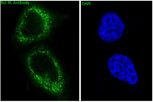
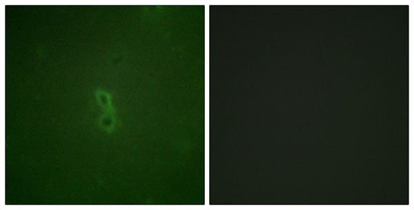
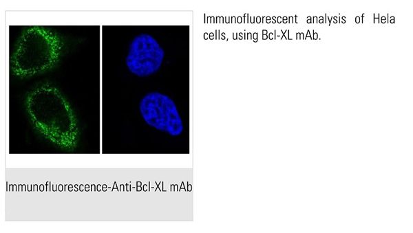
 IF (Immunofluorescence)
(AAA31068 staining NIH/3T3 cells by ICC/IF. Cells were fixed with PFA and permeabilized in 0.1% saponin prior to blocking in 10% serum for 45 minutes at 37 degree C. The primary antibody was diluted 1/400 and incubated with the sample for 1 hour at 37 degree C. A Alexa Fluor 594 conjugated goat polyclonal to rabbit IgG (H+L), diluted 1/600 was used as secondary antibody.)
IF (Immunofluorescence)
(AAA31068 staining NIH/3T3 cells by ICC/IF. Cells were fixed with PFA and permeabilized in 0.1% saponin prior to blocking in 10% serum for 45 minutes at 37 degree C. The primary antibody was diluted 1/400 and incubated with the sample for 1 hour at 37 degree C. A Alexa Fluor 594 conjugated goat polyclonal to rabbit IgG (H+L), diluted 1/600 was used as secondary antibody.)
Rabbit BCL-XL Polyclonal Antibody | anti-BCL-XL antibody
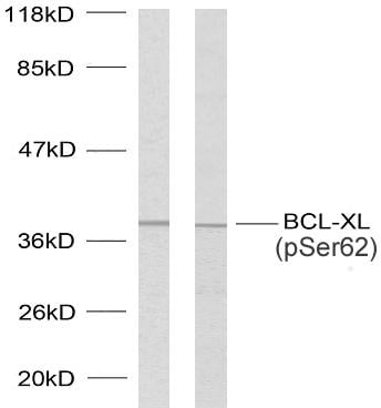
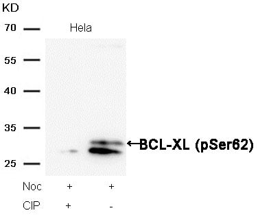
Phospho-BCL-XL (Thr47) Antibody
Phosphate buffered saline, pH 7.4, 150mM NaCl, 0.02% sodium azide and 50% glycerol.
IHC: 1:50-1:200
IF/ICC: 1:100-1:500
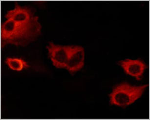
IF (Immunofluorescence)
(AAA31068 staining NIH/3T3 cells by ICC/IF. Cells were fixed with PFA and permeabilized in 0.1% saponin prior to blocking in 10% serum for 45 minutes at 37 degree C. The primary antibody was diluted 1/400 and incubated with the sample for 1 hour at 37 degree C. A Alexa Fluor 594 conjugated goat polyclonal to rabbit IgG (H+L), diluted 1/600 was used as secondary antibody.)
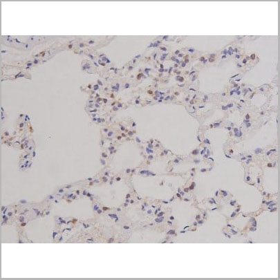
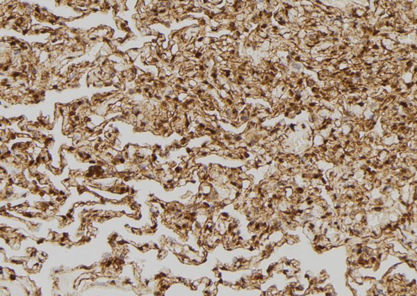
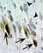
IHC (Immunohistochemistry)
(AAA31068 at 1/200 staining Rat lung tissue sections by IHC-P. The tissue was formaldehyde fixed and a heat mediated antigen retrieval step in citrate buffer was performed. The tissue was then blocked and incubated with the antibody for 1.5 hours at 22 degree C. An HRP conjugated goat anti-rabbit antibody was used as the secondary.)
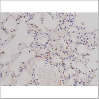
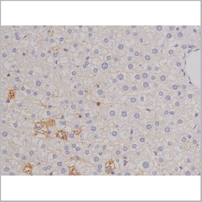
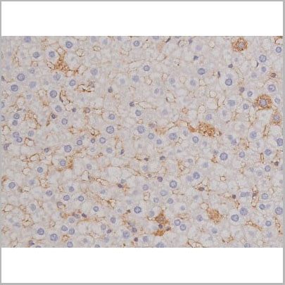
IHC (Immunohistochemistry)
(AAA31068 at 1/200 staining Rat liver tissue sections by IHC-P. The tissue was formaldehyde fixed and a heat mediated antigen retrieval step in citrate buffer was performed. The tissue was then blocked and incubated with the antibody for 1.5 hours at 22 degree C. An HRP conjugated goat anti-rabbit antibody was used as the secondary.)
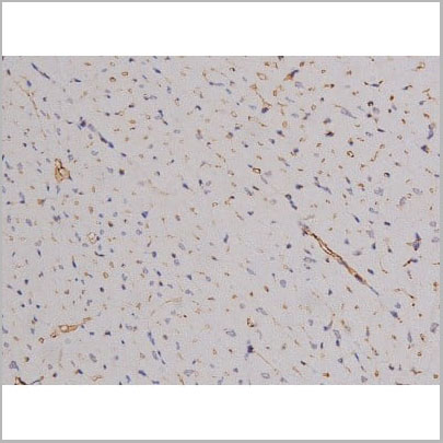
IHC (Immunohistochemistry)
(AAA31068 at 1/200 staining Rat heart tissue sections by IHC-P. The tissue was formaldehyde fixed and a heat mediated antigen retrieval step in citrate buffer was performed. The tissue was then blocked and incubated with the antibody for 1.5 hours at 22 degree C. An HRP conjugated goat anti-rabbit antibody was used as the secondary.)
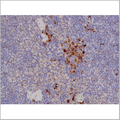
IHC (Immunohistchemistry)
(AAA31068 at 1/200 staining Mouse spleen tissue sections by IHC-P. The tissue was formaldehyde fixed and a heat mediated antigen retrieval step in citrate buffer was performed. The tissue was then blocked and incubated with the antibody for 1.5 hours at 22 degree C. An HRP conjugated goat anti-rabbit antibody was used as the secondary.)
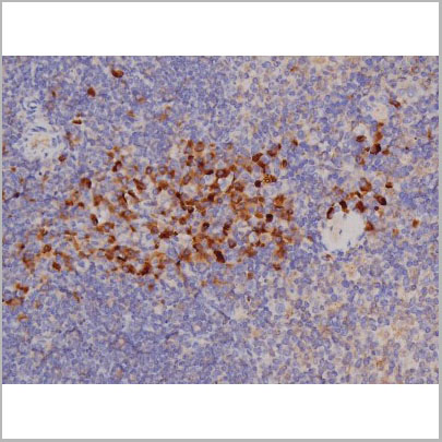
IHC (Immunohistochemistry)
(AAA31068 at 1/200 staining Mouse spleen tissue sections by IHC-P. The tissue was formaldehyde fixed and a heat mediated antigen retrieval step in citrate buffer was performed. The tissue was then blocked and incubated with the antibody for 1.5 hours at 22 degree C. An HRP conjugated goat anti-rabbit antibody was used as the secondary.)
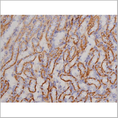
IHC (Immunohistochemistry)
(AAA31068 at 1/200 staining Mouse kidney tissue sections by IHC-P. The tissue was formaldehyde fixed and a heat mediated antigen retrieval step in citrate buffer was performed. The tissue was then blocked and incubated with the antibody for 1.5 hours at 22 degree C. An HRP conjugated goat anti-rabbit antibody was used as the secondary.)
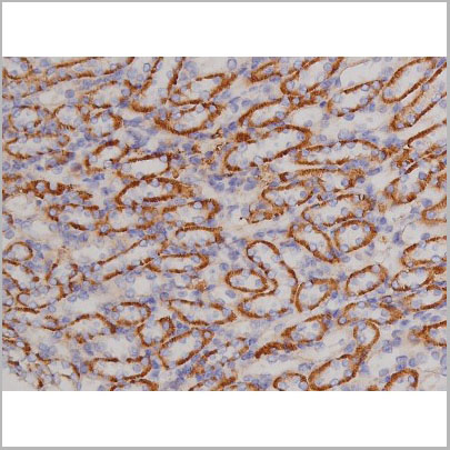
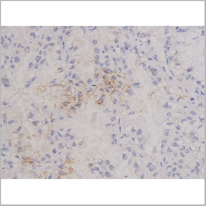
IHC (Immunohistchemistry)
(AAA31068 at 1/200 staining Mouse kidney tissue sections by IHC-P. The tissue was formaldehyde fixed and a heat mediated antigen retrieval step in citrate buffer was performed. The tissue was then blocked and incubated with the antibody for 1.5 hours at 22 degree C. An HRP conjugated goat anti-rabbit antibody was used as the secondary.)
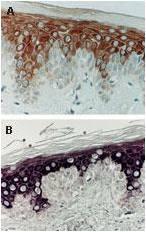
IHC (Immunohistochemistry)
(AAA31068 at 1/200 staining Human lung cancer tissue sections by IHC-P. The tissue was formaldehyde fixed and a heat mediated antigen retrieval step in citrate buffer was performed. The tissue was then blocked and incubated with the antibody for 1.5 hours at 22 degree C. An HRP conjugated goat anti-rabbit antibody was used as the secondary.)
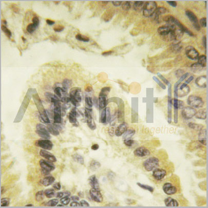
IHC (Immunohistochemistry)
(AAA31068 at 1/100 staining human colon carcinoma tissues sections by IHC-P. The tissue was formaldehyde fixed and a heat mediated antigen retrieval step in citrate buffer was performed. The tissue was then blocked and incubated with the antibody for 1.5 hou)
IHC (Immunohistochemistry)
(AAA31068 at 1/100 staining human colon carcinoma tissues sections by IHC-P. The tissue was formaldehyde fixed and a heat mediated antigen retrieval step in citrate buffer was performed. The tissue was then blocked and incubated with the antibody for 1.5 hou)
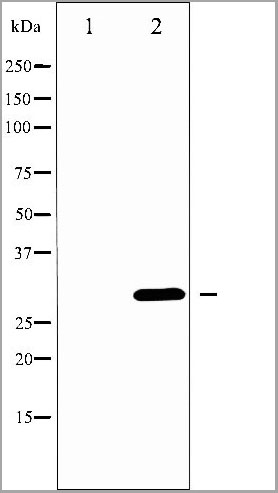
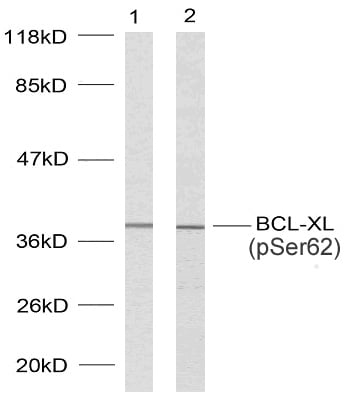
WB (Western Blot)
(Western blot analysis of BCL-XL phosphorylation expression in UV treated 293 whole cell lysates, The lane on the left is treated with the antigen-specific peptide.)
WB (Western Blot)
(Western blot analysis of BCL-XL phosphorylation expression in UV treated 293 whole cell lysates, The lane on the left is treated with the antigen-specific peptide.)
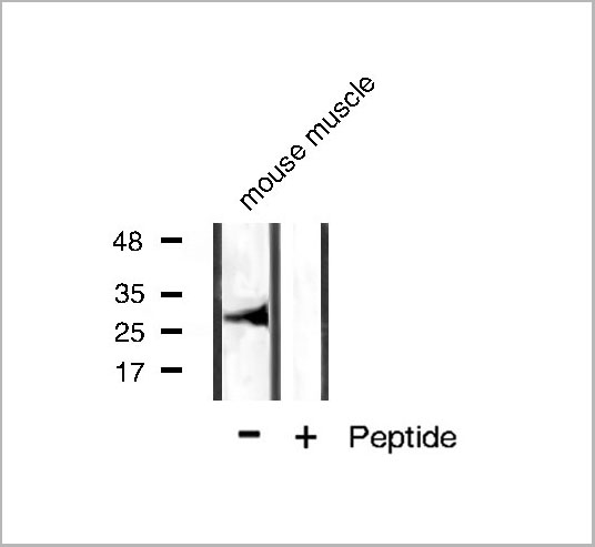
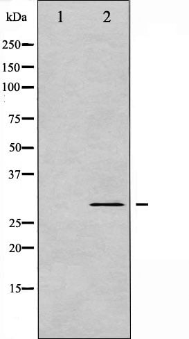
WB (Western Blot)
(Western blot analysis of BCL-XL phosphorylation expression in mouse muscle tissue lysates, The lane on the right is treated with the antigen-specific peptide.)
WB (Western Blot)
(Western blot analysis of BCL-XL phosphorylation expression in mouse muscle tissue lysates, The lane on the right is treated with the antigen-specific peptide.)
Function: Potent inhibitor of cell death. Inhibits activation of caspases. Appears to regulate cell death by blocking the voltage-dependent anion channel (VDAC) by binding to it and preventing the release of the caspase activator, CYC1, from the mitochondrial membrane. Also acts as a regulator of G2 checkpoint and progression to cytokinesis during mitosis.
Subunit Structure: Homodimer. Isoform Bcl-X(L) forms heterodimers with BAX, BAK or BCL2. Heterodimerization with BAX does not seem to be required for anti-apoptotic activity. Interacts with BCL2L11. Interacts with BAD. Interacts (isoform Bcl-X(L)) with SIVA1 (isoform 1); the interaction inhibits the anti-apoptotic activity. Interacts with BECN1 and PGAM5. Isoform Bcl-X(L) interacts with IKZF3. Interacts with HEBP2. Isoform Bcl-X(L) interacts with RTL10/BOP. Interacts with p53/TP53 and BBC3; interaction with BBC3 disrupts the interaction with p53/TP53. Isoform Bcl-X(L) interacts with DNM1L and CLTA; DNM1L and BCL2L1 isoform BCL-X(L) may form a complex in synaptic vesicles that also contains clathrin and MFF. Interacts with ATP5F1A and ATP5F1B; the interactions mediate the association of isoform Bcl-X(L) with the mitochondrial membrane ATP synthase F1F0 ATP synthase. Interacts with VDAC1 (PubMed:25296756). Isoform Bcl-X(L) interacts (via the loop between motifs BH4 and BH3) with NLRP1 (via LRR repeats), but not with NLRP2, NLRP3, NLRP4, PYCARD, nor MEFV (PubMed:17418785). Interacts with BCL2L11 (via BH3) (PubMed:27013495).
Post-translational Modifications: Proteolytically cleaved by caspases during apoptosis. The cleaved protein, lacking the BH4 motif, has pro-apoptotic activity. Phosphorylated on Ser-62 by CDK1. This phosphorylation is partial in normal mitotic cells, but complete in G2-arrested cells upon DNA-damage, thus promoting subsequent apoptosis probably by triggering caspases-mediated proteolysis. Phosphorylated by PLK3, leading to regulate the G2 checkpoint and progression to cytokinesis during mitosis. Phosphorylation at Ser-49 appears during the S phase and G2, disappears rapidly in early mitosis during prometaphase, metaphase and early anaphase, and re-appears during telophase and cytokinesis.
Similarity: The BH4 motif is required for anti-apoptotic activity. The BH1 and BH2 motifs are required for both heterodimerization with other Bcl-2 family members and for repression of cell death.The loop between motifs BH4 and BH3 is required for the interaction with NLRP1. Belongs to the Bcl-2 family.
NCBI and Uniprot Product Information
Predicted: 27 kDa
Customer Reviews
Loading reviews...
Share Your Experience
Similar Products


Product Notes
The BCL-XL bcl2l1 (Catalog #AAA31068) is an Antibody produced from Rabbit and is intended for research purposes only. The product is available for immediate purchase. The Phospho-BCL-XL (Thr47) Antibody reacts with Human, Mouse, Rat and may cross-react with other species as described in the data sheet. AAA Biotech's BCL-XL can be used in a range of immunoassay formats including, but not limited to, WB (Western Blot), IHC (Immunohistochemistry), IF (Immunofluorescence), ICC (Immunocytochemistry), ELISA. WB: 1:500-1:2000 IHC: 1:50-1:200 IF/ICC: 1:100-1:500. Researchers should empirically determine the suitability of the BCL-XL bcl2l1 for an application not listed in the data sheet. Researchers commonly develop new applications and it is an integral, important part of the investigative research process. It is sometimes possible for the material contained within the vial of "BCL-XL, Polyclonal Antibody" to become dispersed throughout the inside of the vial, particularly around the seal of said vial, during shipment and storage. We always suggest centrifuging these vials to consolidate all of the liquid away from the lid and to the bottom of the vial prior to opening. Please be advised that certain products may require dry ice for shipping and that, if this is the case, an additional dry ice fee may also be required.Precautions
All products in the AAA Biotech catalog are strictly for research-use only, and are absolutely not suitable for use in any sort of medical, therapeutic, prophylactic, in-vivo, or diagnostic capacity. By purchasing a product from AAA Biotech, you are explicitly certifying that said products will be properly tested and used in line with industry standard. AAA Biotech and its authorized distribution partners reserve the right to refuse to fulfill any order if we have any indication that a purchaser may be intending to use a product outside of our accepted criteria.Disclaimer
Though we do strive to guarantee the information represented in this datasheet, AAA Biotech cannot be held responsible for any oversights or imprecisions. AAA Biotech reserves the right to adjust any aspect of this datasheet at any time and without notice. It is the responsibility of the customer to inform AAA Biotech of any product performance issues observed or experienced within 30 days of receipt of said product. To see additional details on this or any of our other policies, please see our Terms & Conditions page.Item has been added to Shopping Cart
If you are ready to order, navigate to Shopping Cart and get ready to checkout.