Rabbit Bestrophin Polyclonal Antibody | anti-BEST1 antibody
Bestrophin Antibody BIOTIN-Conjugated
Gene Names
BEST1; ARB; BMD; BEST; RP50; VMD2; TU15B
Applications
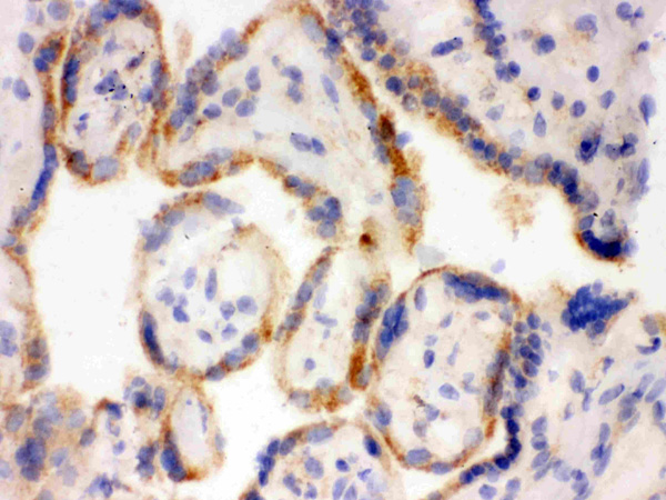
Western Blot, Immunoprecipitation, Immunohistochemistry, Immunofluorescence, ELISA
Synonyms
Bestrophin, Antibody; Bestrophin Antibody BIOTIN-Conjugated; ARB antibody, BEST antibody, Best macular dystrophy antibody, Best1 V1 Delta2 antibody, BMD antibody, RP50 antibody, TU15B antibody Vitelliform mascular dystrophy 2 antibody.; anti-BEST1 antibody
Host
Rabbit
Clonality
Polyclonal
Specificity
This peptide will label the bestrophin 1-1, -2, -3, -4, -5 isoforms that are produced by alternate splicing of the C-Terminal end.
Form/Format
Affinity purified immunoglobulins
Sequence Length
585
Applicable Applications for anti-BEST1 antibody
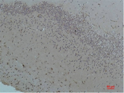
WB (Western Blot), IP (Immunoprecipitation), IHC (Immunohistochemistry), IF (Immunofluorescence), ELISA, CM (Confocal Microscopy)
Immunogen
Synthetic peptide taken within amino acid region 500-585 on human Bestrophin-1 isoform 1 protein.
Cross Reactivity
Human
Determinant
N-epitope
Preparation and Storage
Store at -20°C for long term storage.
Related Product Information for anti-BEST1 antibody
BIOTIN-Conjugated Bestrophin 1 Antibody N-epitope
Forms calcium sensitive chloride channels. Highly permeable to bicarbonate.
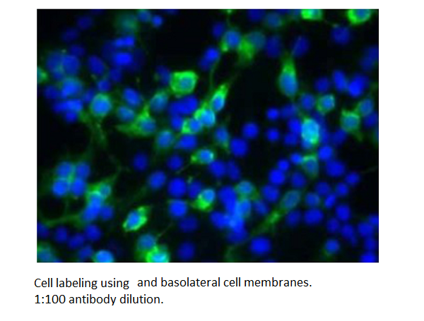
Bestrophin is a 68 kDa basolateral plasma membrane protein expressed in retinal pigment epithelial cells (RPE). Bestrophin is encoded by the VMD2 gene, which is mutated in BEST macular dystrophy, a disease characterized by a depressed light peak due to alteration in the chloride channel activity and changes in the electroculogram (EOG). Recently it was proposed that bestrophin is chloride channel responsible for generating the light peak. It has been shown that Bestrophin interacts with beta catalytic subunit of protein phosphatase 2A (PP2Ac). Such Protein interaction between bestrophin and PP2AC ant the structural subunit of PP2A, PR65 was confirmed by reciprocal immunoprecipitation. The interaction between PP2Ac and the Bestrophin takes place near the Carboxy-terminal end of the protein. Okadic acid induces the phosphorylation of Bestrophin in vitro. Bestrophin also serves in the signal Okadic acid induces the phosphorylation of Bestrophin in virtro. Bestrophin also serves in the signal transduction pathway that modulates the light peak of the EOG, that is regulated by phosphorylation of Bestrophin that in turn is regulated by protein phosphatase 2A (PP2A).
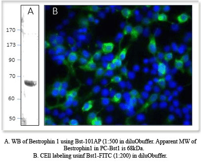
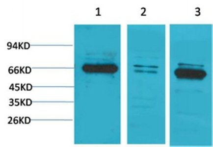
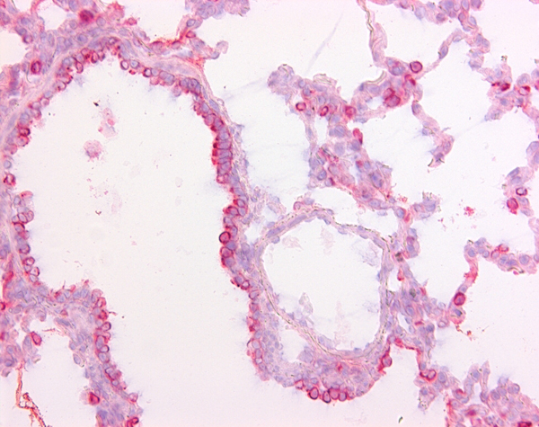
The Anti- Bestrophin- selective antibodies were generated against observed sequences near the N-terminal, mid region or the C-terminal end of the proteins that are unique to Bestrophin protein. The Bestrophin-selective antibodies were affinity purified against inmobilized antigen based affinity chromatography and are represented as epitope-specific antibodies. Antigenic blocking peptides (P-BEST.3) and western blot positive control (PC-BEST) in ready to use SDS-sample buffer are available. The polyclonal antibody strongly labels a 65 kDa protein in RPE cell extracts.
Forms calcium sensitive chloride channels. Highly permeable to bicarbonate.
Bestrophin is a 68 kDa basolateral plasma membrane protein expressed in retinal pigment epithelial cells (RPE). Bestrophin is encoded by the VMD2 gene, which is mutated in BEST macular dystrophy, a disease characterized by a depressed light peak due to alteration in the chloride channel activity and changes in the electroculogram (EOG). Recently it was proposed that bestrophin is chloride channel responsible for generating the light peak. It has been shown that Bestrophin interacts with beta catalytic subunit of protein phosphatase 2A (PP2Ac). Such Protein interaction between bestrophin and PP2AC ant the structural subunit of PP2A, PR65 was confirmed by reciprocal immunoprecipitation. The interaction between PP2Ac and the Bestrophin takes place near the Carboxy-terminal end of the protein. Okadic acid induces the phosphorylation of Bestrophin in vitro. Bestrophin also serves in the signal Okadic acid induces the phosphorylation of Bestrophin in virtro. Bestrophin also serves in the signal transduction pathway that modulates the light peak of the EOG, that is regulated by phosphorylation of Bestrophin that in turn is regulated by protein phosphatase 2A (PP2A).
The Anti- Bestrophin- selective antibodies were generated against observed sequences near the N-terminal, mid region or the C-terminal end of the proteins that are unique to Bestrophin protein. The Bestrophin-selective antibodies were affinity purified against inmobilized antigen based affinity chromatography and are represented as epitope-specific antibodies. Antigenic blocking peptides (P-BEST.3) and western blot positive control (PC-BEST) in ready to use SDS-sample buffer are available. The polyclonal antibody strongly labels a 65 kDa protein in RPE cell extracts.
NCBI and Uniprot Product Information
NCBI GI #
NCBI GeneID
NCBI Accession #
NCBI GenBank Nucleotide #
NCBI Official Full Name
bestrophin-1 isoform 1
NCBI Official Synonym Full Names
bestrophin 1
NCBI Official Symbol
BEST1
NCBI Official Synonym Symbols
ARB; BMD; BEST; RP50; VMD2; TU15B
NCBI Protein Information
bestrophin-1
UniProt Protein Name
Bestrophin-1
UniProt Gene Name
BEST1
UniProt Synonym Gene Names
VMD2
UniProt Entry Name
BEST1_HUMAN
Customer Reviews
Loading reviews...
Share Your Experience
Similar Products


Product Notes
The BEST1 best1 (Catalog #AAA75844) is an Antibody produced from Rabbit and is intended for research purposes only. The product is available for immediate purchase. AAA Biotech's Bestrophin can be used in a range of immunoassay formats including, but not limited to, WB (Western Blot), IP (Immunoprecipitation), IHC (Immunohistochemistry), IF (Immunofluorescence), ELISA, CM (Confocal Microscopy). Researchers should empirically determine the suitability of the BEST1 best1 for an application not listed in the data sheet. Researchers commonly develop new applications and it is an integral, important part of the investigative research process. It is sometimes possible for the material contained within the vial of "Bestrophin, Polyclonal Antibody" to become dispersed throughout the inside of the vial, particularly around the seal of said vial, during shipment and storage. We always suggest centrifuging these vials to consolidate all of the liquid away from the lid and to the bottom of the vial prior to opening. Please be advised that certain products may require dry ice for shipping and that, if this is the case, an additional dry ice fee may also be required.Precautions
All products in the AAA Biotech catalog are strictly for research-use only, and are absolutely not suitable for use in any sort of medical, therapeutic, prophylactic, in-vivo, or diagnostic capacity. By purchasing a product from AAA Biotech, you are explicitly certifying that said products will be properly tested and used in line with industry standard. AAA Biotech and its authorized distribution partners reserve the right to refuse to fulfill any order if we have any indication that a purchaser may be intending to use a product outside of our accepted criteria.Disclaimer
Though we do strive to guarantee the information represented in this datasheet, AAA Biotech cannot be held responsible for any oversights or imprecisions. AAA Biotech reserves the right to adjust any aspect of this datasheet at any time and without notice. It is the responsibility of the customer to inform AAA Biotech of any product performance issues observed or experienced within 30 days of receipt of said product. To see additional details on this or any of our other policies, please see our Terms & Conditions page.Item has been added to Shopping Cart
If you are ready to order, navigate to Shopping Cart and get ready to checkout.