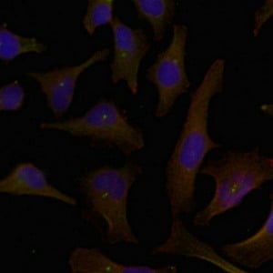
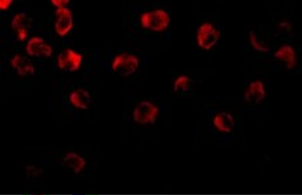
 IF (Immunofluorescence)
(AAA327156 staining Hela cells by IF/ICC. The sample were fixed with PFA and permeabilized in 0.1% Triton X-100, then blocked in 10% serum for 45 minutes at 25 degree C. The primary antibody was diluted at 1/200 and incubated with the sample for 1 hour at 37 degree C. An Alexa Fluor 594 conjugated goat anti-rabbit IgG (H+L) antibody, diluted at 1/600, was used as secondary antibody.)
IF (Immunofluorescence)
(AAA327156 staining Hela cells by IF/ICC. The sample were fixed with PFA and permeabilized in 0.1% Triton X-100, then blocked in 10% serum for 45 minutes at 25 degree C. The primary antibody was diluted at 1/200 and incubated with the sample for 1 hour at 37 degree C. An Alexa Fluor 594 conjugated goat anti-rabbit IgG (H+L) antibody, diluted at 1/600, was used as secondary antibody.)
Rabbit DOK1 Polyclonal Antibody | anti-DOK1 antibody
DOK1 Antibody
Phosphate buffered saline, pH 7.4, 150mM NaCl, 0.02% sodium azide and 50% glycerol.
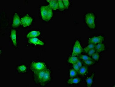
IF (Immunofluorescence)
(AAA327156 staining Hela cells by IF/ICC. The sample were fixed with PFA and permeabilized in 0.1% Triton X-100, then blocked in 10% serum for 45 minutes at 25 degree C. The primary antibody was diluted at 1/200 and incubated with the sample for 1 hour at 37 degree C. An Alexa Fluor 594 conjugated goat anti-rabbit IgG (H+L) antibody, diluted at 1/600, was used as secondary antibody.)
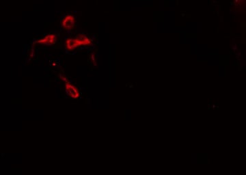
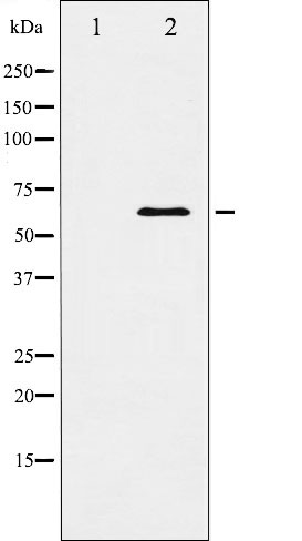
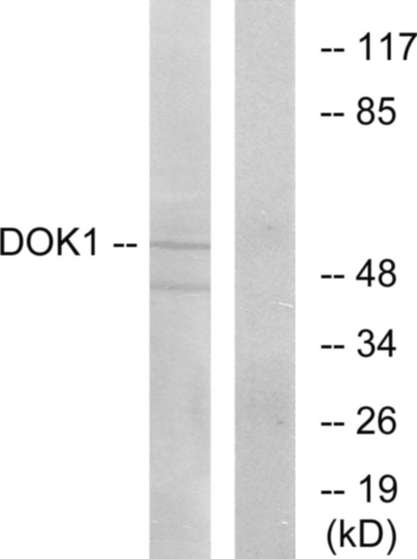
WB (Western Blot)
(Western blot analysis of Jurkat whole cell lysates, using DOK1 Antibody. The lane on the left is treated with the antigen-specific peptide.)
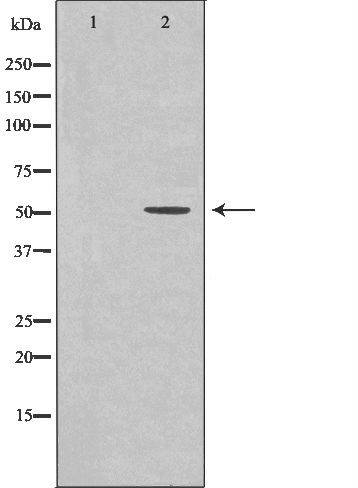
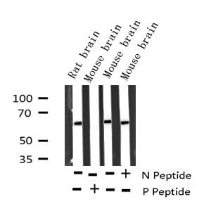
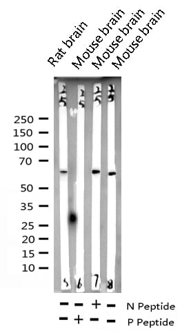
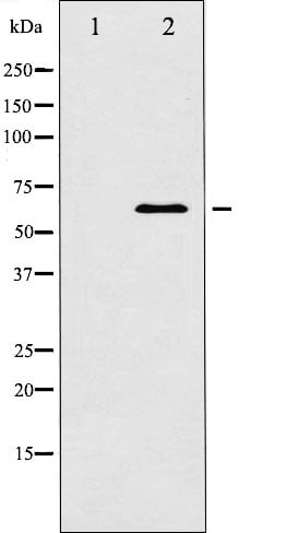
WB (Western Blot)
(Western blot analysis of Jurkat whole cell lysates, using DOK1 Antibody. The lane on the left is treated with the antigen-specific peptide.)
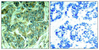
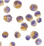
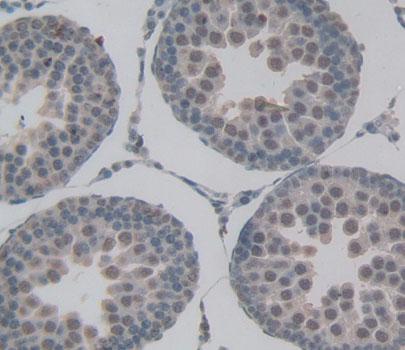
IHC (Immunohistochemistry)
(AAA327156 at 1/100 staining Mouse pancreas tissue by IHC-P. The sample was formaldehyde fixed and a heat mediated antigen retrieval step in citrate buffer was performed. The sample was then blocked and incubated with the antibody for 1.5 hours at 22 degree C. An HRP conjugated goat anti-rabbit antibody was used as the secondary.)
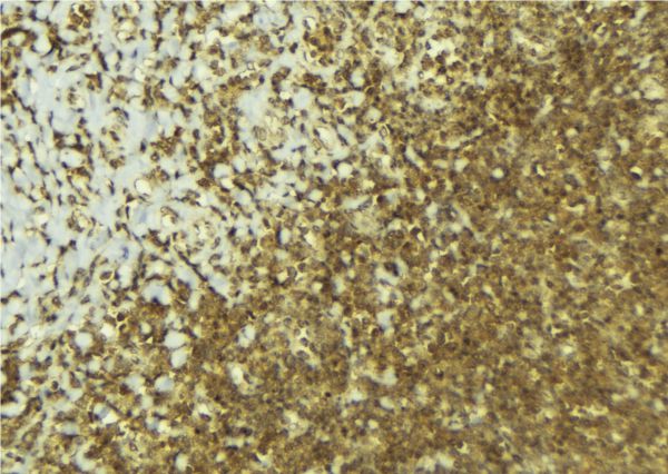
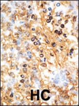
Function: DOK proteins are enzymatically inert adaptor or scaffolding proteins. They provide a docking platform for the assembly of multimolecular signaling complexes. DOK1 appears to be a negative regulator of the insulin signaling pathway. Modulates integrin activation by competing with talin for the same binding site on ITGB3.
Subunit Structure: Interacts with ABL1 (By similarity). Interacts with RasGAP and INPP5D/SHIP1. Interacts directly with phosphorylated ITGB3. Interacts with SRMS (via the SH2 and SH3 domains).
Post-translational Modifications: Constitutively tyrosine-phosphorylated. Phosphorylated by TEC (By similarity). Phosphorylated by LYN (By similarity). Phosphorylated on tyrosine residues by the insulin receptor kinase. Results in the negative regulation of the insulin signaling pathway. Phosphorylated on tyrosine residues by SRMS.
Similarity: The PTB domain mediates receptor interaction. Belongs to the DOK family. Type A subfamily.
NCBI and Uniprot Product Information
Predicted: 53 kDa
Customer Reviews
Loading reviews...
Share Your Experience
Similar Products


Product Notes
The DOK1 dok1 (Catalog #AAA327156) is an Antibody produced from Rabbit and is intended for research purposes only. The product is available for immediate purchase. The DOK1 Antibody reacts with Human, Mouse, Rat and may cross-react with other species as described in the data sheet. AAA Biotech's DOK1 can be used in a range of immunoassay formats including, but not limited to, ELISA, ICC (Immunocytochemistry), IF (Immunofluorescence), IHC (Immunohistochemistry), WB (Western Blot). Researchers should empirically determine the suitability of the DOK1 dok1 for an application not listed in the data sheet. Researchers commonly develop new applications and it is an integral, important part of the investigative research process. It is sometimes possible for the material contained within the vial of "DOK1, Polyclonal Antibody" to become dispersed throughout the inside of the vial, particularly around the seal of said vial, during shipment and storage. We always suggest centrifuging these vials to consolidate all of the liquid away from the lid and to the bottom of the vial prior to opening. Please be advised that certain products may require dry ice for shipping and that, if this is the case, an additional dry ice fee may also be required.Precautions
All products in the AAA Biotech catalog are strictly for research-use only, and are absolutely not suitable for use in any sort of medical, therapeutic, prophylactic, in-vivo, or diagnostic capacity. By purchasing a product from AAA Biotech, you are explicitly certifying that said products will be properly tested and used in line with industry standard. AAA Biotech and its authorized distribution partners reserve the right to refuse to fulfill any order if we have any indication that a purchaser may be intending to use a product outside of our accepted criteria.Disclaimer
Though we do strive to guarantee the information represented in this datasheet, AAA Biotech cannot be held responsible for any oversights or imprecisions. AAA Biotech reserves the right to adjust any aspect of this datasheet at any time and without notice. It is the responsibility of the customer to inform AAA Biotech of any product performance issues observed or experienced within 30 days of receipt of said product. To see additional details on this or any of our other policies, please see our Terms & Conditions page.Item has been added to Shopping Cart
If you are ready to order, navigate to Shopping Cart and get ready to checkout.