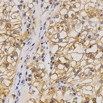
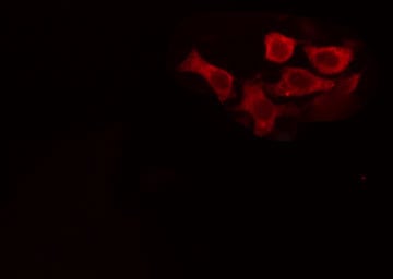
 IHC (Immunohistochemistry)
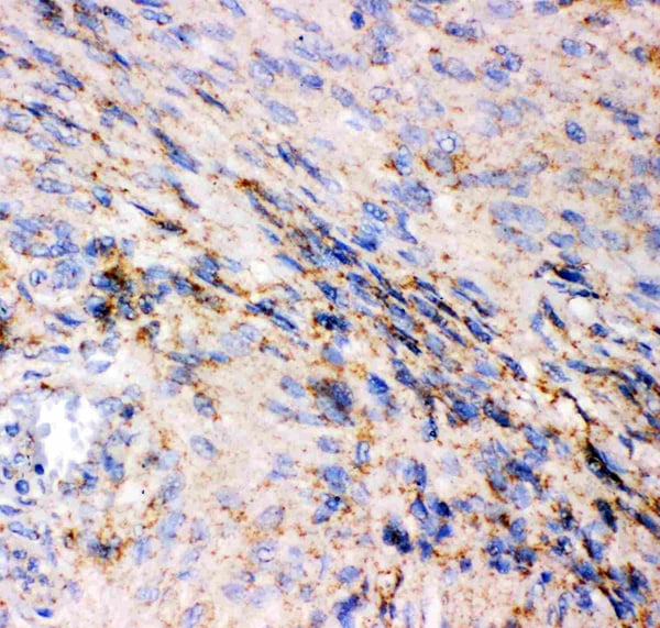
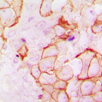
(Figure 5. IHC analysis of GRIA1 using anti-GRIA1 antibody (AAA46057).GRIA1 was detected in paraffin-embedded section of Human Meningeoma Tissue. Heat mediated antigen retrieval was performed in citrate buffer (pH6, epitope retrieval solution) for 20 mins. The tissue section was blocked with 10% goat serum. The tissue section was then incubated with 1ug/ml rabbit anti-GRIA1 Antibody (AAA46057) overnight at 4 degree C. Biotinylated goat anti-rabbit IgG was used as secondary antibody and incubated for 30 minutes at 37 degree C. The tissue section was developed using Strepavidin-Biotin-Complex (SABC) with DAB as the chromogen.)
IHC (Immunohistochemistry)
(Figure 5. IHC analysis of GRIA1 using anti-GRIA1 antibody (AAA46057).GRIA1 was detected in paraffin-embedded section of Human Meningeoma Tissue. Heat mediated antigen retrieval was performed in citrate buffer (pH6, epitope retrieval solution) for 20 mins. The tissue section was blocked with 10% goat serum. The tissue section was then incubated with 1ug/ml rabbit anti-GRIA1 Antibody (AAA46057) overnight at 4 degree C. Biotinylated goat anti-rabbit IgG was used as secondary antibody and incubated for 30 minutes at 37 degree C. The tissue section was developed using Strepavidin-Biotin-Complex (SABC) with DAB as the chromogen.)
Rabbit Glutamate receptor 1 Polyclonal Antibody | anti-GRIA1 antibody
Anti-GRIA1 Antibody
Each vial contains 5mg BSA, 0.9mg NaCl, 0.2mg Na2HPO4, 0.05mg NaN3.
IHC (Immunohistochemistry)
(Figure 5. IHC analysis of GRIA1 using anti-GRIA1 antibody (AAA46057).GRIA1 was detected in paraffin-embedded section of Human Meningeoma Tissue. Heat mediated antigen retrieval was performed in citrate buffer (pH6, epitope retrieval solution) for 20 mins. The tissue section was blocked with 10% goat serum. The tissue section was then incubated with 1ug/ml rabbit anti-GRIA1 Antibody (AAA46057) overnight at 4 degree C. Biotinylated goat anti-rabbit IgG was used as secondary antibody and incubated for 30 minutes at 37 degree C. The tissue section was developed using Strepavidin-Biotin-Complex (SABC) with DAB as the chromogen.)
IHC (Immunohistochemistry)
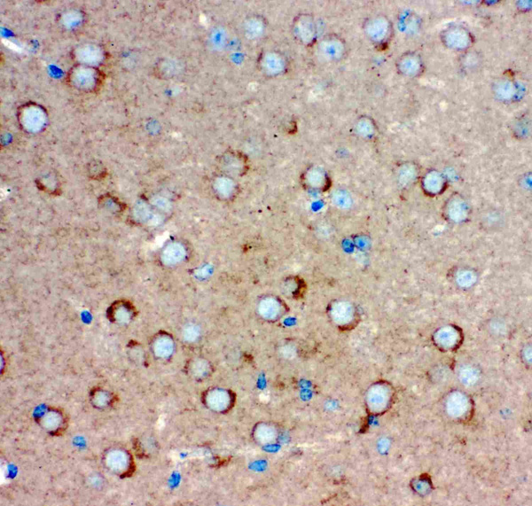
(Figure 4. IHC analysis of GRIA1 using anti-GRIA1 antibody (AAA46057).GRIA1 was detected in paraffin-embedded section of Rat Brain Tissue. Heat mediated antigen retrieval was performed in citrate buffer (pH6, epitope retrieval solution) for 20 mins. The tissue section was blocked with 10% goat serum. The tissue section was then incubated with 1ug/ml rabbit anti-GRIA1 Antibody (AAA46057) overnight at 4 degree C. Biotinylated goat anti-rabbit IgG was used as secondary antibody and incubated for 30 minutes at 37 degree C. The tissue section was developed using Strepavidin-Biotin-Complex (SABC) with DAB as the chromogen.)
IHC (Immunohistochemistry)
(Figure 4. IHC analysis of GRIA1 using anti-GRIA1 antibody (AAA46057).GRIA1 was detected in paraffin-embedded section of Rat Brain Tissue. Heat mediated antigen retrieval was performed in citrate buffer (pH6, epitope retrieval solution) for 20 mins. The tissue section was blocked with 10% goat serum. The tissue section was then incubated with 1ug/ml rabbit anti-GRIA1 Antibody (AAA46057) overnight at 4 degree C. Biotinylated goat anti-rabbit IgG was used as secondary antibody and incubated for 30 minutes at 37 degree C. The tissue section was developed using Strepavidin-Biotin-Complex (SABC) with DAB as the chromogen.)
IHC (Immunohistochemisry)
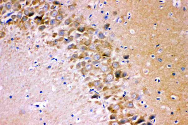
(Figure 3. IHC analysis of GRIA1 using anti-GRIA1 antibody (AAA46057).GRIA1 was detected in paraffin-embedded section of Mouse Brain Tissue. Heat mediated antigen retrieval was performed in citrate buffer (pH6, epitope retrieval solution) for 20 mins. The tissue section was blocked with 10% goat serum. The tissue section was then incubated with 1ug/ml rabbit anti-GRIA1 Antibody (AAA46057) overnight at 4 degree C. Biotinylated goat anti-rabbit IgG was used as secondary antibody and incubated for 30 minutes at 37 degree C. The tissue section was developed using Strepavidin-Biotin-Complex (SABC) with DAB as the chromogen.)
IHC (Immunohistochemisry)
(Figure 3. IHC analysis of GRIA1 using anti-GRIA1 antibody (AAA46057).GRIA1 was detected in paraffin-embedded section of Mouse Brain Tissue. Heat mediated antigen retrieval was performed in citrate buffer (pH6, epitope retrieval solution) for 20 mins. The tissue section was blocked with 10% goat serum. The tissue section was then incubated with 1ug/ml rabbit anti-GRIA1 Antibody (AAA46057) overnight at 4 degree C. Biotinylated goat anti-rabbit IgG was used as secondary antibody and incubated for 30 minutes at 37 degree C. The tissue section was developed using Strepavidin-Biotin-Complex (SABC) with DAB as the chromogen.)
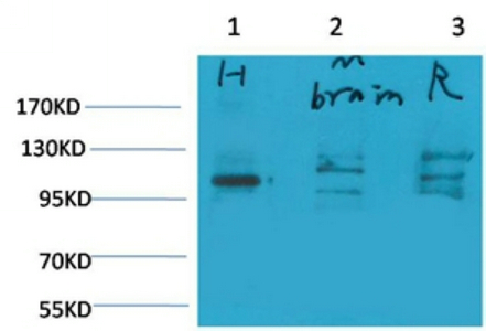
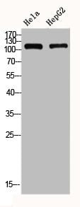
WB (Western Blot)
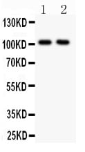
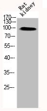
(Figure 2. Western blot analysis of GRIA1 using anti-GRIA1 antibody (AAA46057).Electrophoresis was performed on a 5-20% SDS-PAGE gel at 70V (Stacking gel) / 90V (Resolving gel) for 2-3 hours. The sample well of each lane was loaded with 50ug of sample under reducing conditions.Lane 1: Rat Brain Tissue Lysate,Lane 2: Mouse Brain Tissue Lysate.After Electrophoresis, proteins were transferred to a Nitrocellulose membrane at 150mA for 50-90 minutes. Blocked the membrane with 5% Non-fat Milk/ TBS for 1.5 hour at RT. The membrane was incubated with rabbit anti-GRIA1 antigen affinity purified polyclonal antibody at 0.5ug/mL overnight at 4 degree C, then washed with TBS-0.1%Tween 3 times with 5 minutes each and probed with a goat anti-rabbit IgG-HRP secondary antibody at a dilution of 1:10000 for 1.5 hour at RT. The signal is developed using an Enhanced Chemiluminescent detection (ECL) kit with Tanon 5200 system. A specific band was detected for GRIA1 at approximately 101KD. The expected band size for GRIA1 is at 101KD.)
WB (Western Blot)
(Figure 2. Western blot analysis of GRIA1 using anti-GRIA1 antibody (AAA46057).Electrophoresis was performed on a 5-20% SDS-PAGE gel at 70V (Stacking gel) / 90V (Resolving gel) for 2-3 hours. The sample well of each lane was loaded with 50ug of sample under reducing conditions.Lane 1: Rat Brain Tissue Lysate,Lane 2: Mouse Brain Tissue Lysate.After Electrophoresis, proteins were transferred to a Nitrocellulose membrane at 150mA for 50-90 minutes. Blocked the membrane with 5% Non-fat Milk/ TBS for 1.5 hour at RT. The membrane was incubated with rabbit anti-GRIA1 antigen affinity purified polyclonal antibody at 0.5ug/mL overnight at 4 degree C, then washed with TBS-0.1%Tween 3 times with 5 minutes each and probed with a goat anti-rabbit IgG-HRP secondary antibody at a dilution of 1:10000 for 1.5 hour at RT. The signal is developed using an Enhanced Chemiluminescent detection (ECL) kit with Tanon 5200 system. A specific band was detected for GRIA1 at approximately 101KD. The expected band size for GRIA1 is at 101KD.)
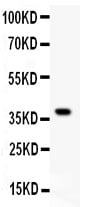
WB (Western Blot)
(Figure 1. Western blot analysis of GRIA1 using anti-GRIA1 antibody (AAA46057).Electrophoresis was performed on a 5-20% SDS-PAGE gel at 70V (Stacking gel) / 90V (Resolving gel) for 2-3 hours.lane 1: Recombinant Human GRIA1 Protein 0.5ng.After Electrophoresis, proteins were transferred to a Nitrocellulose membrane at 150mA for 50-90 minutes. Blocked the membrane with 5% Non-fat Milk/ TBS for 1.5 hour at RT. The membrane was incubated with rabbit anti-GRIA1 antigen affinity purified polyclonal antibody at 0.5ug/mL overnight at 4 degree C, then washed with TBS-0.1%Tween 3 times with 5 minutes each and probed with a goat anti-rabbit IgG-HRP secondary antibody at a dilution of 1:10000 for 1.5 hour at RT. The signal is developed using an Enhanced Chemiluminescent detection (ECL) kit with Tanon 5200 system. A specific band was detected for GRIA1 at approximately 40KD. The expected band size for GRIA1 is at 40KD.)
Background: GLUR1, Glutamate receptor 1, is a protein that in humans is encoded by the GLUR1 gene. GLUR1 mRNA is widely expressed in human brain. Glutamate receptors are the predominant excitatory neurotransmitter receptors in the mammalian brain and are activated in a variety of normal neurophysiologic processes. The classification of glutamate receptors is based on their activation by different pharmacologic agonists. The GRIA1 belongs to a family of alpha-amino-3-hydroxy-5-methyl-4-isoxazole propionate (AMPA) receptors. Each of the members (GRIA1-4) include flip and flop isoforms generated by alternative RNA splicing. The receptor subunits encoded by each isoform vary in their signal transduction properties. The isoform presented here is the flop isoform. In situ hybridization experiments showed that human GRIA1 mRNA is present in granule and pyramidal cells in the hippocampal formation.
NCBI and Uniprot Product Information
Customer Reviews
Loading reviews...
Share Your Experience
Similar Products

Product Notes
The GRIA1 gria1 (Catalog #AAA46057) is an Antibody produced from Rabbit and is intended for research purposes only. The product is available for immediate purchase. The Anti-GRIA1 Antibody reacts with Human, Mouse, Rat. No cross reactivity with other proteins. and may cross-react with other species as described in the data sheet. AAA Biotech's Glutamate receptor 1 can be used in a range of immunoassay formats including, but not limited to, IHC (Immunohistochemistry), WB (Western Blot). Researchers should empirically determine the suitability of the GRIA1 gria1 for an application not listed in the data sheet. Researchers commonly develop new applications and it is an integral, important part of the investigative research process. It is sometimes possible for the material contained within the vial of "Glutamate receptor 1, Polyclonal Antibody" to become dispersed throughout the inside of the vial, particularly around the seal of said vial, during shipment and storage. We always suggest centrifuging these vials to consolidate all of the liquid away from the lid and to the bottom of the vial prior to opening. Please be advised that certain products may require dry ice for shipping and that, if this is the case, an additional dry ice fee may also be required.Precautions
All products in the AAA Biotech catalog are strictly for research-use only, and are absolutely not suitable for use in any sort of medical, therapeutic, prophylactic, in-vivo, or diagnostic capacity. By purchasing a product from AAA Biotech, you are explicitly certifying that said products will be properly tested and used in line with industry standard. AAA Biotech and its authorized distribution partners reserve the right to refuse to fulfill any order if we have any indication that a purchaser may be intending to use a product outside of our accepted criteria.Disclaimer
Though we do strive to guarantee the information represented in this datasheet, AAA Biotech cannot be held responsible for any oversights or imprecisions. AAA Biotech reserves the right to adjust any aspect of this datasheet at any time and without notice. It is the responsibility of the customer to inform AAA Biotech of any product performance issues observed or experienced within 30 days of receipt of said product. To see additional details on this or any of our other policies, please see our Terms & Conditions page.Item has been added to Shopping Cart
If you are ready to order, navigate to Shopping Cart and get ready to checkout.