


 FCM (Flow Cytometry)
(Figure 6. Flow Cytometry analysis of A431 cells using anti-HIF-1 alpha/HIF1A antibody (AAA19213).Overlay histogram showing A431 cells stained with AAA19213 (Blue line). The cells were blocked with 10% normal goat serum. And then incubated with rabbit anti-HIF-1 alpha/HIF1A Antibody (AAA19213, 1μg/1x106 cells) for 30 min at 20 degree C. DyLight®488 conjugated goat anti-rabbit IgG (5-10μg/1x106 cells) was used as secondary antibody for 30 minutes at 20 degree C. Isotype control antibody (Green line) was rabbit IgG (1μg/1x106) used under the same conditions. Unlabelled sample (Red line) was also used as a control.)
FCM (Flow Cytometry)
(Figure 6. Flow Cytometry analysis of A431 cells using anti-HIF-1 alpha/HIF1A antibody (AAA19213).Overlay histogram showing A431 cells stained with AAA19213 (Blue line). The cells were blocked with 10% normal goat serum. And then incubated with rabbit anti-HIF-1 alpha/HIF1A Antibody (AAA19213, 1μg/1x106 cells) for 30 min at 20 degree C. DyLight®488 conjugated goat anti-rabbit IgG (5-10μg/1x106 cells) was used as secondary antibody for 30 minutes at 20 degree C. Isotype control antibody (Green line) was rabbit IgG (1μg/1x106) used under the same conditions. Unlabelled sample (Red line) was also used as a control.)
Rabbit HIF-1 alpha/HIF1A Polyclonal Antibody | anti-HIF1A antibody
Anti-HIF-1 alpha/HIF1A Antibody
IHC-P: 2-5ug/ml|Human, Mouse, Rat|
FC/FACS/FCM: 1-3ug/1x106 cells|Human|
Direct ELISA: 0.1-0.5ug/ml|Human|
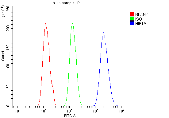
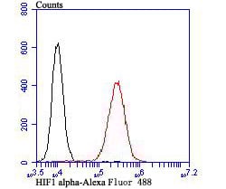
FCM (Flow Cytometry)
(Figure 6. Flow Cytometry analysis of A431 cells using anti-HIF-1 alpha/HIF1A antibody (AAA19213).Overlay histogram showing A431 cells stained with AAA19213 (Blue line). The cells were blocked with 10% normal goat serum. And then incubated with rabbit anti-HIF-1 alpha/HIF1A Antibody (AAA19213, 1μg/1x106 cells) for 30 min at 20 degree C. DyLight®488 conjugated goat anti-rabbit IgG (5-10μg/1x106 cells) was used as secondary antibody for 30 minutes at 20 degree C. Isotype control antibody (Green line) was rabbit IgG (1μg/1x106) used under the same conditions. Unlabelled sample (Red line) was also used as a control.)
IHC (Immunohistochemistry)
(Figure 5. IHC analysis of HIF-1 alpha/HIF1A using anti-HIF-1 alpha/HIF1A antibody (AAA19213).HIF-1 alpha/HIF1A was detected in paraffin-embedded section of rat intestine tissue. Heat mediated antigen retrieval was performed in EDTA buffer (pH8. 0, epitope retrieval solution). The tissue section was blocked with 10% goat serum. The tissue section was then incubated with 2μg/ml rabbit anti-HIF-1 alpha/HIF1A Antibody (AAA19213) overnight at 4 degree C. Biotinylated goat anti-rabbit IgG was used as secondary antibody and incubated for 30 minutes at 37 degree C. The tissue section was developed using Strepavidin-Biotin-Complex (SABC) (Catalog # with DAB as the chromogen.)
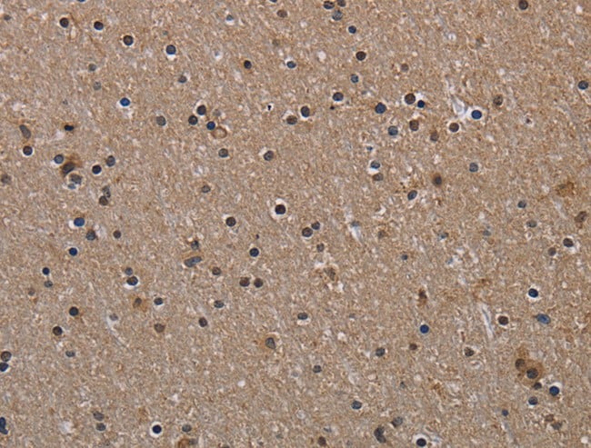
IHC (Immunohistochemistry)
(Figure 4. IHC analysis of HIF-1 alpha/HIF1A using anti-HIF-1 alpha/HIF1A antibody (AAA19213).HIF-1 alpha/HIF1A was detected in paraffin-embedded section of mouse intestine tissue. Heat mediated antigen retrieval was performed in EDTA buffer (pH8. 0, epitope retrieval solution). The tissue section was blocked with 10% goat serum. The tissue section was then incubated with 2μg/ml rabbit anti-HIF-1 alpha/HIF1A Antibody (AAA19213) overnight at 4 degree C. Biotinylated goat anti-rabbit IgG was used as secondary antibody and incubated for 30 minutes at 37 degree C. The tissue section was developed using Strepavidin-Biotin-Complex (SABC) (Catalog # with DAB as the chromogen.)
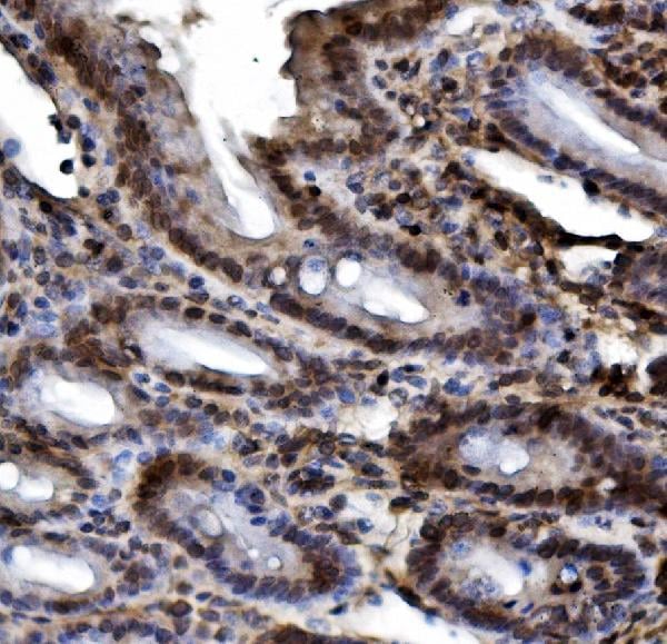
IHC (Immunohistochemistry)
(Figure 3. IHC analysis of HIF-1 alpha/HIF1A using anti-HIF-1 alpha/HIF1A antibody (AAA19213).HIF-1 alpha/HIF1A was detected in paraffin-embedded section of human rectal cancer tissue. Heat mediated antigen retrieval was performed in EDTA buffer (pH8. 0, epitope retrieval solution). The tissue section was blocked with 10% goat serum. The tissue section was then incubated with 2μg/ml rabbit anti-HIF-1 alpha/HIF1A Antibody (AAA19213) overnight at 4 degree C. Biotinylated goat anti-rabbit IgG was used as secondary antibody and incubated for 30 minutes at 37 degree C. The tissue section was developed using Strepavidin-Biotin-Complex (SABC) (Catalog # with DAB as the chromogen.)
IHC (Immunohistochemistry)
(Figure 2. IHC analysis of HIF-1 alpha/HIF1A using anti-HIF-1 alpha/HIF1A antibody (AAA19213).HIF-1 alpha/HIF1A was detected in paraffin-embedded section of human mammary cancer tissue. Heat mediated antigen retrieval was performed in EDTA buffer (pH8. 0, epitope retrieval solution). The tissue section was blocked with 10% goat serum. The tissue section was then incubated with 2μg/ml rabbit anti-HIF-1 alpha/HIF1A Antibody (AAA19213) overnight at 4 degree C. Biotinylated goat anti-rabbit IgG was used as secondary antibody and incubated for 30 minutes at 37 degree C. The tissue section was developed using Strepavidin-Biotin-Complex (SABC) (Catalog # with DAB as the chromogen.)
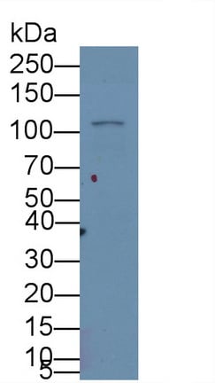
WB (Western Blot)
(Figure 1. Western blot analysis of HIF-1 alpha/HIF1A using anti-HIF-1 alpha/HIF1A antibody (AAA19213).Electrophoresis was performed on a 5-20% SDS-PAGE gel at 70V (Stacking gel) / 90V (Resolving gel) for 2-3 hours. The sample well of each lane was loaded with 30ug of sample under reducing conditions.Lane 1: human A431 whole cell lysatesLane 2: human HEPG2 whole cell lysatesLane 3: human U20S whole cell lysatesLane 4: rat PC-12 whole cell lysatesLane 5: mouse thymus tissue lysates.After Electrophoresis, proteins were transferred to a Nitrocellulose membrane at 150mA for 50-90 minutes. Blocked the membrane with 5% Non-fat Milk/ TBS for 1. 5 hour at RT. The membrane was incubated with rabbit anti-HIF-1 alpha/HIF1A antigen affinity purified polyclonal antibody (Catalog # AAA19213) at 0. 5 μg/mL overnight at 4 degree C, then washed with TBS-0. 1%Tween 3 times with 5 minutes each and probed with a goat anti-rabbit IgG-HRP secondary antibody at a dilution of 1:5000 for 1. 5 hour at RT. The signal is developed using an Enhanced Chemiluminescent detection (ECL) kit (Catalog # with Tanon 5200 system. A specific band was detected for HIF-1 alpha/HIF1A at approximately 93KD. The expected band size for HIF-1 alpha/HIF1A is at 93KD.)
2. Elson, D. A.; Thurston, G.; Huang, L. E.; Ginzinger, D. G.; McDonald, D. M.; Johnson, R. S.; Arbeit, J. M. : Induction of hypervascularity without leakage or inflammation in transgenic mice overexpressing hypoxia-inducible factor-1-alpha. Genes Dev. 15: 2520-2532, 2001.
3. Koshiji, M.; To, K. K. -W.; Hammer, S.; Kumamoto, K.; Harris, A. L.; Modrich, P.; Huang, L. E. : HIF-1-alpha induces genetic instability by transcriptionally downregulating MutS-alpha expression. Molec. Cell 17: 793-803, 2005.
4. Ivan, M.; Kondo, K.; Yang, H.; Kim, W.; Valiando, J.; Ohh, M.; Salic, A.; Asara, J. M.; Lane, W. S.; Kaelin, W. G., Jr. : HIF-alpha targeted for VHL-mediated destruction by proline hydroxylation: implications for O(2) sensing. Science 292: 464-468, 2001.
NCBI and Uniprot Product Information
Customer Reviews
Loading reviews...
Share Your Experience
Similar Products


Product Notes
The HIF1A hif1a (Catalog #AAA19213) is an Antibody produced from Rabbit and is intended for research purposes only. The product is available for immediate purchase. The Anti-HIF-1 alpha/HIF1A Antibody reacts with Human, Mouse, Rat and may cross-react with other species as described in the data sheet. AAA Biotech's HIF-1 alpha/HIF1A can be used in a range of immunoassay formats including, but not limited to, WB (Western Blot), IHC (Immunohistochemistry), FCM/FACS (Flow Cytometry), ELISA (Direct ELISA). WB: 0.25-0.5ug/ml|Human, Mouse, Rat| IHC-P: 2-5ug/ml|Human, Mouse, Rat| FC/FACS/FCM: 1-3ug/1x106 cells|Human| Direct ELISA: 0.1-0.5ug/ml|Human|. Researchers should empirically determine the suitability of the HIF1A hif1a for an application not listed in the data sheet. Researchers commonly develop new applications and it is an integral, important part of the investigative research process. It is sometimes possible for the material contained within the vial of "HIF-1 alpha/HIF1A, Polyclonal Antibody" to become dispersed throughout the inside of the vial, particularly around the seal of said vial, during shipment and storage. We always suggest centrifuging these vials to consolidate all of the liquid away from the lid and to the bottom of the vial prior to opening. Please be advised that certain products may require dry ice for shipping and that, if this is the case, an additional dry ice fee may also be required.Precautions
All products in the AAA Biotech catalog are strictly for research-use only, and are absolutely not suitable for use in any sort of medical, therapeutic, prophylactic, in-vivo, or diagnostic capacity. By purchasing a product from AAA Biotech, you are explicitly certifying that said products will be properly tested and used in line with industry standard. AAA Biotech and its authorized distribution partners reserve the right to refuse to fulfill any order if we have any indication that a purchaser may be intending to use a product outside of our accepted criteria.Disclaimer
Though we do strive to guarantee the information represented in this datasheet, AAA Biotech cannot be held responsible for any oversights or imprecisions. AAA Biotech reserves the right to adjust any aspect of this datasheet at any time and without notice. It is the responsibility of the customer to inform AAA Biotech of any product performance issues observed or experienced within 30 days of receipt of said product. To see additional details on this or any of our other policies, please see our Terms & Conditions page.Frequently Asked Questions
What is HIF-1 alpha used as a marker for in hypoxia or cellular stress studies?
HIF-1 alpha (Hypoxia-Inducible Factor 1-alpha) is the primary marker for cellular response to low oxygen levels. Under normal conditions, it is rapidly degraded, but it stabilizes and accumulates during hypoxia. Researchers use the AAA Biotech HIF-1 alpha antibody to study how cells adapt to metabolic stress, oxygen deprivation, and various pathological states like ischemia.
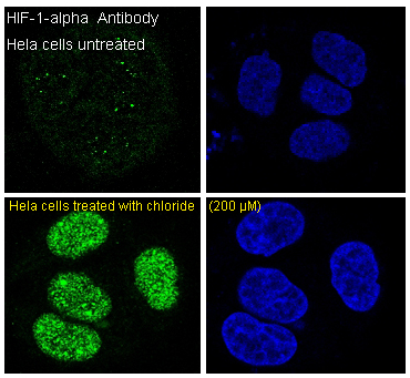
Can this HIF-1 alpha antibody be used for Western blot or immunofluorescence?
Yes, this antibody is validated for both Western blot and immunofluorescence (IF). Western blotting is typically used to observe the stabilization and protein levels of HIF-1 alpha in cell lysates. Immunofluorescence allows researchers to visualize the nuclear translocation of HIF-1 alpha, which is a hallmark of its activation as a transcription factor in AAA Biotech-supported research.
How does HIF-1 alpha expression change under low oxygen conditions?
Under low oxygen conditions (hypoxia), the enzymatic degradation of HIF-1 alpha is inhibited. This leads to a rapid accumulation of the protein within the cytoplasm and its subsequent translocation into the nucleus. By using a specific antibody from AAA Biotech, scientists can track this increase in expression, which triggers the transcription of genes involved in glycolysis and angiogenesis.
Is HIF-1 alpha involved in cancer and metabolic research?
HIF-1 alpha is a critical focus in cancer research because tumors often exist in hypoxic environments. It promotes tumor survival, angiogenesis, and metabolic reprogramming. AAA Biotech provides HIF-1 alpha antibodies to help researchers investigate how this protein contributes to tumor progression, therapy resistance, and the shift from oxidative phosphorylation to glycolysis in cancer cells.
Can this antibody detect endogenous HIF-1 alpha in human or animal samples?
Yes, the HIF-1 alpha antibody from AAA Biotech is designed to detect endogenous levels of the protein in a variety of samples, including human and common laboratory animal models. Because HIF-1 alpha is highly conserved, this antibody is a reliable tool for studying natural physiological responses to oxygen stress across different species without the need for overexpression.
How does fixation or treatment method affect HIF-1 alpha staining quality?
HIF-1 alpha is a highly labile protein. Fixation methods, such as using 4% paraformaldehyde, are crucial for preserving the protein's localization, especially in the nucleus. Because it degrades so quickly upon re-oxygenation, samples must be processed or fixed immediately. AAA Biotech recommends optimized protocols to ensure that the staining reflects the true hypoxic state of the cells.
Item has been added to Shopping Cart
If you are ready to order, navigate to Shopping Cart and get ready to checkout.