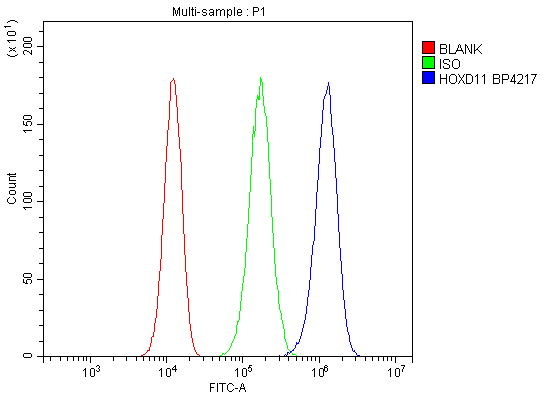
 FCM (Flow Cytometry)
(Figure 9. Flow Cytometry analysis of RAW264. 7 cells using anti-HOXD11 antibody (AAA19334).Overlay histogram showing RAW264. 7 cells stained with AAA19334 (Blue line). The cells were blocked with 10% normal goat serum. And then incubated with rabbit anti-HOXD11 Antibody (AAA19334, 1μg/1x106 cells) for 30 min at 20 degree C. DyLight®488 conjugated goat anti-rabbit IgG (5-10μg/1x106 cells) was used as secondary antibody for 30 minutes at 20 degree C. Isotype control antibody (Green line) was rabbit IgG (1μg/1x106) used under the same conditions. Unlabelled sample (Red line) was also used as a control.)
FCM (Flow Cytometry)
(Figure 9. Flow Cytometry analysis of RAW264. 7 cells using anti-HOXD11 antibody (AAA19334).Overlay histogram showing RAW264. 7 cells stained with AAA19334 (Blue line). The cells were blocked with 10% normal goat serum. And then incubated with rabbit anti-HOXD11 Antibody (AAA19334, 1μg/1x106 cells) for 30 min at 20 degree C. DyLight®488 conjugated goat anti-rabbit IgG (5-10μg/1x106 cells) was used as secondary antibody for 30 minutes at 20 degree C. Isotype control antibody (Green line) was rabbit IgG (1μg/1x106) used under the same conditions. Unlabelled sample (Red line) was also used as a control.)
Rabbit HOXD11 Polyclonal Antibody | anti-HOXD11 antibody
Anti-HOXD11 Antibody
IHC-P: Concentration: 2-5ug/ml ; Tested Species: Human
ICC/IF: Concentration: 5ug/ml ; Tested Species: Human
FC: Concentration: 1-3ug/1x106 cell ; Tested Species: Human, Mouse, Rat
Test Species: In-house tested species with positive results.
Other applications have not been tested.
Optimal dilutions should be determined by end users.
FCM (Flow Cytometry)
(Figure 9. Flow Cytometry analysis of RAW264. 7 cells using anti-HOXD11 antibody (AAA19334).Overlay histogram showing RAW264. 7 cells stained with AAA19334 (Blue line). The cells were blocked with 10% normal goat serum. And then incubated with rabbit anti-HOXD11 Antibody (AAA19334, 1μg/1x106 cells) for 30 min at 20 degree C. DyLight®488 conjugated goat anti-rabbit IgG (5-10μg/1x106 cells) was used as secondary antibody for 30 minutes at 20 degree C. Isotype control antibody (Green line) was rabbit IgG (1μg/1x106) used under the same conditions. Unlabelled sample (Red line) was also used as a control.)
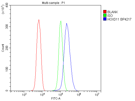
FCM (Flow Cytometry)
(Figure 8. Flow Cytometry analysis of NRK cells using anti-HOXD11 antibody (AAA19334).Overlay histogram showing NRK cells stained with AAA19334 (Blue line). The cells were blocked with 10% normal goat serum. And then incubated with rabbit anti-HOXD11 Antibody (AAA19334, 1μg/1x106 cells) for 30 min at 20 degree C. DyLight®488 conjugated goat anti-rabbit IgG (5-10μg/1x106 cells) was used as secondary antibody for 30 minutes at 20 degree C. Isotype control antibody (Green line) was rabbit IgG (1μg/1x106) used under the same conditions. Unlabelled sample (Red line) was also used as a control.)
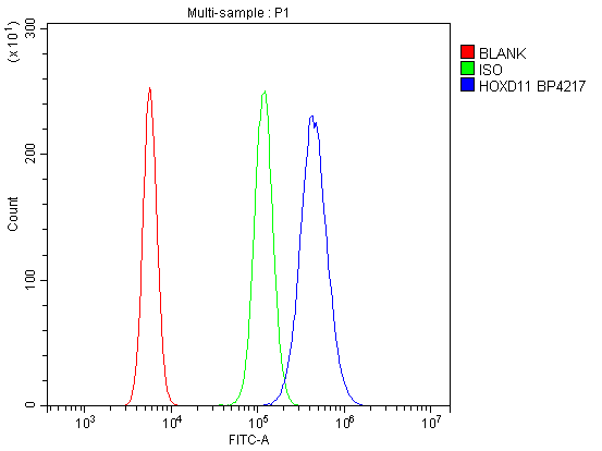
FCM (Flow Cytometry)
(Figure 7. Flow Cytometry analysis of 293T cells using anti-HOXD11 antibody (AAA19334).Overlay histogram showing 293T cells stained with AAA19334 (Blue line). The cells were blocked with 10% normal goat serum. And then incubated with rabbit anti-HOXD11 Antibody (AAA19334, 1μg/1x106 cells) for 30 min at 20 degree C. DyLight®488 conjugated goat anti-rabbit IgG (5-10μg/1x106 cells) was used as secondary antibody for 30 minutes at 20 degree C. Isotype control antibody (Green line) was rabbit IgG (1μg/1x106) used under the same conditions. Unlabelled sample (Red line) was also used as a control.)
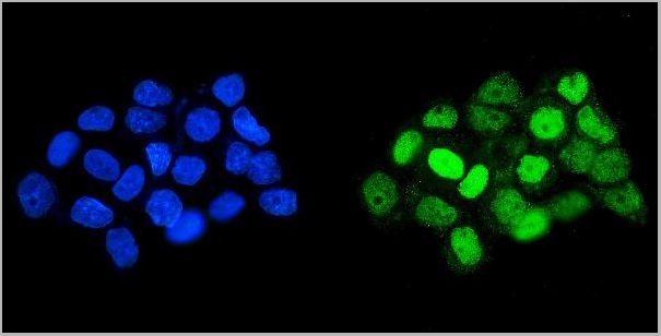
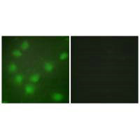
IF (Immunofluorescence)
(Figure 6. IF analysis of HOXD11 using anti- HOXD11 antibody (AAA19334).HOXD11 was detected in immunocytochemical section of A431 cells. Enzyme antigen retrieval was performed using IHC enzyme antigen retrieval reagent for 15 mins. The cells were blocked with 10% goat serum. And then incubated with 5μg/mL rabbit anti-HOXD11 Antibody (AAA19334) overnight at 4 degree C. DyLight®488 Conjugated Goat Anti-Rabbit IgG was used as secondary antibody at 1:100 dilution and incubated for 30 minutes at 37 degree C. The section was counterstained with DAPI. Visualize using a fluorescence microscope and filter sets appropriate for the label used.)
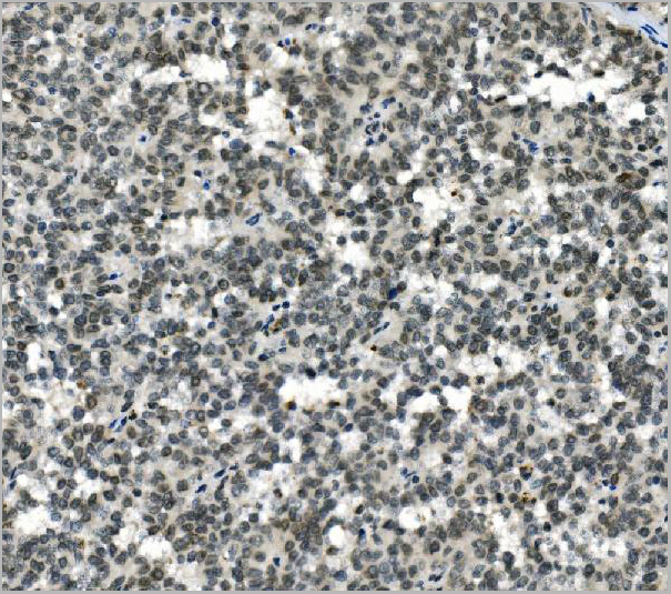
IHC (Immunohistochemistry)
(Figure 5. IHC analysis of HOXD11 using anti-HOXD11 antibody (AAA19334).HOXD11 was detected in paraffin-embedded section of human testicular cancer tissue. Heat mediated antigen retrieval was performed in EDTA buffer (pH8. 0, epitope retrieval solution). The tissue section was blocked with 10% goat serum. The tissue section was then incubated with 2μg/ml rabbit anti-HOXD11 Antibody (AAA19334) overnight at 4 degree C. Biotinylated goat anti-rabbit IgG was used as secondary antibody and incubated for 30 minutes at 37 degree C. The tissue section was developed using Strepavidin-Biotin-Complex (SABC) (Catalog # with DAB as the chromogen.)
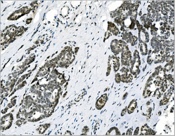
IHC (Immunohistochemistry)
(Figure 4. IHC analysis of HOXD11 using anti-HOXD11 antibody (AAA19334).HOXD11 was detected in paraffin-embedded section of human skin cancer tissue. Heat mediated antigen retrieval was performed in EDTA buffer (pH8. 0, epitope retrieval solution). The tissue section was blocked with 10% goat serum. The tissue section was then incubated with 2μg/ml rabbit anti-HOXD11 Antibody (AAA19334) overnight at 4 degree C. Biotinylated goat anti-rabbit IgG was used as secondary antibody and incubated for 30 minutes at 37 degree C. The tissue section was developed using Strepavidin-Biotin-Complex (SABC) (Catalog # with DAB as the chromogen.)
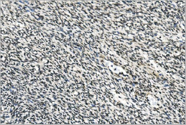
IHC (Immunohistochemistry)
(Figure 3. IHC analysis of HOXD11 using anti-HOXD11 antibody (AAA19334).HOXD11 was detected in paraffin-embedded section of human ovarian cancer tissue. Heat mediated antigen retrieval was performed in EDTA buffer (pH8. 0, epitope retrieval solution). The tissue section was blocked with 10% goat serum. The tissue section was then incubated with 2μg/ml rabbit anti-HOXD11 Antibody (AAA19334) overnight at 4 degree C. Biotinylated goat anti-rabbit IgG was used as secondary antibody and incubated for 30 minutes at 37 degree C. The tissue section was developed using Strepavidin-Biotin-Complex (SABC) (Catalog # with DAB as the chromogen.)
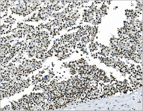
IHC (Immunohistochemistry)
(Figure 2. IHC analysis of HOXD11 using anti-HOXD11 antibody (AAA19334).HOXD11 was detected in paraffin-embedded section of human melanoma tissue. Heat mediated antigen retrieval was performed in EDTA buffer (pH8. 0, epitope retrieval solution). The tissue section was blocked with 10% goat serum. The tissue section was then incubated with 2μg/ml rabbit anti-HOXD11 Antibody (AAA19334) overnight at 4 degree C. Biotinylated goat anti-rabbit IgG was used as secondary antibody and incubated for 30 minutes at 37 degree C. The tissue section was developed using Strepavidin-Biotin-Complex (SABC) (Catalog # with DAB as the chromogen.)
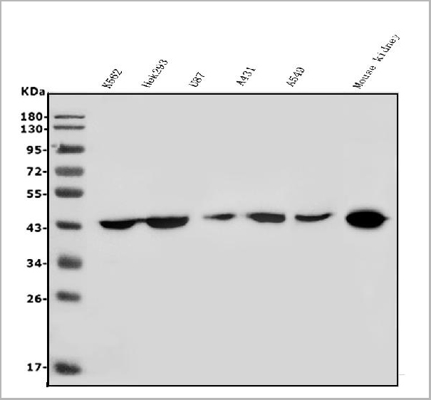
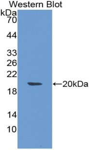
WB (Western Blot)
(Figure 1. Western blot analysis of HOXD11 using anti-HOXD11 antibody (AAA19334).Electrophoresis was performed on a 5-20% SDS-PAGE gel at 70V (Stacking gel) / 90V (Resolving gel) for 2-3 hours. The sample well of each lane was loaded with 50ug of sample under reducing conditions.Lane 1: human K562 whole cell lysatesLane 2: human Hek293 whole cell lysatesLane 3: human U87 whole cell lysatesLane 4: human A431 whole cell lysatesLane 5: human A549 whole cell lysatesLane 6: mouse kidney tissue lysates.After Electrophoresis, proteins were transferred to a Nitrocellulose membrane at 150mA for 50-90 minutes. Blocked the membrane with 5% Non-fat Milk/ TBS for 1. 5 hour at RT. The membrane was incubated with rabbit anti-HOXD11 antigen affinity purified polyclonal antibody (Catalog # AAA19334) at 0. 5 μg/mL overnight at 4 degree C, then washed with TBS-0. 1%Tween 3 times with 5 minutes each and probed with a goat anti-rabbit IgG-HRP secondary antibody at a dilution of 1:5000 for 1. 5 hour at RT. The signal is developed using an Enhanced Chemiluminescent detection (ECL) kit (Catalog # with Tanon 5200 system. A specific band was detected for HOXD11 at approximately 45KD. The expected band size for HOXD11 is at 45KD.)
2. Johnson, R. L., Tabin, C. J. Molecular models for vertebrate limb development. Cell 90: 979-990, 1997.
3. Kmita, M., Fraudeau, N., Herault, Y., Duboule, D. Serial deletions and duplications suggest a mechanism for the collinearity of Hoxd genes in limbs. Nature 420: 145-150, 2002.
NCBI and Uniprot Product Information
Customer Reviews
Loading reviews...
Share Your Experience
Similar Products
Product Notes
The HOXD11 hoxd11 (Catalog #AAA19334) is an Antibody produced from Rabbit and is intended for research purposes only. The product is available for immediate purchase. The Anti-HOXD11 Antibody reacts with Human, Mouse, Rat and may cross-react with other species as described in the data sheet. AAA Biotech's HOXD11 can be used in a range of immunoassay formats including, but not limited to, WB (Western Blot), IHC (Immunohistochemistry), ICC (Immunocytochemistry), IF (Immunofluorescence), FCM/FACS (Flow Cytometry). WB: Concentration: 0.25-0.5ug/ml ; Tested Species: Human, Mouse IHC-P: Concentration: 2-5ug/ml ; Tested Species: Human ICC/IF: Concentration: 5ug/ml ; Tested Species: Human FC: Concentration: 1-3ug/1x106 cell ; Tested Species: Human, Mouse, Rat Test Species: In-house tested species with positive results. Other applications have not been tested. Optimal dilutions should be determined by end users. Researchers should empirically determine the suitability of the HOXD11 hoxd11 for an application not listed in the data sheet. Researchers commonly develop new applications and it is an integral, important part of the investigative research process. It is sometimes possible for the material contained within the vial of "HOXD11, Polyclonal Antibody" to become dispersed throughout the inside of the vial, particularly around the seal of said vial, during shipment and storage. We always suggest centrifuging these vials to consolidate all of the liquid away from the lid and to the bottom of the vial prior to opening. Please be advised that certain products may require dry ice for shipping and that, if this is the case, an additional dry ice fee may also be required.Precautions
All products in the AAA Biotech catalog are strictly for research-use only, and are absolutely not suitable for use in any sort of medical, therapeutic, prophylactic, in-vivo, or diagnostic capacity. By purchasing a product from AAA Biotech, you are explicitly certifying that said products will be properly tested and used in line with industry standard. AAA Biotech and its authorized distribution partners reserve the right to refuse to fulfill any order if we have any indication that a purchaser may be intending to use a product outside of our accepted criteria.Disclaimer
Though we do strive to guarantee the information represented in this datasheet, AAA Biotech cannot be held responsible for any oversights or imprecisions. AAA Biotech reserves the right to adjust any aspect of this datasheet at any time and without notice. It is the responsibility of the customer to inform AAA Biotech of any product performance issues observed or experienced within 30 days of receipt of said product. To see additional details on this or any of our other policies, please see our Terms & Conditions page.Item has been added to Shopping Cart
If you are ready to order, navigate to Shopping Cart and get ready to checkout.