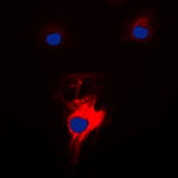
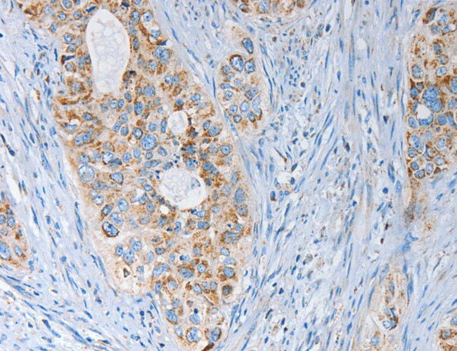
 IF (Immunofluorescence)
(AAA324023 staining HepG2 by IF/ICC. The sample were fixed with PFA and permeabilized in 0.1% Triton X-100, then blocked in 10% serum for 45 minutes at 25 degree C. The primary antibody was diluted at 1/200 and incubated with the sample for 1 hour at 37 degree C. An Alexa Fluor 594 conjugated goat anti-rabbit IgG (H+L) Ab, diluted at 1/600, was used as the secondary antibody.)
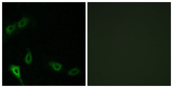
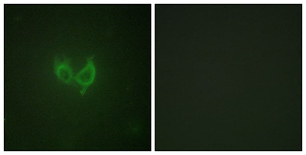
IF (Immunofluorescence)
(AAA324023 staining HepG2 by IF/ICC. The sample were fixed with PFA and permeabilized in 0.1% Triton X-100, then blocked in 10% serum for 45 minutes at 25 degree C. The primary antibody was diluted at 1/200 and incubated with the sample for 1 hour at 37 degree C. An Alexa Fluor 594 conjugated goat anti-rabbit IgG (H+L) Ab, diluted at 1/600, was used as the secondary antibody.)
Rabbit MARK2 Polyclonal Antibody | anti-MARK2 antibody
MARK2 Antibody
Gene Names
MARK2; EMK1; EMK-1; PAR-1; Par1b; Par-1b
Reactivity
Human, Mouse, Rat
Applications
ELISA, Immunocytochemistry, Immunofluorescence, Western Blot
Purity
The antiserum was purified by peptide affinity chromatography using SulfoLink Coupling Resin.
Synonyms
MARK2, Antibody; MARK2 Antibody; ELKL motif kinase 1; ELKL motif kinase; EMK-1; EMK1; MAP/microtubule affinity regulating kinase 2; MAP/microtubule affinity-regulating kinase 2; Mark2; MARK2_HUMAN; MGC99619; PAR 1; Par 1b; PAR1 homolog; Par1b; Ser/Thr protein kinase PAR 1B; Serine/threonine protein kinase EMK; Serine/threonine protein kinase MARK2; Serine/threonine-protein kinase MARK2; anti-MARK2 antibody
Host
Rabbit
Reactivity
Human, Mouse, Rat
Clonality
Polyclonal
Isotype
IgG
Specificity
MARK2 antibody detects endogenous levels of total MARK2
Purity/Purification
The antiserum was purified by peptide affinity chromatography using SulfoLink Coupling Resin.
Form/Format
Liquid
Phosphate buffered saline, pH 7.4, 150mM NaCl, 0.02% sodium azide and 50% glycerol.
Phosphate buffered saline, pH 7.4, 150mM NaCl, 0.02% sodium azide and 50% glycerol.
Concentration
1mg/ml (varies by lot)
Sequence Length
788
Applicable Applications for anti-MARK2 antibody
ELISA, ICC (Immunocytochemistry), IF (Immunofluorescence), WB (Western Blot)
Immunogen
A synthesized peptide
Subcellular Location
Cell Membrane. Phosphorylated by PRKCZ in polarized epithelial cells, resulting in an interaction with YWHAZ which promotes relocation from the lateral to the apical membrane.
Tissue Specificity
High levels of expression in heart, brain, skeletal muscle and pancreas, lower levels observed in lung, liver and kidney.
Conjugation
Unconjugated
Preparation and Storage
Store at -20 degree C. Stable for 12 months from date of receipt.
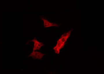
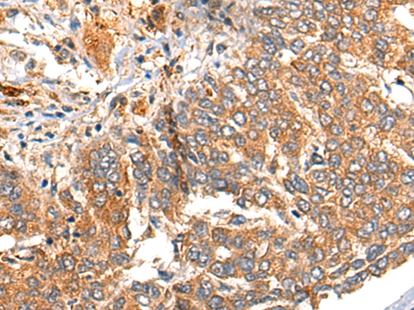
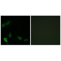
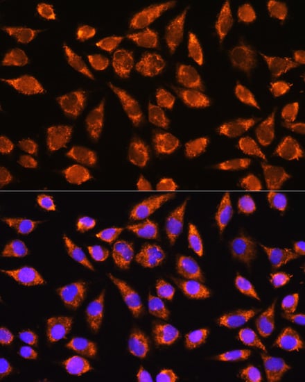
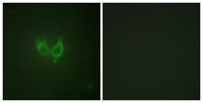
IF (Immunofluorescence)
(AAA324023 staining HepG2 by IF/ICC. The sample were fixed with PFA and permeabilized in 0.1% Triton X-100, then blocked in 10% serum for 45 minutes at 25 degree C. The primary antibody was diluted at 1/200 and incubated with the sample for 1 hour at 37 degree C. An Alexa Fluor 594 conjugated goat anti-rabbit IgG (H+L) Ab, diluted at 1/600, was used as the secondary antibody.)
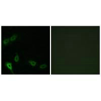
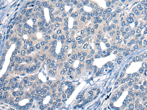
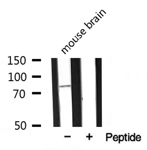
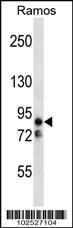
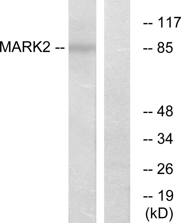
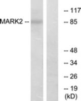
WB (Western Blot)
(Western blot analysis of extracts from mouse Brian, using MARK2 antibody.)
WB (Western Blot)
(Western blot analysis of extracts from mouse Brian, using MARK2 antibody.)
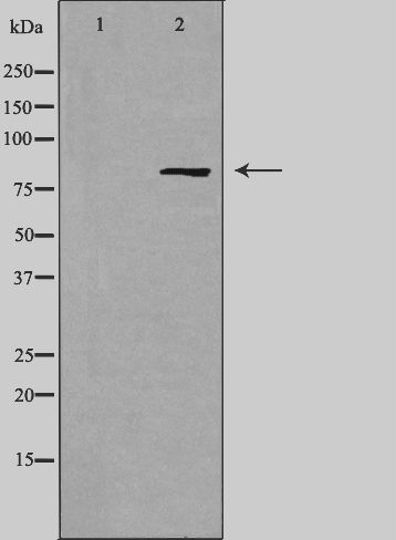
WB (Western Blot)
(Western blot analysis of extracts from COS-7 cells, using MARK2 antibody.)
WB (Western Blot)
(Western blot analysis of extracts from COS-7 cells, using MARK2 antibody.)
Related Product Information for anti-MARK2 antibody
Function: Serine/threonine-protein kinase involved in cell polarity and microtubule dynamics regulation. Phosphorylates CRTC2/TORC2, DCX, HDAC7, KIF13B, MAP2, MAP4, MAPT/TAU, and RAB11FIP2. Plays a key role in cell polarity by phosphorylating the microtubule-associated proteins MAP2, MAP4 and MAPT/TAU at KXGS motifs, causing detachment from microtubules, and their disassembly. Regulates epithelial cell polarity by phosphorylating RAB11FIP2. Involved in the regulation of neuronal migration through its dual activities in regulating cellular polarity and microtubule dynamics, possibly by phosphorylating and regulating DCX. Regulates axogenesis by phosphorylating KIF13B, promoting interaction between KIF13B and 14-3-3 and inhibiting microtubule-dependent accumulation of KIF13B. Also required for neurite outgrowth and establishment of neuronal polarity. Regulates localization and activity of some histone deacetylases by mediating phosphorylation of HDAC7, promoting subsequent interaction between HDAC7 and 14-3-3 and export from the nucleus. Also acts as a positive regulator of the Wnt signaling pathway, probably by mediating phosphorylation of dishevelled proteins (DVL1, DVL2 and/or DVL3). Modulates the developmental decision to build a columnar versus a hepatic epithelial cell apparently by promoting a switch from a direct to a transcytotic mode of apical protein delivery. Essential for the asymmetric development of membrane domains of polarized epithelial cells.
Subunit Structure: Homodimer. Interacts with PAK5; leading to inhibit the protein kinase activity (By similarity). Interacts with MTCL1 isoform 1; the interaction is direct and increases MARK2 microtubule-binding ability. Interacts (when phosphorylated at Thr-596) with YWHAZ. In case of infection, interacts with H.pylori CagA, leading to inhibit kinase activity and junctional and polarity defects.
Post-translational Modifications: Autophosphorylated. Phosphorylated at Thr-208 by STK11/LKB1 in complex with STE20-related adapter-alpha (STRADA) pseudo kinase and CAB39. Phosphorylation at Thr-208 by TAOK1 activates the kinase activity, leading to phosphorylation and detachment of MAPT/TAU from microtubules. Phosphorylation at Ser-212 by GSK3-beta (GSK3B) inhibits the kinase activity. Phosphorylation by CaMK1 promotes activity and is required to promote neurite outgrowth. Phosphorylation at Thr-596 by PRKCZ/aPKC in polarized epithelial cells inhibits the kinase activity and promotes binding to 14-3-3 protein YWHAZ, leading to relocation from cell membrane to cytoplasm.
Similarity: The UBA domain does not seem to bind ubiquitin and ubiquitin-like and might play a role in regulating the enzyme conformation and localization. Activation of the kinase activity following phosphorylation at Thr-208 is accompanied by a conformational change that alters the orientation of the UBA domain with respect to the catalytic domain.The KA1 domain mediates binding to phospholipids and targeting to membranes. Belongs to the protein kinase superfamily. CAMK Ser/Thr protein kinase family. SNF1 subfamily.
NCBI and Uniprot Product Information
NCBI GI #
NCBI GeneID
NCBI Accession #
NCBI GenBank Nucleotide #
Molecular Weight
Observed: 85 kD
Predicted: 88 kDa
Predicted: 88 kDa
NCBI Official Full Name
serine/threonine-protein kinase MARK2 isoform d
NCBI Official Synonym Full Names
microtubule affinity regulating kinase 2
NCBI Official Symbol
MARK2
NCBI Official Synonym Symbols
EMK1; EMK-1; PAR-1; Par1b; Par-1b
NCBI Protein Information
serine/threonine-protein kinase MARK2
UniProt Protein Name
Serine/threonine-protein kinase MARK2
UniProt Gene Name
MARK2
UniProt Synonym Gene Names
EMK-1; Par-1b; Par1b
Customer Reviews
Loading reviews...
Share Your Experience
Similar Products


Product Notes
The MARK2 mark2 (Catalog #AAA324023) is an Antibody produced from Rabbit and is intended for research purposes only. The product is available for immediate purchase. The MARK2 Antibody reacts with Human, Mouse, Rat and may cross-react with other species as described in the data sheet. AAA Biotech's MARK2 can be used in a range of immunoassay formats including, but not limited to, ELISA, ICC (Immunocytochemistry), IF (Immunofluorescence), WB (Western Blot). Researchers should empirically determine the suitability of the MARK2 mark2 for an application not listed in the data sheet. Researchers commonly develop new applications and it is an integral, important part of the investigative research process. It is sometimes possible for the material contained within the vial of "MARK2, Polyclonal Antibody" to become dispersed throughout the inside of the vial, particularly around the seal of said vial, during shipment and storage. We always suggest centrifuging these vials to consolidate all of the liquid away from the lid and to the bottom of the vial prior to opening. Please be advised that certain products may require dry ice for shipping and that, if this is the case, an additional dry ice fee may also be required.Precautions
All products in the AAA Biotech catalog are strictly for research-use only, and are absolutely not suitable for use in any sort of medical, therapeutic, prophylactic, in-vivo, or diagnostic capacity. By purchasing a product from AAA Biotech, you are explicitly certifying that said products will be properly tested and used in line with industry standard. AAA Biotech and its authorized distribution partners reserve the right to refuse to fulfill any order if we have any indication that a purchaser may be intending to use a product outside of our accepted criteria.Disclaimer
Though we do strive to guarantee the information represented in this datasheet, AAA Biotech cannot be held responsible for any oversights or imprecisions. AAA Biotech reserves the right to adjust any aspect of this datasheet at any time and without notice. It is the responsibility of the customer to inform AAA Biotech of any product performance issues observed or experienced within 30 days of receipt of said product. To see additional details on this or any of our other policies, please see our Terms & Conditions page.Item has been added to Shopping Cart
If you are ready to order, navigate to Shopping Cart and get ready to checkout.