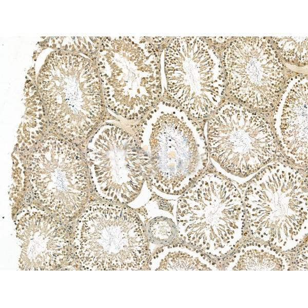
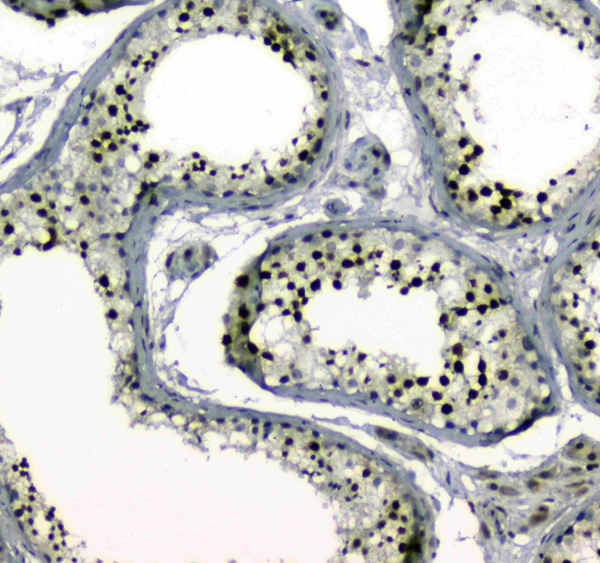
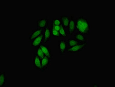
 IHC (Immunohistochemisry)
(AAA328898 at 1/100 staining Mouse testis tissue by IHC-P. The sample was formaldehyde fixed and a heat mediated antigen retrieval step in citrate buffer was performed. The sample was then blocked and incubated with the primary antibody at 4°C overnight. An HRP conjugated anti-Rabbit antibody was used as the secondary antibody.)
IHC (Immunohistochemisry)
(AAA328898 at 1/100 staining Mouse testis tissue by IHC-P. The sample was formaldehyde fixed and a heat mediated antigen retrieval step in citrate buffer was performed. The sample was then blocked and incubated with the primary antibody at 4°C overnight. An HRP conjugated anti-Rabbit antibody was used as the secondary antibody.)
Rabbit MDC1 Polyclonal Antibody | anti-MDC1 antibody
MDC1 Antibody
Predicted Reactivity: Pig(100%), Bovine(88%), Horse(100%), Sheep(100%), Rabbit(88%), Dog(100%)
Predicted Reactivity: Pig(100%), Bovine(88%), Horse(100%), Sheep(100%), Rabbit(88%), Dog(100%)
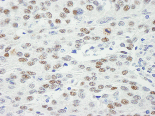
IHC (Immunohistochemisry)
(AAA328898 at 1/100 staining Mouse testis tissue by IHC-P. The sample was formaldehyde fixed and a heat mediated antigen retrieval step in citrate buffer was performed. The sample was then blocked and incubated with the primary antibody at 4°C overnight. An HRP conjugated anti-Rabbit antibody was used as the secondary antibody.)
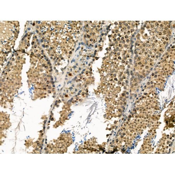
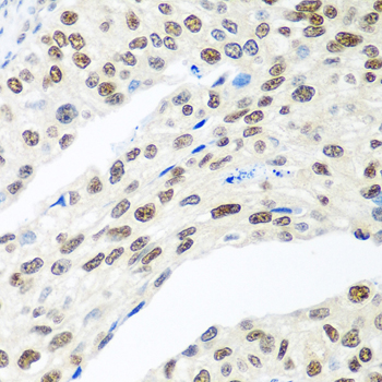
IHC (Immunohiostchemistry)
(AAA328898 at 1/100 staining Rat testis tissue by IHC-P. The sample was formaldehyde fixed and a heat mediated antigen retrieval step in citrate buffer was performed. The sample was then blocked and incubated with the primary antibody at 4°C overnight. An HRP conjugated anti-Rabbit antibody was used as the secondary antibody.)
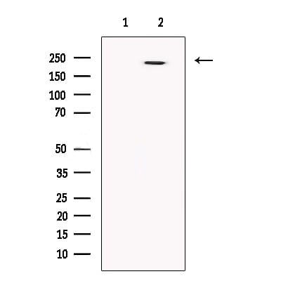
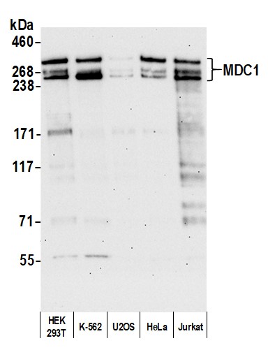
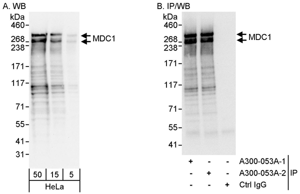
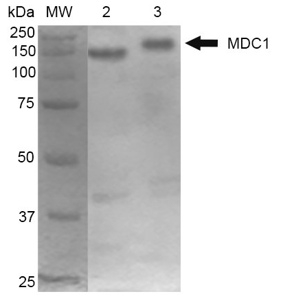
WB (Western Blot)
(Western blot analysis of extracts from HEK293, using MDC1 Antibody. The lane on the left was treated with blocking peptide.)
WB (Western Blot)
(Western blot analysis of extracts from HEK293, using MDC1 Antibody. The lane on the left was treated with blocking peptide.)
Post Translational Modifications: Phosphorylated upon exposure to ionizing radiation (IR), ultraviolet radiation (UV), and hydroxyurea (HU). Phosphorylation in response to IR requires ATM, NBN, and possibly CHEK2. Also phosphorylated during the G2/M phase of the cell cycle and during activation of the mitotic spindle checkpoint. Phosphorylation at Thr-4 by ATM stabilizes and enhances homodimerization via the FHA domain.Sumoylation at Lys-1840 by PIAS4 following DNA damage promotes ubiquitin-mediated degradation.Ubiquitinated by RNF4, leading to proteasomal degradation; undergoes 'Lys-48'-linked polyubiquitination.
Subcellular Location: Nucleus. Chromosome. Note: Associated with chromatin. Relocalizes to discrete nuclear foci following DNA damage, this requires 'Ser-139' phosphorylation of H2AX. Colocalizes with APTX at sites of DNA double-strand breaks.
Tissue Specificity: Highly expressed in testis.
Subunit Structure: Homodimer. Interacts with several proteins involved in the DNA damage response, although not all these interactions may be direct. Interacts with H2AX, which requires phosphorylation of H2AX on 'Ser-139'. Interacts with the MRN complex, composed of MRE11, RAD50, and NBN. Interacts with CHEK2, which requires ATM-mediated phosphorylation of 'Thr-68' within the FHA domain of CHEK2. Interacts constitutively with the BRCA1-BARD1 complex, SMC1A and TP53BP1. Interacts with ATM and FANCD2, and these interactions are reduced upon DNA damage. Also interacts with the PRKDC complex, composed of XRCC6/KU70, XRCC5/KU80 and PRKDC/XRCC7. This interaction may be required for PRKDC autophosphorylation, which is essential for DNA double strand break (DSB) repair. When phosphorylated by ATM, interacts with RNF8 (via FHA domain). Interacts with CEP164. When phosphorylated, interacts with APTX (via FHA-like domain).
Similarity: Tandemly repeated BRCT domains are characteristic of proteins involved in DNA damage signaling. In MDC1, these repeats are required for localization to chromatin which flanks sites of DNA damage marked by 'Ser-139' phosphorylation of H2AX.
NCBI and Uniprot Product Information
Predicted Molecular Weight: (Calculated)227kDa.
Customer Reviews
Loading reviews...
Share Your Experience
Similar Products


Product Notes
The MDC1 mdc1 (Catalog #AAA328898) is an Antibody produced from Rabbit and is intended for research purposes only. The product is available for immediate purchase. The MDC1 Antibody reacts with Human, Mouse, Rat Predicted Reactivity: Pig(100%), Bovine(88%), Horse(100%), Sheep(100%), Rabbit(88%), Dog(100%) and may cross-react with other species as described in the data sheet. AAA Biotech's MDC1 can be used in a range of immunoassay formats including, but not limited to, ELISA. Researchers should empirically determine the suitability of the MDC1 mdc1 for an application not listed in the data sheet. Researchers commonly develop new applications and it is an integral, important part of the investigative research process. It is sometimes possible for the material contained within the vial of "MDC1, Polyclonal Antibody" to become dispersed throughout the inside of the vial, particularly around the seal of said vial, during shipment and storage. We always suggest centrifuging these vials to consolidate all of the liquid away from the lid and to the bottom of the vial prior to opening. Please be advised that certain products may require dry ice for shipping and that, if this is the case, an additional dry ice fee may also be required.Precautions
All products in the AAA Biotech catalog are strictly for research-use only, and are absolutely not suitable for use in any sort of medical, therapeutic, prophylactic, in-vivo, or diagnostic capacity. By purchasing a product from AAA Biotech, you are explicitly certifying that said products will be properly tested and used in line with industry standard. AAA Biotech and its authorized distribution partners reserve the right to refuse to fulfill any order if we have any indication that a purchaser may be intending to use a product outside of our accepted criteria.Disclaimer
Though we do strive to guarantee the information represented in this datasheet, AAA Biotech cannot be held responsible for any oversights or imprecisions. AAA Biotech reserves the right to adjust any aspect of this datasheet at any time and without notice. It is the responsibility of the customer to inform AAA Biotech of any product performance issues observed or experienced within 30 days of receipt of said product. To see additional details on this or any of our other policies, please see our Terms & Conditions page.Item has been added to Shopping Cart
If you are ready to order, navigate to Shopping Cart and get ready to checkout.