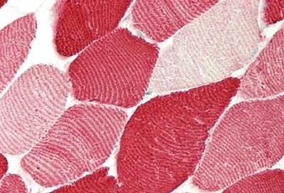
 IHC (Immunohistochemistry)
(AAA290898 staining MYL1 in human heart tissue sections by Immunohistochemistry (IHC-P - paraformaldehyde-fixed, paraffin-embedded sections). Tissue was fixed with formaldehyde and blocked with 3% BSA for 0. 5 hour at room temperature; antigen retrieval was by heat mediation with a citrate buffer (pH6). Samples were incubated with primary antibody (1/25) for 1 hours at 37°C. A undiluted biotinylated goat polyvalent antibody was used as the secondary antibody.)
IHC (Immunohistochemistry)
(AAA290898 staining MYL1 in human heart tissue sections by Immunohistochemistry (IHC-P - paraformaldehyde-fixed, paraffin-embedded sections). Tissue was fixed with formaldehyde and blocked with 3% BSA for 0. 5 hour at room temperature; antigen retrieval was by heat mediation with a citrate buffer (pH6). Samples were incubated with primary antibody (1/25) for 1 hours at 37°C. A undiluted biotinylated goat polyvalent antibody was used as the secondary antibody.)
Rabbit anti-Human, Mouse MYL1 Polyclonal Antibody | anti-MYL1 antibody
MYL1 Antibody (Center)
For long term storage store at -20 degree C in small aliquots to prevent freeze-thaw cycles.
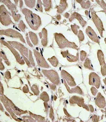
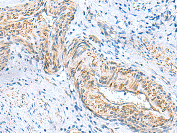
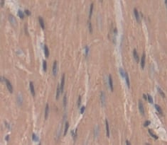
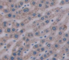
IHC (Immunohistochemistry)
(AAA290898 staining MYL1 in human heart tissue sections by Immunohistochemistry (IHC-P - paraformaldehyde-fixed, paraffin-embedded sections). Tissue was fixed with formaldehyde and blocked with 3% BSA for 0. 5 hour at room temperature; antigen retrieval was by heat mediation with a citrate buffer (pH6). Samples were incubated with primary antibody (1/25) for 1 hours at 37°C. A undiluted biotinylated goat polyvalent antibody was used as the secondary antibody.)
IF (Immunofluorescence)
(Immunofluorescent analysis of 4% paraformaldehyde-fixed, 0. 1% Triton X-100 permeabilized Hela cells labeling MYL1 with AAA290898 at 1/25 dilution, followed by Dylight® 488-conjugated goat anti-Rabbit IgG secondary antibody at 1/200 dilution (green). Immunofluorescence image showing Cytoplasm and weak nucleus staining on Hela cell line. The nuclear counter stain is DAPI (blue).)
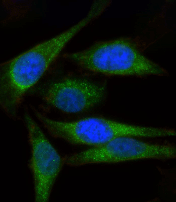
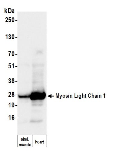
WB (Western Blot)
(All lanes : Anti-MYL1 Antibody (Center) at 1:2000 dilutionLane 1: Human heart lysateLane 2: Human skeletal muslce lysateLane 3: Mouse skeletal muscle lysateLane 4: Rat skeletal muscle lysateLysates/proteins at 20 ug per lane.SecondaryGoat Anti-Rabbit IgG, (H+L), Peroxidase conjugated at 1/10000 dilution.Predicted band size : 21 kDaBlocking/Dilution buffer: 5% NFDM/TBST.)
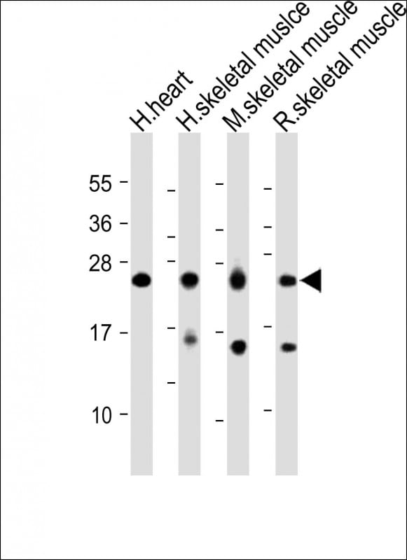
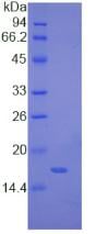
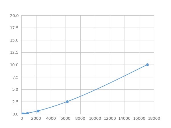
FCM/FACS (Flow Cytometry)
(Overlay histogram showing Hela cells stained with AAA290898 (green line). The cells were fixed with 2% paraformaldehyde (10 min) and then permeabilized with 90% methanol for 10 min. The cells were then icubated in 2% bovine serum albumin to block non-specific protein-protein interactions followed by the antibody (AAA290898, 1:25 dilution) for 60 min at 37 degree C. The secondary antibody used was Goat-Anti-Rabbit IgG, DyLight 488 Conjugated Highly Cross-Adsorbed(OE188374) at 1/200 dilution for 40 min at 37 degree C. Isotype control antibody (blue line) was rabbit IgG1 (1mug/1x10^6 cells) used under the same conditions. Acquisition of >10, 000 events was performed.)
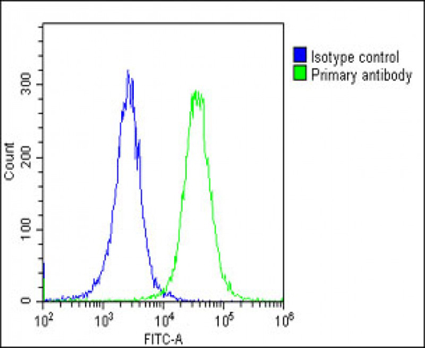
NCBI and Uniprot Product Information
Customer Reviews
Loading reviews...
Share Your Experience
Similar Products


Product Notes
The MYL1 myl1 (Catalog #AAA290898) is an Antibody produced from Rabbit and is intended for research purposes only. The product is available for immediate purchase. The MYL1 Antibody (Center) reacts with Human, Mouse and may cross-react with other species as described in the data sheet. AAA Biotech's MYL1 can be used in a range of immunoassay formats including, but not limited to, ELISA, WB (Western Blot), FCM/FACS (Flow Cytometry), IHC (Immunohistochemistry). Researchers should empirically determine the suitability of the MYL1 myl1 for an application not listed in the data sheet. Researchers commonly develop new applications and it is an integral, important part of the investigative research process. It is sometimes possible for the material contained within the vial of "MYL1, Polyclonal Antibody" to become dispersed throughout the inside of the vial, particularly around the seal of said vial, during shipment and storage. We always suggest centrifuging these vials to consolidate all of the liquid away from the lid and to the bottom of the vial prior to opening. Please be advised that certain products may require dry ice for shipping and that, if this is the case, an additional dry ice fee may also be required.Precautions
All products in the AAA Biotech catalog are strictly for research-use only, and are absolutely not suitable for use in any sort of medical, therapeutic, prophylactic, in-vivo, or diagnostic capacity. By purchasing a product from AAA Biotech, you are explicitly certifying that said products will be properly tested and used in line with industry standard. AAA Biotech and its authorized distribution partners reserve the right to refuse to fulfill any order if we have any indication that a purchaser may be intending to use a product outside of our accepted criteria.Disclaimer
Though we do strive to guarantee the information represented in this datasheet, AAA Biotech cannot be held responsible for any oversights or imprecisions. AAA Biotech reserves the right to adjust any aspect of this datasheet at any time and without notice. It is the responsibility of the customer to inform AAA Biotech of any product performance issues observed or experienced within 30 days of receipt of said product. To see additional details on this or any of our other policies, please see our Terms & Conditions page.Item has been added to Shopping Cart
If you are ready to order, navigate to Shopping Cart and get ready to checkout.