

Rabbit anti-Human NEK2 Polyclonal Antibody | anti-NEK2 antibody
NEK2 Antibody (Center)
IHC~~1:100
WB~~1:1000
WB (Western Blot)
(Western blot analysis of anti-NEK2 Antibody (Center) in HL60 cell line lysates (35ug/lane). NEK2(arrow) was detected using the purified Pab.)
WB (Western Blot)
(Western blot analysis of lysates from 293T, Hela, Jurkat, K562, MCF-7, and mouse NIH/3T3 cell line (from left to right), using NEK2 Antibody (C410). AAA28694 was diluted at 1:1000 at each lane. A goat anti-rabbit IgG H&L(HRP) at 1:5000 dilution was used as the secondary antibody. Lysates at 35ug per lane.)
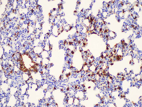
IHC (Immunohistochemistry)
(Immunohistochemical analysis of paraffin-embedded M. brain section using NEK2 Antibody(Center). AAA28694 was diluted at 1:100 dilution. A peroxidase-conjugated goat anti-rabbit IgG at 1:400 dilution was used as the secondary antibody, followed by DAB staining.)
IHC (Immunohistochemistry)
(Immunohistochemical analysis of paraffin-embedded R. brain section using NEK2 Antibody(Center). AAA28694 was diluted at 1:100 dilution. A peroxidase-conjugated goat anti-rabbit IgG at 1:400 dilution was used as the secondary antibody, followed by DAB staining.)
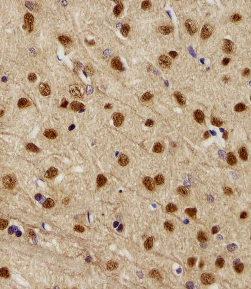
IHC (Immunohistochemistry)
(Immunohistochemical analysis of paraffin-embedded H. brain section using NEK2 Antibody(Center). AAA28694 was diluted at 1:100 dilution. A peroxidase-conjugated goat anti-rabbit IgG at 1:400 dilution was used as the secondary antibody, followed by DAB staining.)
IF (Immunofluorescence)
(Fluorescent image of U251 cells stained with hNEK2-C410. AAA28694 was diluted at 1:25 dilution. An Alexa Fluor 488-conjugated goat anti-rabbit lgG at 1:400 dilution was used as the secondary antibody (green). Cytoplasmic actin was counterstained with Alexa Fluor 555 conjugated with Phalloidin (red).)