

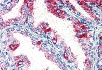
 IHC (Immunohistochemistry)
(AAA322386 at 1/100 staining Mouse testis tissue by IHC-P. The sample was formaldehyde fixed and a heat mediated antigen retrieval step in citrate buffer was performed. The sample was then blocked and incubated with the antibody for 1.5 hours at 22 degree C. An HRP conjugated goat anti-rabbit antibody was used as the secondary.)
IHC (Immunohistochemistry)
(AAA322386 at 1/100 staining Mouse testis tissue by IHC-P. The sample was formaldehyde fixed and a heat mediated antigen retrieval step in citrate buffer was performed. The sample was then blocked and incubated with the antibody for 1.5 hours at 22 degree C. An HRP conjugated goat anti-rabbit antibody was used as the secondary.)
Rabbit PKC zeta Polyclonal Antibody | anti-PRKCZ antibody
PKC zeta Antibody
Phosphate buffered saline, pH 7.4, 150mM NaCl, 0.02% sodium azide and 50% glycerol.
IHC (Immunohistochemistry)
(AAA322386 at 1/100 staining Mouse testis tissue by IHC-P. The sample was formaldehyde fixed and a heat mediated antigen retrieval step in citrate buffer was performed. The sample was then blocked and incubated with the antibody for 1.5 hours at 22 degree C. An HRP conjugated goat anti-rabbit antibody was used as the secondary.)
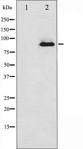
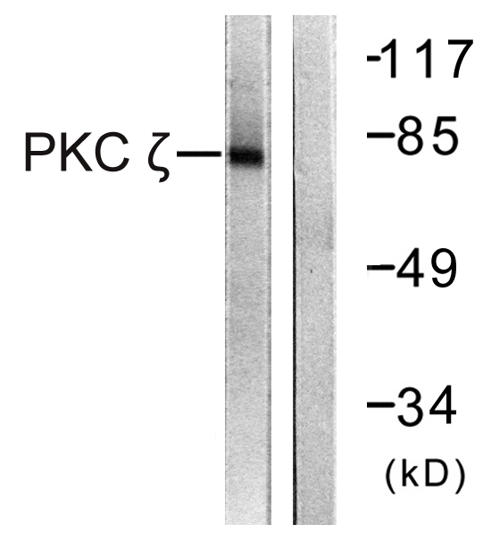
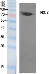
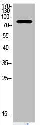
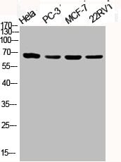
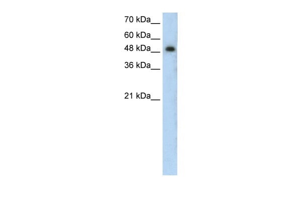
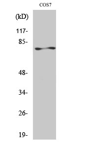
WB (Western Blot)
(Western blot analysis of PKC zeta expression in PMA treated COS7 whole cell lysates, The lane on the left is treated with the antigen-specific peptide.)
WB (Western Blot)
(Western blot analysis of PKC zeta expression in PMA treated COS7 whole cell lysates, The lane on the left is treated with the antigen-specific peptide.)
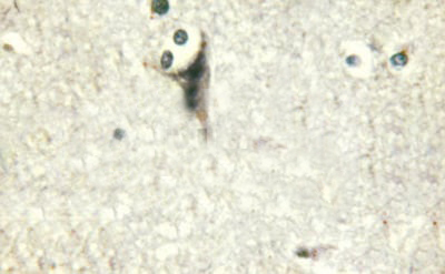
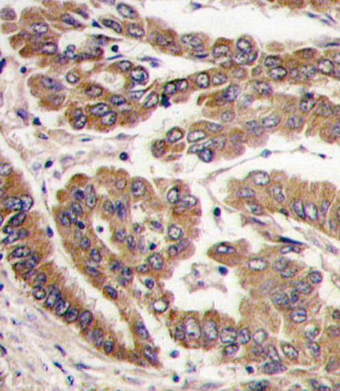
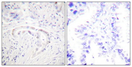
IHC (Immunohistochemisry)
(AAA322386 at 1/100 staining human brain tissues sections by IHC-P. The tissue was formaldehyde fixed and a heat mediated antigen retrieval step in citrate buffer was performed. The tissue was then blocked and incubated with the antibody for 1.5 hours at 64 degree C)
IHC (Immunohistochemisry)
(AAA322386 at 1/100 staining human brain tissues sections by IHC-P. The tissue was formaldehyde fixed and a heat mediated antigen retrieval step in citrate buffer was performed. The tissue was then blocked and incubated with the antibody for 1.5 hours at 64 degree C)
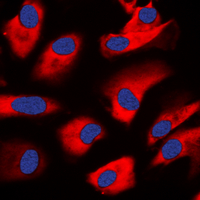
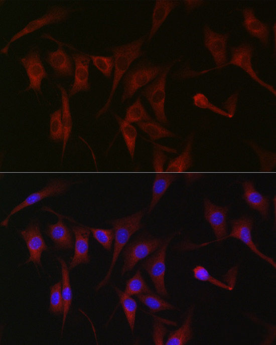
IF (Immunofluorescence)
(AAA322386 staining NIH-3T3 by IF/ICC. The sample were fixed with PFA and permeabilized in 0.1% Triton X-100, then blocked in 10% serum for 45 minutes at 25 degree C. The primary antibody was diluted at 1/200 and incubated with the sample for 1 hour at 37 degree C. An Alexa Fluor 594 conjugated goat anti-rabbit IgG (H+L) Ab, diluted at 1/600, was used as the secondary antibody.)
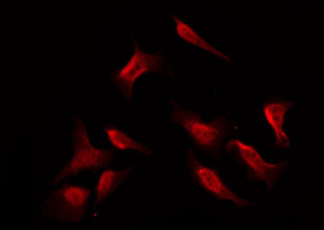
IF (Immunofluorescence)
(AAA322386 staining HeLa cells by IF/ICC. The sample were fixed with PFA and permeabilized in 0.1% Triton X-100, then blocked in 10% serum for 45 minutes at 25 degree C. The primary antibody was diluted at 1/200 and incubated with the sample for 1 hour at 37 degree C. An Alexa Fluor 594 conjugated goat anti-rabbit IgG (H+L) antibody, diluted at 1/600, was used as secondary antibody.)
Function: Calcium-and diacylglycerol-independent serine/threonine-protein kinase that functions in phosphatidylinositol 3-kinase (PI3K) pathway and mitogen-activated protein (MAP) kinase cascade, and is involved in NF-kappa-B activation, mitogenic signaling, cell proliferation, cell polarity, inflammatory response and maintenance of long-term potentiation (LTP). Upon lipopolysaccharide (LPS) treatment in macrophages, or following mitogenic stimuli, functions downstream of PI3K to activate MAP2K1/MEK1-MAPK1/ERK2 signaling cascade independently of RAF1 activation. Required for insulin-dependent activation of AKT3, but may function as an adapter rather than a direct activator. Upon insulin treatment may act as a downstream effector of PI3K and contribute to the activation of translocation of the glucose transporter SLC2A4/GLUT4 and subsequent glucose transport in adipocytes. In EGF-induced cells, binds and activates MAP2K5/MEK5-MAPK7/ERK5 independently of its kinase activity and can activate JUN promoter through MEF2C. Through binding with SQSTM1/p62, functions in interleukin-1 signaling and activation of NF-kappa-B with the specific adapters RIPK1 and TRAF6. Participates in TNF-dependent transactivation of NF-kappa-B by phosphorylating and activating IKBKB kinase, which in turn leads to the degradation of NF-kappa-B inhibitors. In migrating astrocytes, forms a cytoplasmic complex with PARD6A and is recruited by CDC42 to function in the establishment of cell polarity along with the microtubule motor and dynein. In association with FEZ1, stimulates neuronal differentiation in PC12 cells. In the inflammatory response, is required for the T-helper 2 (Th2) differentiation process, including interleukin production, efficient activation of JAK1 and the subsequent phosphorylation and nuclear translocation of STAT6. May be involved in development of allergic airway inflammation (asthma), a process dependent on Th2 immune response. In the NF-kappa-B-mediated inflammatory response, can relieve SETD6-dependent repression of NF-kappa-B target genes by phosphorylating the RELA subunit at 'Ser-311'. Necessary and sufficient for LTP maintenance in hippocampal CA1 pyramidal cells. In vein endothelial cells treated with the oxidant peroxynitrite, phosphorylates STK11 leading to nuclear export of STK11, subsequent inhibition of PI3K/Akt signaling, and increased apoptosis. Phosphorylates VAMP2 in vitro (PubMed:17313651).
Subunit Structure: Forms a ternary complex with SQSTM1 and KCNAB2. Forms another ternary complex with SQSTM1 and GABRR3. Forms a complex with SQSTM1 and MAP2K5 (By similarity). Interacts with PARD6A, PARD6B, PARD6G and SQSTM1. Part of a complex with PARD3, PARD6A or PARD6B or PARD6G and CDC42 or RAC1. Interacts with ADAP1/CENTA1. Forms a ternary complex composed of SQSTM1 and PAWR. Interacts directly with SQSTM1 (Probable). Interacts with IKBKB. Interacts (via the protein kinase domain) with WWC1. Forms a tripartite complex with WWC1 and DDR1, but predominantly in the absence of collagen. Component of the Par polarity complex, composed of at least phosphorylated PRKCZ, PARD3 and TIAM1. Interacts with PDPK1 (via N-terminal region). Interacts with WDFY2 (via WD repeats 1-3) (PubMed:16792529). Interacts with VAMP2 (PubMed:17313651). Forms a complex with WDFY2 and VAMP2 (PubMed:17313651).
Post-translational Modifications: CDH5 is required for its phosphorylation at Thr-410. Phosphorylated by protein kinase PDPK1; phosphorylation is inhibited by the apoptotic C-terminal cleavage product of PKN2. Phosphorylation at Thr-410 by PI3K activates the kinase.
Similarity: The PB1 domain mediate mutually exclusive interactions with SQSTM1 and PARD6B.The C1 domain does not bind the diacylglycerol (DAG). Belongs to the protein kinase superfamily. AGC Ser/Thr protein kinase family. PKC subfamily.
NCBI and Uniprot Product Information
Predicted: 68 kDa
Customer Reviews
Loading reviews...
Share Your Experience
Similar Products


Product Notes
The PRKCZ prkcz (Catalog #AAA322386) is an Antibody produced from Rabbit and is intended for research purposes only. The product is available for immediate purchase. The PKC zeta Antibody reacts with Human, Mouse, Rat and may cross-react with other species as described in the data sheet. AAA Biotech's PKC zeta can be used in a range of immunoassay formats including, but not limited to, ELISA, ICC (Immunocytochemistry), IF (Immunofluorescence), IHC (Immunohistochemistry), WB (Western Blot). Researchers should empirically determine the suitability of the PRKCZ prkcz for an application not listed in the data sheet. Researchers commonly develop new applications and it is an integral, important part of the investigative research process. It is sometimes possible for the material contained within the vial of "PKC zeta, Polyclonal Antibody" to become dispersed throughout the inside of the vial, particularly around the seal of said vial, during shipment and storage. We always suggest centrifuging these vials to consolidate all of the liquid away from the lid and to the bottom of the vial prior to opening. Please be advised that certain products may require dry ice for shipping and that, if this is the case, an additional dry ice fee may also be required.Precautions
All products in the AAA Biotech catalog are strictly for research-use only, and are absolutely not suitable for use in any sort of medical, therapeutic, prophylactic, in-vivo, or diagnostic capacity. By purchasing a product from AAA Biotech, you are explicitly certifying that said products will be properly tested and used in line with industry standard. AAA Biotech and its authorized distribution partners reserve the right to refuse to fulfill any order if we have any indication that a purchaser may be intending to use a product outside of our accepted criteria.Disclaimer
Though we do strive to guarantee the information represented in this datasheet, AAA Biotech cannot be held responsible for any oversights or imprecisions. AAA Biotech reserves the right to adjust any aspect of this datasheet at any time and without notice. It is the responsibility of the customer to inform AAA Biotech of any product performance issues observed or experienced within 30 days of receipt of said product. To see additional details on this or any of our other policies, please see our Terms & Conditions page.Item has been added to Shopping Cart
If you are ready to order, navigate to Shopping Cart and get ready to checkout.