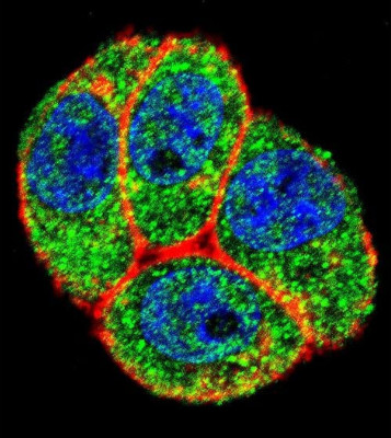
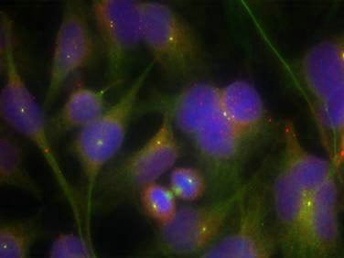
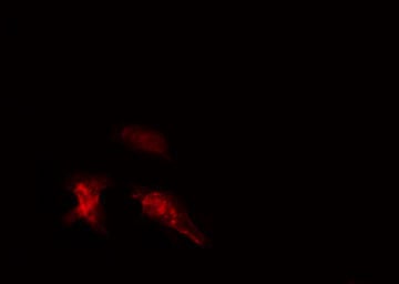
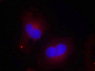
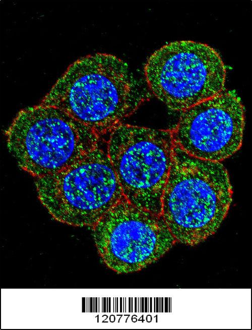
 IF (Immunofluorescence)
(Confocal immunofluorescent analysis of PTK2 Antibody (Center) with Hela cell followed by Alexa Fluor 488-conjugated goat anti-rabbit lgG (green). Actin filaments have been labeled with Alexa Fluor 555 phalloidin (red).DAPI was used to stain the cell nuclear (blue).)
IF (Immunofluorescence)
(Confocal immunofluorescent analysis of PTK2 Antibody (Center) with Hela cell followed by Alexa Fluor 488-conjugated goat anti-rabbit lgG (green). Actin filaments have been labeled with Alexa Fluor 555 phalloidin (red).DAPI was used to stain the cell nuclear (blue).)
Rabbit PTK2 Polyclonal Antibody | anti-PTK2 antibody
PTK2 Antibody (Center)
Predicted: Chicken, Mouse, Rat
Predicted: Chicken, Mouse, Rat
IF (Immunofluorescence)
(Confocal immunofluorescent analysis of PTK2 Antibody (Center) with Hela cell followed by Alexa Fluor 488-conjugated goat anti-rabbit lgG (green). Actin filaments have been labeled with Alexa Fluor 555 phalloidin (red).DAPI was used to stain the cell nuclear (blue).)
IF (Immunofluorescence)
(Confocal immunofluorescent analysis of PTK2 Antibody (Center) with Hela cell followed by Alexa Fluor 488-conjugated goat anti-rabbit lgG (green). Actin filaments have been labeled with Alexa Fluor 555 phalloidin (red).DAPI was used to stain the cell nuclear (blue).)
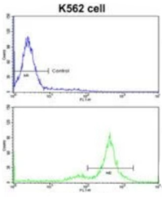
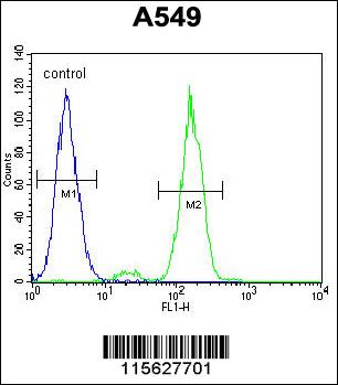
FCM/FACS (Flow Cytometry)
(Flow cytometric analysis of K562 cells using PTK2 Antibody (Center)(bottom histogram) compared to a negative control cell (top histogram)FITC-conjugated goat-anti-rabbit secondary antibodies were used for the analysis.)
FCM/FACS (Flow Cytometry)
(Flow cytometric analysis of K562 cells using PTK2 Antibody (Center)(bottom histogram) compared to a negative control cell (top histogram)FITC-conjugated goat-anti-rabbit secondary antibodies were used for the analysis.)
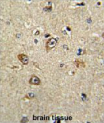
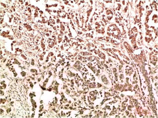
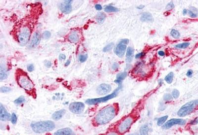
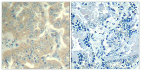
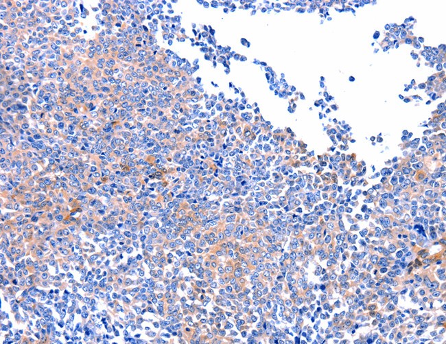
IHC (Immunohistochemisry)
(Formalin-fixed and paraffin-embedded human brain tissue reacted with PTK2 Antibody (Center), which was peroxidase-conjugated to the secondary antibody, followed by DAB staining. This data demonstrates the use of this antibody for immunohistochemistry; clinical relevance has not been evaluated.)
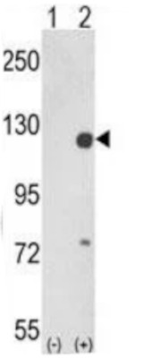
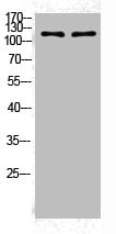
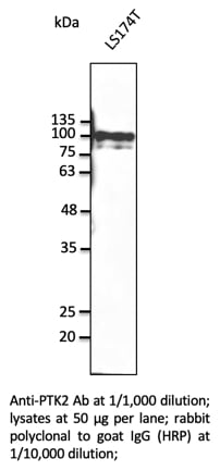
WB (Western Blot)
(Western blot analysis of PTK2 (arrow) using rabbit polyclonal PTK2 Antibody (Center). 293 cell lysates (2 ug/lane) either nontransfected (Lane 1) or transiently transfected with the PTK2 gene (Lane 2).)
WB (Western Blot)
(Western blot analysis of PTK2 (arrow) using rabbit polyclonal PTK2 Antibody (Center). 293 cell lysates (2 ug/lane) either nontransfected (Lane 1) or transiently transfected with the PTK2 gene (Lane 2).)
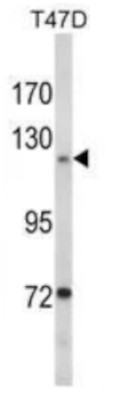
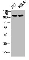
WB (Western Blot)
(Western blot analysis of PTK2 Antibody (Center) in T47D cell line lysates (35ug/lane). PTK2 (arrow) was detected using the purified Pab.)
WB (Western Blot)
(Western blot analysis of PTK2 Antibody (Center) in T47D cell line lysates (35ug/lane). PTK2 (arrow) was detected using the purified Pab.)
Polte,T.R. et.al., Proc. Natl. Acad. Sci. U.S.A. 92 (23), 10678-10682 (1995)
Gervais,F.G., et.al., J. Biol. Chem. 273 (27), 17102-17108 (1998)
NCBI and Uniprot Product Information
Customer Reviews
Loading reviews...
Share Your Experience
Similar Products


Product Notes
The PTK2 ptk2 (Catalog #AAA288401) is an Antibody produced from Rabbit and is intended for research purposes only. The product is available for immediate purchase. The immunogen sequence is 396-423. The PTK2 Antibody (Center) reacts with Human Predicted: Chicken, Mouse, Rat and may cross-react with other species as described in the data sheet. AAA Biotech's PTK2 can be used in a range of immunoassay formats including, but not limited to, IF (Immunofluorescence), FCM/FACS (Flow Cytometry), IHC (Immunohistochemistry), ELISA, WB (Western Blot). Researchers should empirically determine the suitability of the PTK2 ptk2 for an application not listed in the data sheet. Researchers commonly develop new applications and it is an integral, important part of the investigative research process. It is sometimes possible for the material contained within the vial of "PTK2, Polyclonal Antibody" to become dispersed throughout the inside of the vial, particularly around the seal of said vial, during shipment and storage. We always suggest centrifuging these vials to consolidate all of the liquid away from the lid and to the bottom of the vial prior to opening. Please be advised that certain products may require dry ice for shipping and that, if this is the case, an additional dry ice fee may also be required.Precautions
All products in the AAA Biotech catalog are strictly for research-use only, and are absolutely not suitable for use in any sort of medical, therapeutic, prophylactic, in-vivo, or diagnostic capacity. By purchasing a product from AAA Biotech, you are explicitly certifying that said products will be properly tested and used in line with industry standard. AAA Biotech and its authorized distribution partners reserve the right to refuse to fulfill any order if we have any indication that a purchaser may be intending to use a product outside of our accepted criteria.Disclaimer
Though we do strive to guarantee the information represented in this datasheet, AAA Biotech cannot be held responsible for any oversights or imprecisions. AAA Biotech reserves the right to adjust any aspect of this datasheet at any time and without notice. It is the responsibility of the customer to inform AAA Biotech of any product performance issues observed or experienced within 30 days of receipt of said product. To see additional details on this or any of our other policies, please see our Terms & Conditions page.Item has been added to Shopping Cart
If you are ready to order, navigate to Shopping Cart and get ready to checkout.